Im 1st year mbbs student at Hngu university [ Bhagyoday medical college Kadi ] in exam imp questions list i saw one question is 8 mark question is HMP shunt & it's significance, significance of nadph give a answer of this question easy to understand English language,when I read this question answer I understand easily, give answer this type when I read this question answer first time so much answer I understand easily in first time and when i write this in exam my sir will give me full out of full marks and easy to read and understand,answer give as 1 st year mbbs [ Biochemistry ]and easy to understand and write Thank you

Reading File
Finding Sources
Finding Sources
Finding Sources
Finding Sources
Reading File
Reading File
Reading File
Reading File
Reading File
Reading File
Reading File
Reading File
Reading File
Loading Image
I now have everything needed. Let me write the complete, student-friendly 8-mark exam answer.

HMP Shunt (Hexose Monophosphate Pathway) - Its Significance & Significance of NADPH

[8 Mark Answer - 1st Year MBBS Biochemistry]

INTRODUCTION / DEFINITION

The HMP Shunt (also called the Hexose Monophosphate Shunt, Pentose Phosphate Pathway, or Phosphogluconate Pathway) is an alternative pathway for oxidation of glucose that runs parallel to glycolysis.
  • It starts with Glucose-6-phosphate (just like glycolysis)
  • But instead of producing ATP, it produces NADPH and Ribose-5-phosphate
  • It does NOT produce any ATP
  • All reactions take place in the cytosol
Simple way to remember: HMP Shunt = a "side road" that branches off from glycolysis, but leads to NADPH and ribose instead of energy.

TWO PHASES OF HMP SHUNT

PHASE 1 - Oxidative Phase (Irreversible)

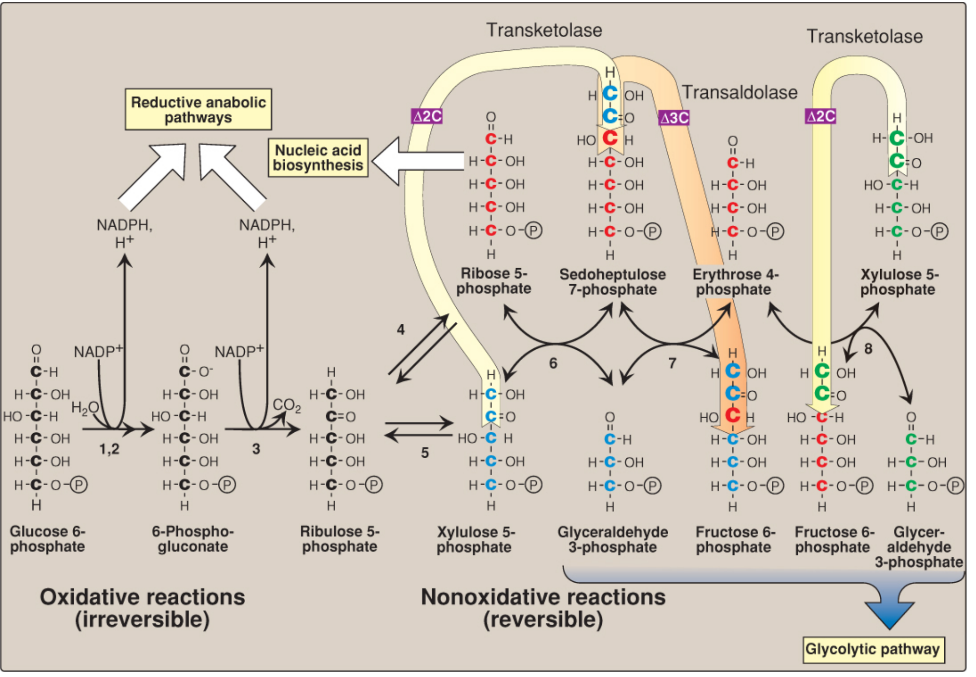
This is the main productive phase. It converts Glucose-6-phosphate into Ribulose-5-phosphate, releasing CO2 and producing 2 NADPH per glucose-6-phosphate.
Step-by-step reactions:
StepSubstrateEnzymeProductCoenzyme
1Glucose-6-phosphateGlucose-6-phosphate dehydrogenase (G6PD)6-PhosphogluconolactoneNADP+ → NADPH
26-PhosphogluconolactoneGluconolactone hydrolase6-Phosphogluconate-
36-Phosphogluconate6-Phosphogluconate dehydrogenaseRibulose-5-phosphate + CO2NADP+ → NADPH
Key points of Phase 1:
  • G6PD catalyzes the first, rate-limiting (committed) step
  • 2 NADPH are produced per molecule of Glucose-6-phosphate
  • 1 CO2 is released
  • NADPH itself inhibits G6PD (feedback inhibition) - so when NADPH is needed, the pathway speeds up

PHASE 2 - Non-Oxidative Phase (Reversible)

This phase interconverts sugars of different carbon lengths (3C, 4C, 5C, 6C, 7C sugars).
Key enzymes:
  • Ribose-5-phosphate isomerase - converts Ribulose-5-phosphate → Ribose-5-phosphate (needed for DNA/RNA)
  • Transketolase (requires TPP / Vitamin B1 as cofactor) - transfers 2-carbon units between sugars
  • Transaldolase - transfers 3-carbon units between sugars
Final products of non-oxidative phase:
  • Fructose-6-phosphate and Glyceraldehyde-3-phosphate (both enter glycolysis)
This phase is reversible - it can also work backwards to produce Ribose-5-phosphate from glycolytic intermediates even without the oxidative phase (important in muscles that have low G6PD activity).

PATHWAY DIAGRAM

HMP Shunt / Pentose Phosphate Pathway diagram showing oxidative (irreversible) and non-oxidative (reversible) phases
Lippincott's Illustrated Biochemistry 8e - Pentose Phosphate Pathway

SIGNIFICANCE OF HMP SHUNT

1. Production of NADPH (Most Important)

  • HMP shunt is the main source of NADPH in the body
  • NADPH is essential for many biosynthetic reactions (detailed below)

2. Production of Ribose-5-phosphate

  • Ribose-5-phosphate is needed to make nucleotides (ATP, GTP) and nucleic acids (DNA and RNA)
  • All rapidly dividing cells need this (bone marrow, skin, intestine)

3. Connects to Glycolysis

  • The non-oxidative phase produces Fructose-6-phosphate and Glyceraldehyde-3-phosphate, which enter glycolysis directly
  • So this pathway acts as a bridge between glucose oxidation and glycolysis

4. Active Tissues

The pathway is very active in:
  • Liver - fatty acid and cholesterol synthesis
  • Adipose tissue - fat synthesis
  • Adrenal cortex, testes, ovaries, placenta - steroid hormone synthesis
  • Red blood cells (RBCs) - protection against oxidative damage
  • Lactating mammary gland - fat synthesis for milk
  • Low activity in: skeletal muscle

SIGNIFICANCE OF NADPH

NADPH (Nicotinamide Adenine Dinucleotide Phosphate - Reduced) is the most important product of HMP shunt. It is used as a reducing agent in many vital reactions.

1. Fatty Acid Synthesis (Lipogenesis)

  • NADPH is essential for synthesis of fatty acids
  • Used by fatty acid synthase enzyme
  • Active in liver, adipose tissue, mammary gland

2. Cholesterol and Steroid Hormone Synthesis

  • NADPH is required for synthesis of cholesterol
  • Cholesterol is converted to steroid hormones (cortisol, aldosterone, estrogen, testosterone) - all need NADPH
  • Active in: adrenal cortex, gonads, placenta

3. Protection of Red Blood Cells (RBCs) from Hemolysis

  • This is the most clinically important function of NADPH
  • H2O2 (hydrogen peroxide) is a toxic oxidant produced in RBCs
  • NADPH → keeps Glutathione in reduced form (GSH) via Glutathione reductase
  • Reduced glutathione (GSH) then destroys H2O2 via Glutathione peroxidase
  • This protects RBC membrane from oxidative damage and prevents hemolysis
Clinical link: G6PD Deficiency
  • In G6PD deficiency, NADPH cannot be made
  • GSH cannot be regenerated
  • H2O2 accumulates → RBC membrane is damaged → hemolytic anemia
  • Triggered by: primaquine (antimalarial drug), fava beans, infections
  • X-linked recessive inheritance

4. Detoxification of Drugs and Xenobiotics (Drug Metabolism)

  • In liver, NADPH is used by Cytochrome P450 (CYP) enzymes (mixed function oxidases)
  • These enzymes hydroxylate drugs, toxins, and pollutants to make them water-soluble for excretion
  • Reaction: Drug + NADPH + O2 → Drug-OH + NADP+ + H2O

5. Killing of Bacteria by White Blood Cells (Respiratory Burst)

  • Neutrophils and macrophages use NADPH oxidase enzyme
  • NADPH oxidase converts O2 into superoxide (O2-) which kills bacteria
  • This is called the "respiratory burst" or oxidative burst
  • Important defense mechanism against bacterial infections
Clinical link: Chronic Granulomatous Disease (CGD)
  • NADPH oxidase is defective
  • Neutrophils cannot kill bacteria properly
  • Repeated life-threatening infections

6. Synthesis of Nitric Oxide (NO)

  • NO synthase uses NADPH to produce Nitric Oxide (NO) from arginine
  • NO causes vasodilation (widening of blood vessels)
  • Important in blood pressure regulation

7. Regeneration of Vitamin C and Vitamin E (Antioxidants)

  • NADPH helps regenerate these antioxidant vitamins in their active reduced forms

REGULATION OF HMP SHUNT

FactorEffect on HMP Shunt
High NADPHInhibits G6PD (feedback inhibition) - slows pathway
Low NADPH / High NADP+Activates G6PD - speeds pathway up
InsulinUpregulates G6PD gene expression - increases activity

SUMMARY TABLE

FeatureHMP Shunt
Starting materialGlucose-6-phosphate
Main productsNADPH + Ribose-5-phosphate
ATP producedNone
CO2 releasedYes (1 per G6P)
LocationCytosol
Rate-limiting enzymeGlucose-6-phosphate dehydrogenase (G6PD)
Key coenzyme usedNADP+ (not NAD+)
Most active tissuesLiver, adipose, adrenal cortex, RBCs, gonads

EASY MEMORY TIPS

  • "HMP shunt = NADPH factory" - remember it makes NADPH, not ATP
  • "G6PD deficiency = broken NADPH factory = fragile RBCs"
  • NADPH uses: "Fat Steroid Glutathione Detox Burst" = Fatty acids, Steroids, Glutathione (protect RBCs), Detoxification, Burst (respiratory burst in WBCs)
  • Transketolase needs Vitamin B1 (thiamine) - so B1 deficiency = transketolase test used to diagnose it

Sources: Lippincott's Illustrated Reviews: Biochemistry, 8th ed., Chapter 13 | Harper's Illustrated Biochemistry, 32nd ed., Chapter 20

Im 1st year mbbs student at Hngu university [ Bhagyoday medical college Kadi ] in exam imp questions list i saw one question is 5 mark question is fluid mosaic modal give a answer of this question easy to understand English language,when I read this question answer I understand easily, give answer this type when I read this question answer first time so much answer I understand easily in first time and when i write this in exam my sir will give me full out of full marks and easy to read and understand,answer give as 1 st year mbbs [ Biochemistry ]and easy to understand and write Thank you

Reading File
Finding Sources
Finding Sources
Reading File
Reading File
Reading File
Reading File
Reading File
Loading Image
Loading Image
I now have everything needed - both diagrams and complete textbook content. Here is the full, student-friendly 5-mark exam answer:

Fluid Mosaic Model of Cell Membrane

[5 Mark Answer - 1st Year MBBS Biochemistry]

INTRODUCTION

The Fluid Mosaic Model is the most accepted model that explains the structure of the cell (plasma) membrane.
It was proposed by Singer and Nicolson in 1972.
What does the name mean?
  • "Fluid" - the lipid bilayer is not rigid/solid; the lipid molecules and some proteins can move freely sideways (like people moving in a crowd)
  • "Mosaic" - the proteins are scattered throughout the lipid bilayer like tiles in a mosaic pattern

THE DIAGRAM

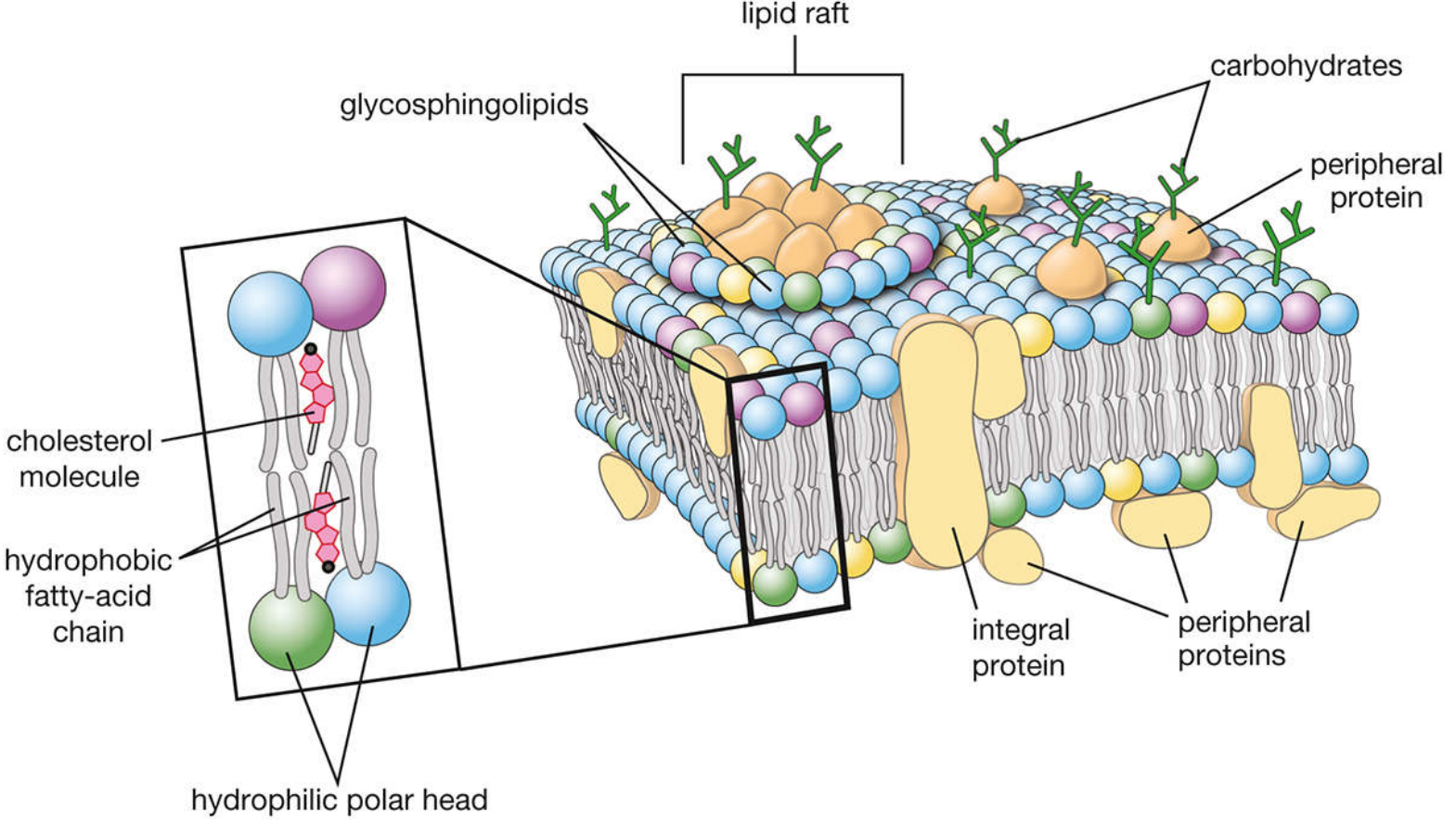
Fluid Mosaic Model showing lipid bilayer with integral proteins, peripheral proteins, carbohydrate chains, and lipid rafts
Modified Fluid Mosaic Model of Cell Membrane (Histology Text & Atlas)
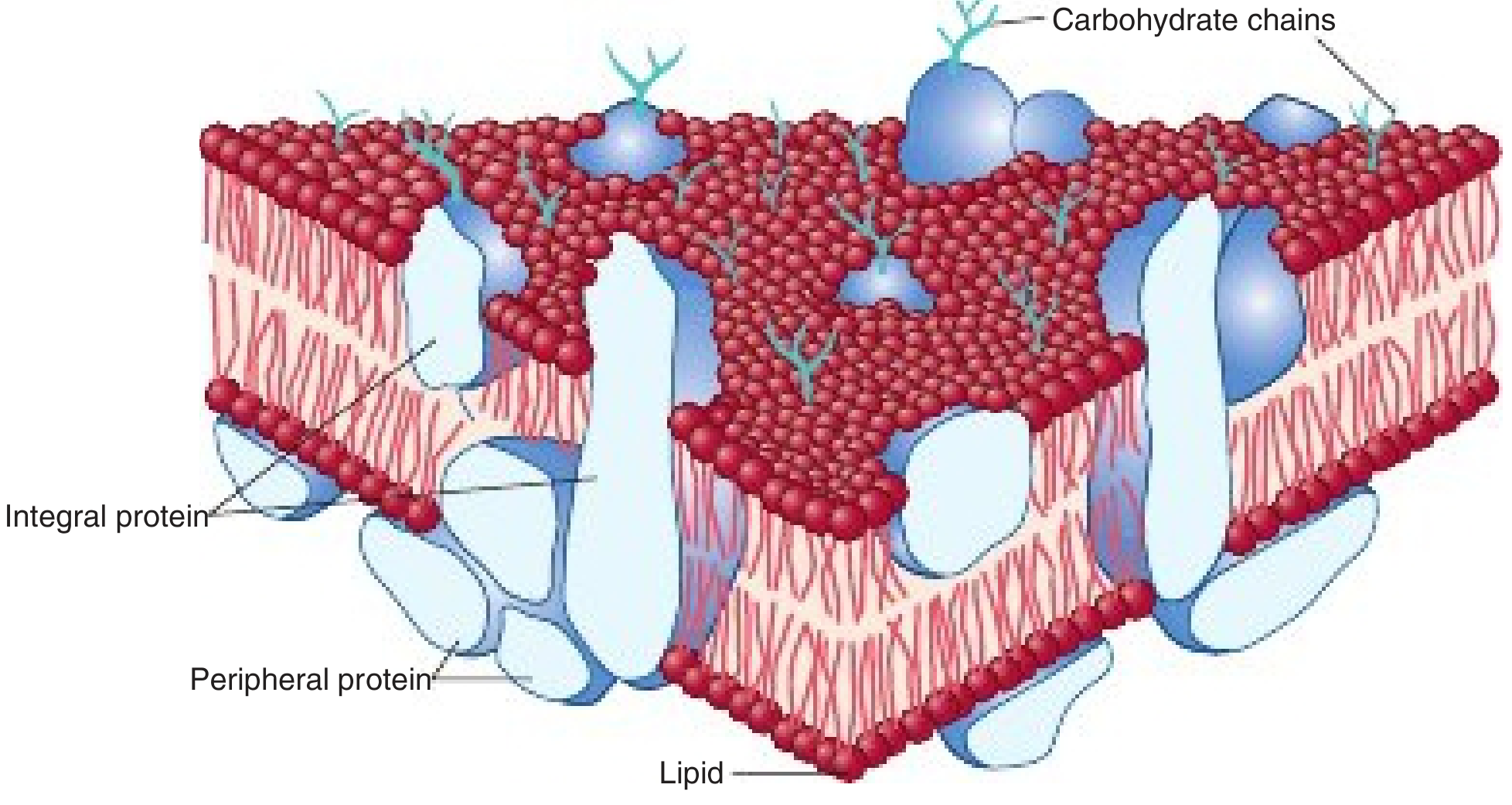
Fluid Mosaic Model - Harper's Biochemistry showing integral and peripheral proteins in lipid bilayer
Fluid Mosaic Model - Harper's Illustrated Biochemistry 32e

COMPONENTS OF THE CELL MEMBRANE

The cell membrane is made of 3 main components:

1. LIPID BILAYER (The Main Framework)

  • The membrane is made of two layers of phospholipid molecules arranged back-to-back
  • Each phospholipid molecule has:
    • A hydrophilic "polar head" (loves water) - faces OUTSIDE (towards ECF and ICF)
    • A hydrophobic "fatty acid tail" (hates water) - faces INSIDE (towards each other)
  • This is called an amphipathic molecule (has both water-loving and water-fearing parts)
  • Cholesterol molecules are also present in between the phospholipids - they help control fluidity of the membrane
Easy analogy: Think of the lipid bilayer like a sandwich - the bread (polar heads) is on the outside touching water, and the filling (fatty acid tails) is on the inside, hidden away from water.

2. PROTEINS (The Mosaic Pattern)

Proteins are embedded in or attached to the lipid bilayer. There are two types:
FeatureIntegral ProteinsPeripheral Proteins
LocationEmbedded deep inside or span the entire bilayer (transmembrane)Loosely attached to the surface (inner or outer) of the membrane
Bond typeHeld by hydrophobic interactions - hard to removeHeld by weak ionic/electrostatic bonds - easier to remove
RemovalNeed detergents to removeRemoved by mild salt solutions
ExamplesIon channels, transport proteins, receptors, Na+-K+ ATPaseSpectrin (in RBCs), G-proteins
Transmembrane proteins span the membrane completely from outside to inside. They are in contact with both extracellular fluid (ECF) and intracellular fluid (ICF).

3. CARBOHYDRATES (The Identity Tags)

  • Short carbohydrate chains (oligosaccharides) are attached to proteins and lipids on the outer surface only
  • When attached to proteins = Glycoproteins
  • When attached to lipids = Glycolipids
  • Together, they form the Glycocalyx (cell coat) on the outer surface
  • Functions of carbohydrates: cell-to-cell recognition, cell adhesion, acting as receptors

KEY PROPERTIES OF THE FLUID MOSAIC MODEL

1. Fluidity

  • The lipid molecules can move laterally (sideways within the same layer) - this is fast
  • Movement across layers ("flip-flop" / transverse movement) is extremely slow
  • Cholesterol regulates fluidity:
    • At high temperature: cholesterol reduces fluidity (prevents too much movement)
    • At low temperature: cholesterol prevents rigidity (prevents freezing/solidification)

2. Asymmetry (Inside is different from Outside)

  • The outer leaflet has: Phosphatidylcholine, Sphingomyelin, Glycolipids, Glycoproteins
  • The inner leaflet has: Phosphatidylserine, Phosphatidylethanolamine
  • Carbohydrates are only on the outer surface - never on the inner surface

3. Selective Permeability

  • Small, nonpolar molecules (O2, CO2, fatty acids) cross freely
  • Polar molecules and ions need protein channels or transporters to cross

LIPID RAFTS (Modern Addition to the Model)

  • Lipid rafts are small, special regions in the membrane that are rich in cholesterol and glycosphingolipids
  • They are more ordered/rigid than the surrounding membrane
  • They act as "platforms" for receptor signaling and protein sorting
  • (This is why it is now called the "Modified Fluid Mosaic Model")

FUNCTIONS OF CELL MEMBRANE (Based on this Model)

FunctionHow
Selective barrierLipid bilayer blocks most polar molecules
TransportIntegral proteins act as channels/pumps
Cell recognitionGlycocalyx (glycoproteins and glycolipids)
Receptor functionIntegral proteins bind hormones, drugs, signals
Cell signalingReceptors + G-proteins on membrane
Structural supportPeripheral proteins link to cytoskeleton

EASY SUMMARY TO REMEMBER

"A fluid, dynamic, lipid bilayer with floating proteins - like icebergs floating in a lipid sea"
  • FLUID = lipids and proteins can move laterally
  • MOSAIC = proteins scattered like mosaic tiles in the bilayer
  • ASYMMETRIC = inside and outside surfaces are different
  • SELECTIVE = only certain things can cross
  • Proposed by Singer & Nicolson, 1972

QUICK RECALL TABLE

FeatureDetail
Proposed bySinger and Nicolson (1972)
Main lipidPhospholipids (bilayer)
Other lipidCholesterol (controls fluidity)
Protein typesIntegral (transmembrane) and Peripheral
Carbohydrates locationOnly outer surface
Fluidity controlCholesterol
Special regionsLipid rafts
PropertyAmphipathic (both hydrophilic and hydrophobic)

Sources: Harper's Illustrated Biochemistry, 32nd ed., Chapter 40 | Costanzo Physiology, 7th ed. | Histology: A Text and Atlas, 7th ed.
This is a shared conversation. Sign in to Orris to start your own chat.