
Everything about monochorionic and dichochorionic twins
monochorionic dichorionic twins placenta chorionicity diagram
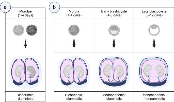
This medical illustration depicts the embryological development and placental classification of dizygotic (Panel a) and monozygotic (Panel b) twin pregnancies. Panel a shows dizygotic twins arising from two separate morulae, resulting in a dichorionic-diamniotic (DCDA) pregnancy with two distinct placentas, chorions, and amniotic sacs. Panel b illustrates the three types of monozygotic twinning based on the timing of zygote cleavage. Splitting at the morula stage (days 1-4) leads to a DCDA pregnancy. Splitting at the early blastocyst stage (days 4-8) results in a monochorionic-diamniotic (MCDA) pregnancy, characterized by a single shared chorion and placenta but two separate amniotic sacs divided by a thin membrane. Splitting at the late blastocyst stage (days 8-12) results in a monochorionic-monoamniotic (MCMA) pregnancy, where both fetuses share a single chorion, placenta, and amniotic sac without a dividing membrane. The diagram highlights the relationship between the timing of embryological division and the resulting chorionicity and amnionicity, which are critical for obstetric management and risk assessment.
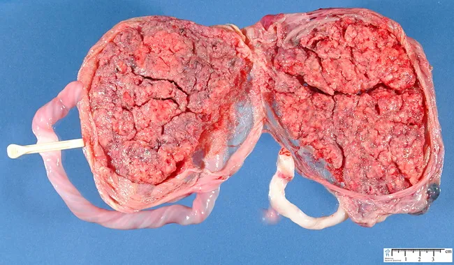
This is a gross pathology photograph of a twin placental specimen illustrating dichorionic diamniotic placentation. The specimen shows two distinct placental masses with rough, lobulated surfaces, separated by a midline tissue interface and paired cord-like structures consistent with separate umbilical cords. The surface texture reveals vascular cores and exposed parenchyma; no obvious infarcts or focal gross lesions are evident in the captured view. The coloration is pink–red with vascular patency visible throughout. A measurement scale is present at the lower margin for size reference. This configuration is compatible with dizygotic twins or monozygotic twins that split early enough to establish two placentas; however, placental dichorionic diamniotic architecture alone cannot reliably determine zygosity. The image provides a clear demonstration of placental architecture, placental margins, and inter-placental tissue that pathologists assess when evaluating twin pregnancies. Clinically, this specimen is relevant to obstetricians and perinatal pathologists for educational purposes, placental pathology review, and maternal-fetal medicine counseling. It supports teaching on chorionicity, amnionicity, and the gross criteria used to infer twin type in placental specimens. This image is suitable for digital teaching modules, literature reviews, and clinical case discussions, helping learners recognize twin placenta morphology, chorionicity criteria, and the limitations of gross assessment today.
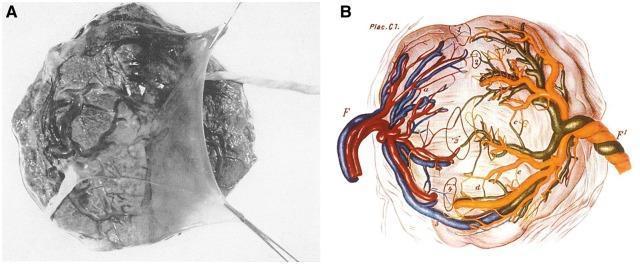
This figure presents the vascular anatomy of a monochorionic twin placenta through a clinical photograph (A) and a detailed anatomical diagram (B). Panel A shows the gross morphology of a single placental disc featuring a dividing amnion tissue membrane and the insertion of two distinct umbilical cords. The chorionic plate surface exhibits a complex network of superficial fetal vessels. Panel B provides a diagrammatic representation of the shared placental circulation, illustrating vascular anastomoses between the two fetal systems. The diagram labels umbilical cords as 'F' and 'F1', representing each twin, and highlights five numbered sites (1-5) of vascular interconnection. Arterial and venous vessels are color-coded (red, blue, and orange) to demonstrate the branching patterns and anastomotic pathways. This educational visual explains the anatomical basis for shared circulation in monozygotic twins, which is clinically significant for understanding conditions such as twin-twin transfusion syndrome (TTTS) and in utero cellular chimerism.
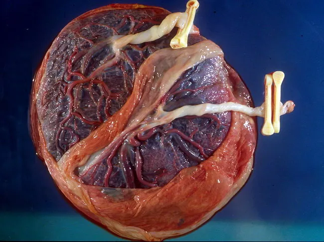
Gross placental specimen from a dichorionic diamniotic twin gestation, presented as a single cross-section showing two distinct placental discs with a zone of partial chorionic plate fusion. The placenta appears rounded and well vascularized, with exposed fetal vasculature and a visible maternal surface, illustrating the vascularized intervillous space and decidual tissue. Each twin’s chorion and amnion are typically separate in DCDA placentas, but this specimen demonstrates greater fusion at the placental margins, creating a continuous placental surface with intermingled villous structures in the fused region. The dual vascular pedicles are evident, and the umbilical cord insertions may be unilateral or near the fusion seam, reflecting the twin gestation pattern. This configuration highlights the complexity of placental contiguity in DCDA pregnancies and raises considerations about potential intertwin vascular anastomoses within the fused area. Notably, the appearance preserves distinguishable cotyledons corresponding to each twin on the maternal surface, while the fused zone represents a morphological variant rather than a complete monochorionic designation. Clinically, such specimens aid in correlating sonographic chorionicity with postnatal placental architecture, inform risk assessment for placental insufficiency or twin-to-twin transfusion in atypical DCDA cases, and provide educational insight into placental development and twin placentation. Aid for education and research.
"monochorionic twins" complications management
"monochorionic twins" AND "twin-twin transfusion"
Monochorionic and Dichorionic Twins: A Complete Overview
1. Definitions and Classification
| Type | Chorions | Amnions | Abbreviation |
|---|---|---|---|
| Diamniotic-Dichorionic | 2 | 2 | DCDA |
| Diamniotic-Monochorionic | 1 | 2 | MCDA |
| Monoamniotic-Monochorionic | 1 | 1 | MCMA |
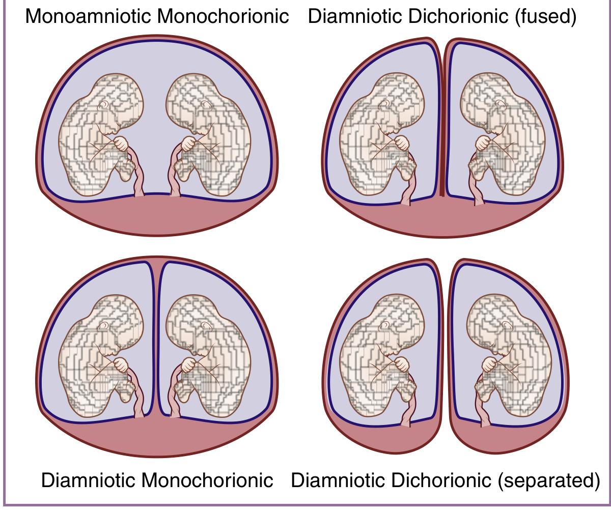
2. Embryological Origin: Timing of Division
| Timing of split | Result |
|---|---|
| Days 1-4 (morula stage) | DCDA (each twin gets its own chorion) |
| Days 4-8 (early blastocyst) | MCDA (shared chorion, separate amnions) |
| Days 8-12 (late blastocyst) | MCMA (shared chorion AND amnion) |
| After day 13 | Conjoined twins |
3. Dichorionic Placentation
Structure
- The intertwin membrane is thick and opaque - composed of two amnion layers separated by fused chorion (4 layers total)
- The twin peak (lambda) sign on ultrasound: a wedge of placental tissue projecting into the base of the intertwin membrane - this is the hallmark of dichorionic placentation in the first trimester
- In virtually all cases, fused DCDA placentas have separate chorionic vascular beds - there are almost no vascular anastomoses between twins
Zygosity
4. Monochorionic Placentation
Structure
- The intertwin membrane (in MCDA) is thin and semitranslucent - composed of only two amnion layers, without interposed chorion (2 layers)
- In MCMA twins, there is no dividing membrane
Vascular Anastomoses
"In contrast to fused dichorionic placentas, almost all monochorionic placentas (>95%) exhibit intertwin vascular anastomoses crossing the intertwin membrane." - Creasy & Resnik's MFM
- Artery-to-Artery (AA) - superficial, bidirectional; compensates for flow imbalances
- Vein-to-Vein (VV) - superficial, bidirectional
- Artery-to-Vein (AV) - deep, at villous capillary level; obligatorily unidirectional; the most clinically important type, underlying TTTS
5. Sonographic Determination of Chorionicity
| Sign | Chorionicity | Timing |
|---|---|---|
| Lambda (twin peak) sign | Dichorionic | T1 > T2 |
| T-sign (thin membrane meets chorionic plate at right angle) | Monochorionic | T1 > T2 |
| Number of placental masses | DC = 2 (or 1 fused); MC = 1 | T1/T2 |
| Fetal sex discordance | Confirms dichorionic (dizygotic) | Any |
| Intertwin membrane thickness | DC = thick (>2mm); MC = thin | T2 |
6. Epidemiology
- Twin pregnancies account for ~3% of all live births; ~30% are monochorionic
- Among spontaneous monozygotic twins: ~70% are MCDA, ~28% are DCDA, ~1-2% are MCMA
- Overall perinatal mortality is approximately 12% in monochorionic twins compared to 2-5% in dichorionic twins, and even higher in MCMA twins - Creasy & Resnik's MFM
7. Complications of Monochorionic Twins (MC-Specific)
A. Twin-to-Twin Transfusion Syndrome (TTTS)
- Occurs in 10-15% of MCDA twins
- Caused by net unidirectional AV anastomoses with insufficient AA compensation - blood shunts from one twin (donor) to the other (recipient)
- Donor twin: hypovolemia, oligohydramnios, growth restriction, "stuck twin"
- Recipient twin: hypervolemia, polyhydramnios, cardiomegaly, hydrops
| Stage | Features |
|---|---|
| I | Oligohydramnios/polyhydramnios discordance; bladder visible in donor |
| II | Absent bladder in donor |
| III | Critically abnormal Dopplers (absent/reversed end-diastolic flow in umbilical artery, ductus venosus, or pulsatile umbilical vein) |
| IV | Hydrops in either twin |
| V | Death of one or both twins |
B. Selective Fetal Growth Restriction (sFGR)
- Affects ~10-15% of MC twins
- Caused by unequal placental sharing (one twin gets a smaller placental territory)
- Three types based on umbilical artery Doppler patterns in the smaller twin:
- Type I: positive end-diastolic flow (best prognosis)
- Type II: absent/reversed end-diastolic flow (worst prognosis, high risk of sudden IUFD)
- Type III: intermittently absent/reversed end-diastolic flow (intermediate, unpredictable)
C. Twin Anemia-Polycythemia Sequence (TAPS)
- Chronic, slow transfusion via small AV anastomoses
- No amniotic fluid discordance (distinguishes it from TTTS)
- One twin becomes severely anemic, the other polycythemic
- Diagnosed by MCA peak systolic velocity (PSV): anemic twin has MCA-PSV >1.5 MoM; polycythemic twin has MCA-PSV <0.8 MoM
- Can occur spontaneously or as a complication after laser treatment of TTTS (post-laser TAPS)
D. Twin Reversed Arterial Perfusion (TRAP) / Acardiac Twin
- Rare (~1 in 35,000 pregnancies), exclusive to MC twins
- One twin (the "pump twin") perfuses a structurally abnormal, acardiac twin via a reversed arterial flow through a large AA anastomosis
- The pump twin is at risk of high-output cardiac failure
- Treatment: radiofrequency ablation or cord coagulation of the acardiac twin's umbilical cord
E. Intrauterine Death of One Twin
"The risk for significant neurologic morbidity is increased after intrauterine death of one fetus in a monochorionic, but not dichorionic, gestation. Abnormal neonatal cranial imaging is noted in 34% of monochorionic twin survivors compared with 16% of dichorionic twin survivors." - Creasy & Resnik's MFM
F. Conjoined Twins
- Occur when splitting is delayed beyond day 13
- Incidence: ~1 in 50,000-100,000 deliveries
- Types (by site of fusion):
- Thoracopagus (40-75%): joined at chest; commonly share heart - worst prognosis
- Omphalopagus (25-33%): joined at abdomen/umbilicus
- Pygopagus (20%): joined at sacrum, face away from each other - usually good prognosis for separation
- Ischiopagus (5%): share common bony pelvis
- Craniopagus (1%): skull/vascular fusion - prognosis depends on degree of vascular sharing
8. Surveillance and Management
Dichorionic Twins
- Ultrasound every 4 weeks from 16 weeks
- Growth scans at 28, 32, 36 weeks
- Lower threshold for delivery: 38 weeks (uncomplicated DCDA)
- No specific surveillance for TTTS/TAPS (no shared circulation)
Monochorionic Diamniotic Twins (MCDA)
- Ultrasound every 2 weeks from 16 weeks for:
- Amniotic fluid discordance (TTTS)
- MCA-PSV (TAPS)
- Growth discordance and Doppler (sFGR)
- Delivery: typically 36-37 weeks (uncomplicated MCDA); earlier if complications
- Fetal echocardiography recommended (cardiac complications from TTTS)
Monochorionic Monoamniotic Twins (MCMA)
- Highest risk group (~10-20% perinatal mortality)
- Risks: cord entanglement (present in virtually all MCMA by mid-gestation), TTTS, TAPS, congenital anomalies
- Inpatient monitoring recommended from ~26-28 weeks
- Delivery: 32-34 weeks by cesarean section
9. First Trimester Screening Considerations
- In dichorionic gestations: each fetus gets a fetus-specific risk for aneuploidy using its own nuchal translucency (NT) + maternal serum markers
- In monochorionic gestations: fetuses are assumed to have identical karyotypes (identical twins), so risk is calculated from the average NT of both fetuses
- NT discordance in MC twins can be a marker for TTTS development
- Cell-free fetal DNA (cfDNA) screening: more complex in twin pregnancies; sensitivity somewhat lower than in singletons
10. Summary Comparison Table
| Feature | Dichorionic (DCDA) | Monochorionic Diamniotic (MCDA) | Monochorionic Monoamniotic (MCMA) |
|---|---|---|---|
| Chorions | 2 | 1 | 1 |
| Amnions | 2 | 2 | 1 |
| Intertwin membrane | Thick, 4-layer (2 amnion + 2 chorion) | Thin, 2-layer (2 amnion only) | Absent |
| US sign | Lambda/twin peak | T-sign | No membrane |
| Vascular anastomoses | Virtually none | >95% have anastomoses | >95%, plus cord entanglement |
| Zygosity | Dizygotic or MZ | Always MZ (rare exceptions) | Always MZ |
| Perinatal mortality | 2-5% | ~12% | ~15-20% |
| TTTS risk | None | 10-15% | Present |
| TAPS risk | None | ~5% spontaneous; ~13% post-laser | Present |
| sFGR risk | Can occur | ~10-15% (MC-specific pattern) | Present |
| Cord entanglement | No | No | ~70-100% |
| Delivery timing | ~38 weeks | 36-37 weeks | 32-34 weeks |
- Creasy & Resnik's Maternal-Fetal Medicine: Principles and Practice (9th ed.) - Chapters on Twin Placentation, Complications of Monochorionic Twins, and Intrauterine Demise
- Robbins, Cotran & Kumar Pathologic Basis of Disease - Twin Placentas section
- Hamer et al. (2024), Acta Obstet Gynecol Scand - laser ablation in TTTS (PMID 38873725)