Define Diabetes, different regimen to control preop DM, patient with uncontrolled diabetes posted for exploratory laprotomy, describe in brief methods to control intra op glucose level 20 mark answer

Reading File
Finding Sources
Finding Sources
Finding Sources
Finding Sources
Finding Sources
Finding Sources
Finding Sources
Reading File
Reading File
Reading File
Reading File
Reading File
Reading File
Reading File
Reading File
I now have comprehensive content from authoritative textbooks (Barash Clinical Anesthesia 9e, Morgan & Mikhail 7e, Current Surgical Therapy 14e). Let me compose the full 20-mark answer.

Diabetes Mellitus: Definition, Perioperative Regimens, and Intraoperative Glucose Control


1. DEFINITION OF DIABETES MELLITUS (3 marks)

Diabetes mellitus (DM) is a chronic metabolic disorder characterized by hyperglycemia resulting from defects in insulin secretion, insulin action, or both. This persistent hyperglycemia leads to impaired metabolism of carbohydrates, fats, and proteins, and over time causes damage to multiple organ systems -- particularly the eyes, kidneys, nerves, heart, and blood vessels.
WHO/ADA Diagnostic Criteria:
  • Fasting plasma glucose >= 126 mg/dL (7.0 mmol/L), or
  • 2-hour plasma glucose >= 200 mg/dL during OGTT, or
  • Random plasma glucose >= 200 mg/dL with symptoms, or
  • HbA1c >= 6.5%
Classification:
TypeMechanism
Type 1 DMAbsolute insulin deficiency - autoimmune destruction of pancreatic beta cells; prone to DKA
Type 2 DMRelative insulin deficiency + insulin resistance; associated with obesity, sedentary lifestyle
Gestational DMFirst diagnosed in pregnancy; glucose intolerance resolves post-partum
Other specific typesMonogenic (MODY), secondary to drugs (steroids), pancreatitis, endocrinopathies
The stress of surgery releases cortisol and catecholamines, which worsen hyperglycemia -- making perioperative glycemic management especially important.

2. PREOPERATIVE REGIMENS TO CONTROL DM (7 marks)

A. Preoperative Assessment

Before elective surgery, diabetic patients should be evaluated 1-2 weeks in advance by the anesthesiologist and ideally an endocrinologist. Key workup includes:
  • HbA1c - target <8% for Type 1, <7% for Type 2 (ADA); if >8% with severe hyperglycemia (>250 mg/dL), delay elective surgery
  • Blood glucose (fasting and random)
  • Serum electrolytes, creatinine/BUN (renal function)
  • 12-lead ECG (silent myocardial ischemia is common due to autonomic neuropathy)
  • Check for end-organ damage: retinopathy, nephropathy, neuropathy, cardiovascular disease
As noted in Morgan & Mikhail's Clinical Anesthesiology (7e), "abnormally elevated HbA1c concentrations identify patients with poor long-term control of blood glucose. These patients are more likely to have hyperglycemia on the day of surgery and have an increased risk of complications, adverse outcomes, and increased costs."

B. Management of Oral Hypoglycemic Agents

Drug ClassPerioperative Management
Metformin (biguanide)Withhold 24-48 hours before surgery; risk of lactic acidosis, especially with contrast use or renal impairment. Restart only when renal/hepatic function confirmed adequate
Sulfonylureas (glibenclamide, glipizide)Withhold on day of surgery (long half-life); risk of prolonged hypoglycemia
Thiazolidinediones (pioglitazone)Withhold on day of surgery; fluid retention risk
DPP-4 inhibitors (sitagliptin)Can usually be continued; low hypoglycemia risk
GLP-1 agonists (liraglutide)Withhold on day of surgery; delayed gastric emptying risk
SGLT-2 inhibitors (empagliflozin, dapagliflozin, canagliflozin)Stop 3 days before surgery (ertugliflozin: 4 days before); high risk of euglycemic DKA from fluid and hormonal changes related to surgery

C. Insulin Regimens

Type 1 DM (absolute insulin deficiency):
  • Must never stop basal insulin - even when fasting, to prevent DKA
  • Reduce basal (long-acting: glargine/detemir) by 50% the night before or morning of surgery
  • Hold rapid-acting/short-acting insulin while NPO (no carbohydrate intake to cover)
  • If blood glucose >180 mg/dL preoperatively, give a correction dose of rapid-acting insulin per sliding scale
Type 2 DM on insulin:
  • Reduce evening long-acting insulin dose by 50-80% the night before
  • Hold morning dose of short-acting insulin if NPO
  • Check blood glucose on arrival; if >180 mg/dL, give supplemental rapid-acting insulin per sliding scale
Common insulin regimens used preoperatively:
  1. Basal-Bolus Regimen - Long-acting insulin (glargine once daily) + rapid-acting insulin (lispro/aspart) with meals + correction sliding scale. Most physiological regimen; preferred for inpatients.
  2. NPH + Regular (Split-Mixed) Regimen - Twice-daily NPH (intermediate-acting) + regular insulin before breakfast and dinner. Traditional approach; less flexible.
  3. Sliding Scale Insulin (SSI) alone - Reactive correction based on blood glucose readings. Not recommended as sole therapy - inadequate and unpredictable.
  4. Continuous Subcutaneous Insulin Infusion (CSII - Insulin Pump) - Delivers continuous basal short-acting insulin + meal boluses. For short procedures, the pump can be continued; for major/longer surgery, switch to IV insulin infusion.
Day-of-surgery checklist:
  • Schedule surgery first case of the day to minimize fasting duration
  • Check blood glucose on arrival to preop area
  • If BG <70 mg/dL: administer IV dextrose
  • If BG >180 mg/dL: supplemental rapid-acting insulin
  • Hold all oral agents; continue basal insulin at reduced dose (Type 1) or hold (Type 2)

3. PATIENT WITH UNCONTROLLED DM POSTED FOR EMERGENCY EXPLORATORY LAPAROTOMY - INTRAOPERATIVE GLUCOSE MANAGEMENT (10 marks)

A. The Problem

Exploratory laparotomy is a major surgical stress. In a patient with uncontrolled diabetes, surgery further worsens hyperglycemia through:
  • Release of stress hormones (cortisol, catecholamines, glucagon, growth hormone)
  • Inhibition of insulin secretion
  • Increased hepatic gluconeogenesis and glycogenolysis
  • Peripheral insulin resistance
Elevated intraoperative glucose levels impair neutrophil chemotaxis and phagocytosis, cause overproduction of reactive oxygen species, impair vascular reactivity, and increase surgical site infection (SSI) risk, wound healing failure, and mortality.

B. Target Blood Glucose Intraoperatively

There is no single consensus, but general recommendations:
SocietyTarget
Society for Ambulatory Anesthesia<180 mg/dL
American Diabetes Association (ADA)140-180 mg/dL
Society of Critical Care Medicine<150 mg/dL
American College of Physicians140-200 mg/dL
All societiesAvoid hypoglycemia (<85 mg/dL)
As stated in Barash Clinical Anesthesia 9e: "There is general consensus that an attempt should be made to control the upper limit of glucose to less than 200 mg/dL."
"Tight control" (BG <110 mg/dL) is NOT recommended - the landmark NICE-SUGAR trial showed increased mortality in critically ill patients with intensive glucose control (80-110 mg/dL) compared with conventional control (<180 mg/dL), primarily due to hypoglycemia.

C. Methods to Control Intraoperative Glucose

1. Intravenous Insulin Infusion (Preferred for Major Surgery)

This is the method of choice for a patient with uncontrolled DM undergoing major abdominal surgery (exploratory laparotomy).
Why IV over subcutaneous? Subcutaneous/IM insulin absorption is unpredictable during surgery due to altered tissue perfusion from anesthesia, hypothermia, and hemodynamic changes. IV insulin offers precise, titratable control.
Preparation:
  • Regular insulin (short-acting) 50 units in 50 mL normal saline = 1 unit/mL concentration
  • Administer via syringe pump through a dedicated IV line (side port)
Starting dose (Morgan & Mikhail 7e formula):
Units/hour = Plasma glucose (mg/dL) ÷ 150
For example: BG 300 mg/dL → start at 2 units/hour
Titration:
  • Adjust infusion rate hourly based on point-of-care glucose measurement
  • Target: maintain BG between 140-180 mg/dL (ADA) or 85-180 mg/dL (Morgan & Mikhail)
Important coadministration:
  • Give separate IV glucose (5% dextrose at 1-1.5 mL/kg/h) to prevent hypoglycemia and provide substrate (do NOT use glucose as the maintenance fluid)
  • Use a separate isotonic non-glucose solution (e.g. Ringer's lactate or normal saline) for volume replacement and intraoperative losses
  • Add 20 mEq KCl per liter to maintenance fluids as insulin drives potassium intracellularly

2. Subcutaneous Sliding Scale Insulin (For Short/Minor Procedures)

  • Appropriate only for short, noninvasive operations in well-controlled diabetics
  • Administer short-acting (regular) insulin SC per sliding scale based on glucose levels
  • Half the usual morning NPH dose SC preoperatively + D5W infusion at 1.5 mL/kg/h
  • Intraoperative hyperglycemia (>180 mg/dL) treated with IV regular insulin boluses
  • 1 unit regular insulin IV lowers BG by approximately 25-30 mg/dL in a 70 kg adult
  • Not suitable for emergency laparotomy (unpredictable absorption)

3. Glucose-Insulin-Potassium (GIK) Infusion

  • Combined infusion containing glucose + insulin + potassium in a single bag
  • Historically used; less flexible because adjusting insulin requires changing the entire bag
  • Concurrent separate infusions of insulin and glucose are preferred as they are more easily adjusted independently

4. Blood Glucose Monitoring Protocol

  • Measure blood glucose every 1 hour during IV insulin infusion
  • Use bedside point-of-care glucometer (or arterial blood gas analyzer if arterial line present)
  • If BG <85 mg/dL (hypoglycemia): stop insulin infusion, give 50 mL of 50% dextrose (D50W) IV - each mL of 50% dextrose raises BG by ~2 mg/dL in a 70 kg patient
  • Document all readings and infusion rate adjustments
  • All signs of hypoglycemia are masked under general anesthesia -- diaphoresis, tachycardia, confusion are absent; monitoring is the only protection

5. Anesthetic Considerations Specific to Uncontrolled Diabetics

  • Airway: Assess for "prayer sign" (stiff joint syndrome due to glycosylation) - inability to oppose palms may predict difficult intubation
  • Gastroparesis: Diabetic autonomic neuropathy causes delayed gastric emptying; treat as full stomach (rapid sequence induction, metoclopramide + antacid preoperatively)
  • Autonomic neuropathy: Risk of post-induction hypotension; have vasopressors ready
  • Fluid management: Avoid dextrose-containing fluids as primary maintenance; use balanced salt solutions
  • Renal function: Monitor urine output; adjust fluid and drug dosing if nephropathy present
  • Wound care: Strict aseptic technique throughout; hyperglycemia promotes SSI

6. Summary Algorithm for Intraoperative Management

On arrival to OT → Check BG
    BG <70 mg/dL → IV Dextrose 50% (25 mL), recheck in 15 min
    BG 70-180 mg/dL → Hourly monitoring, no insulin correction needed
    BG 180-250 mg/dL → Start IV insulin infusion (BG÷150 units/hr)
                        + D5W 1 mL/kg/hr separately
    BG >250 mg/dL → Start insulin infusion, check ketones
                     Give 10 units IV bolus regular insulin, then infusion
                     Monitor electrolytes (K+, Na+)
    
Intraoperatively → Check BG every 1 hour
                → Titrate insulin infusion to keep BG 140-180 mg/dL
                → Add 20 mEq KCl per litre of maintenance fluid
                → Continue through postoperative period in ICU

Summary Table

PhaseAction
PreopHbA1c + BG + ECG; hold oral agents (SGLT2i 3-4 days before); reduce basal insulin 50%; correct severe hyperglycemia
Day of surgeryFirst case; check BG on arrival; supplemental SC insulin if BG >180; IV access
IntraoperativeIV regular insulin infusion (conc: 1 unit/mL); target BG 140-180 mg/dL; separate D5W at 1 mL/kg/h; hourly BG monitoring; add KCl to fluids
PostoperativeContinue IV insulin infusion in ICU; transition to SC basal-bolus when oral intake resumes

Sources:
  • Barash, Cullen & Stoelting's Clinical Anesthesia, 9e - Perioperative Glucose Management, pp. 1780-1782
  • Morgan & Mikhail's Clinical Anesthesiology, 7e - pp. 1412-1415
  • Current Surgical Therapy, 14e - Perioperative Glycemic Control, pp. 1571-1573

Uncontrolled DM POSTED for perianal abscess 20 mark answer

Finding Sources
Finding Sources
Finding Sources
Reading File
Finding Sources
Finding Sources
Finding Sources
Finding Sources
Reading File
Reading File
Reading File
Reading File
Reading File
Searching Images

perianal abscess anatomy spaces classification diagram

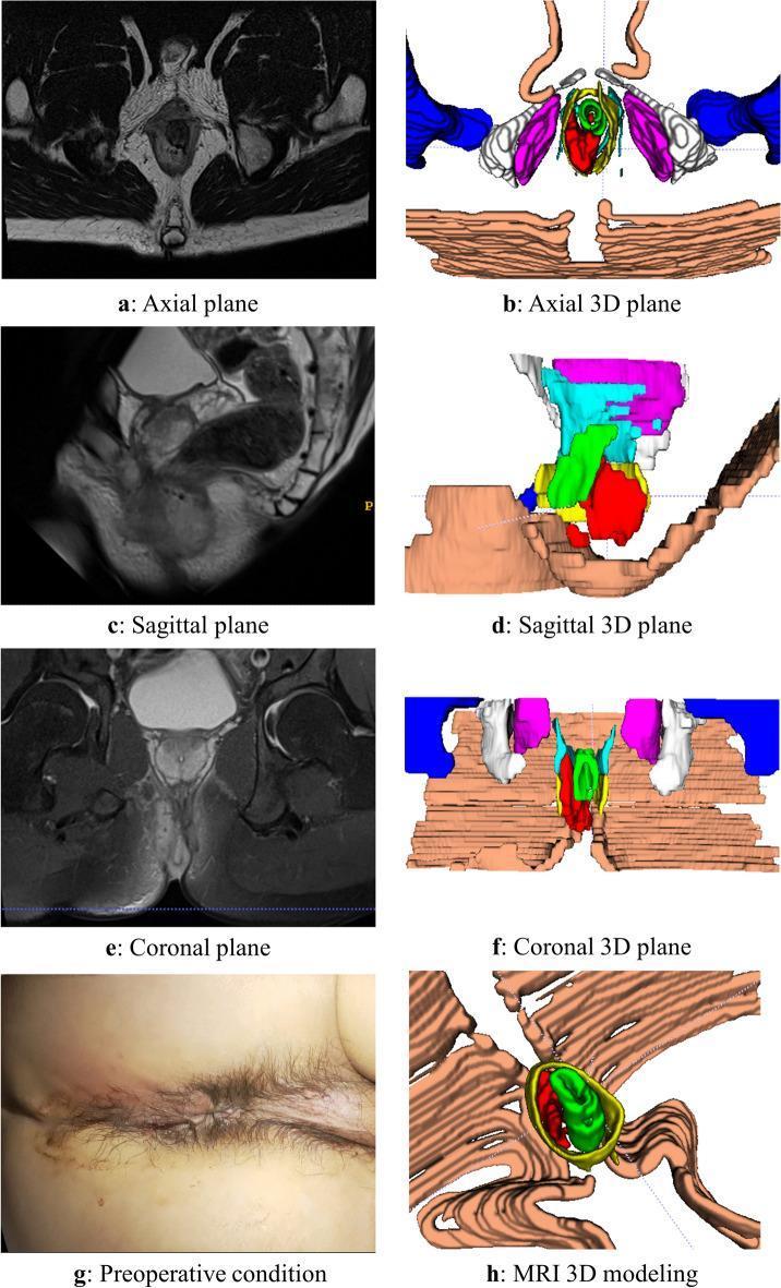
This composite educational image illustrates a case of an intersphincteric anorectal abscess through multi-modal diagnostic views. The panel includes original MRI scans and corresponding color-coded 3D reconstructions across axial (a, b), sagittal (c, d), and coronal (e, f) planes. The MRI images demonstrate the complex anatomy of the perianal region with the abscess appearing as a localized, high-signal intensity lesion on T2-weighted imaging. In the 3D models, the red structure represents the abscess, highlighting its spatial relationship to the anal canal, internal and external sphincters (represented by concentric green and yellow layers), and the levator ani muscle. Panel (g) provides a clinical photograph of the preoperative perianal region, and panel (h) displays a detailed 3D modeling view. This visual aid is designed to teach colorectal surgeons and radiologists the volumetric assessment and anatomical mapping of perianal fistulas and abscesses, emphasizing the value of 3D reconstruction in clarifying tissue demarcations that may be obscured in standard 2D grayscale radiology.

This composite educational image illustrates a case of an intersphincteric anorectal abscess through multi-modal diagnostic views. The panel includes original MRI scans and corresponding color-coded 3D reconstructions across axial (a, b), sagittal (c, d), and coronal (e, f) planes. The MRI images demonstrate the complex anatomy of the perianal region with the abscess appearing as a localized, high-signal intensity lesion on T2-weighted imaging. In the 3D models, the red structure represents the abscess, highlighting its spatial relationship to the anal canal, internal and external sphincters (represented by concentric green and yellow layers), and the levator ani muscle. Panel (g) provides a clinical photograph of the preoperative perianal region, and panel (h) displays a detailed 3D modeling view. This visual aid is designed to teach colorectal surgeons and radiologists the volumetric assessment and anatomical mapping of perianal fistulas and abscesses, emphasizing the value of 3D reconstruction in clarifying tissue demarcations that may be obscured in standard 2D grayscale radiology.

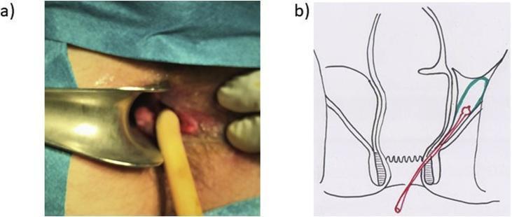
A composite image illustrating the surgical management of a complex perianal abscess. Image (a) is a clinical photograph of a perianal surgical site. A metallic anal retractor is used to visualize the rectal lumen and fistula opening. A yellow, mushroom-tip Pezzer drain is shown inserted through an inter-sphincteric fistula tract to facilitate continuous drainage of deeper infection. Image (b) is a coronal anatomical diagram of the anorectal region. The illustration depicts the rectum, internal and external anal sphincters, and the supralevator space. A green-contoured area highlights a supralevator abscess. A red line represents the Pezzer drain, showing its path originating externally, traversing the inter-sphincteric plane, and terminating within the abscess cavity. The educational focus is on the surgical treatment of supralevator abscesses and the specific placement of self-retaining drains for long-term management of complex fistulous tracts in colorectal surgery.

A composite image illustrating the surgical management of a complex perianal abscess. Image (a) is a clinical photograph of a perianal surgical site. A metallic anal retractor is used to visualize the rectal lumen and fistula opening. A yellow, mushroom-tip Pezzer drain is shown inserted through an inter-sphincteric fistula tract to facilitate continuous drainage of deeper infection. Image (b) is a coronal anatomical diagram of the anorectal region. The illustration depicts the rectum, internal and external anal sphincters, and the supralevator space. A green-contoured area highlights a supralevator abscess. A red line represents the Pezzer drain, showing its path originating externally, traversing the inter-sphincteric plane, and terminating within the abscess cavity. The educational focus is on the surgical treatment of supralevator abscesses and the specific placement of self-retaining drains for long-term management of complex fistulous tracts in colorectal surgery.

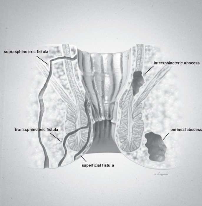
**Imaging Modality:** Medical illustration/Anatomical diagram.

**Anatomical Region:** Coronal cross-section of the anorectal region, including the anal canal, rectum, internal and external anal sphincters, and surrounding perianal soft tissues.

**Observed Pathology:** The illustration classifies various types of anorectal fistulas and abscesses based on their anatomical trajectory and location.

**Characteristic Visual Features:**
*   **Fistula Tracts:** Three distinct epithelial-lined tracks are labeled. A **superficial fistula** is shown confined to the perianal skin and distal anal canal. A **transsphincteric fistula** is depicted crossing both the internal and external sphincter muscles into the ischioanal fossa. A **suprasphincteric fistula** is illustrated looping above the puborectalis muscle before descending to the skin surface.
*   **Abscess Formations:** Two localized collections of fluid/pus are identified. An **intersphincteric abscess** is positioned within the space between the internal and external sphincter muscles. A **perineal (perianal) abscess** is shown as a larger pocket located in the subcutaneous tissue adjacent to the anal verge.

**Key Diagnostic Features:** The diagram emphasizes the relationship between inflammatory tracts and the sphincter complex, following the Parks classification system for fistula-in-ano. Suitable for clinical education on perianal sepsis and surgical planning.

**Imaging Modality:** Medical illustration/Anatomical diagram. **Anatomical Region:** Coronal cross-section of the anorectal region, including the anal canal, rectum, internal and external anal sphincters, and surrounding perianal soft tissues. **Observed Pathology:** The illustration classifies various types of anorectal fistulas and abscesses based on their anatomical trajectory and location. **Characteristic Visual Features:** * **Fistula Tracts:** Three distinct epithelial-lined tracks are labeled. A **superficial fistula** is shown confined to the perianal skin and distal anal canal. A **transsphincteric fistula** is depicted crossing both the internal and external sphincter muscles into the ischioanal fossa. A **suprasphincteric fistula** is illustrated looping above the puborectalis muscle before descending to the skin surface. * **Abscess Formations:** Two localized collections of fluid/pus are identified. An **intersphincteric abscess** is positioned within the space between the internal and external sphincter muscles. A **perineal (perianal) abscess** is shown as a larger pocket located in the subcutaneous tissue adjacent to the anal verge. **Key Diagnostic Features:** The diagram emphasizes the relationship between inflammatory tracts and the sphincter complex, following the Parks classification system for fistula-in-ano. Suitable for clinical education on perianal sepsis and surgical planning.

I now have comprehensive material from multiple authoritative sources. Let me compose the complete 20-mark answer.

Uncontrolled Diabetes Mellitus Posted for Perianal Abscess - 20 Mark Answer


INTRODUCTION

A patient with uncontrolled diabetes mellitus (DM) presenting for incision and drainage (I&D) of a perianal abscess poses a unique anesthetic and surgical challenge. This is an urgent, semi-emergency procedure - the infected septic focus must be drained promptly (unlike purely elective surgery), yet the metabolic derangements of uncontrolled DM must be addressed to reduce perioperative morbidity. The answer integrates surgical anatomy, perioperative diabetic management, anesthetic technique, and postoperative care.

PART 1: PERIANAL ABSCESS - SURGICAL OVERVIEW (4 marks)

Definition and Pathogenesis

A perianal abscess is a collection of pus in the tissue spaces around the rectum and anal canal. Approximately 80% arise from infection of the anal cryptoglandular glands (crypto-glandular theory), where bacteria infect the anal glands at the dentate line, spreading into adjacent tissue spaces. Other causes include skin infections, trauma, Crohn's disease, malignancy, and - importantly - immunosuppression from uncontrolled diabetes.

Why Diabetics Are Uniquely Vulnerable

Uncontrolled hyperglycemia impairs host defense in multiple ways:
  • Inhibits neutrophil chemotaxis and phagocytosis
  • Impairs vascular reactivity (via angiotensin II and nitric oxide synthase pathways)
  • Increases vascular permeability and platelet/leukocyte activation
  • Peripheral neuropathy reduces pain sensation, delaying presentation
  • Microangiopathy reduces tissue perfusion, impairing healing
This leads to more aggressive, extensive, and polymicrobial infections in diabetics. Perianal abscess can rapidly spread to Fournier's gangrene (necrotising fasciitis of the perineum) in uncontrolled diabetics - a life-threatening emergency.

Classification of Anorectal Abscesses (Parks Classification)

Perianal abscess and fistula anatomy diagram showing Parks classification
TypeLocationFrequency
Perianal (subcutaneous)Subcutaneous tissue at anal vergeMost common
IschiorectalIschiorectal fossa; may cross posteriorly = horseshoeCommon
IntersphinctericBetween internal and external sphinctersDifficult to diagnose
SupralevatorAbove levator ani; may extend into pelvisLeast common; most serious

Surgical Principle

Incision and Drainage (I&D) is the definitive treatment - antibiotics alone cannot treat a pus collection. As stated in Schwartz's Principles of Surgery (11e): "Most perianal abscesses can be drained under local anesthesia in the office, clinic, or emergency department. Larger, more complicated abscesses may require drainage in the operating room."
Technique: Cruciate or elliptical incision over the point of maximum fluctuation, close to the anal margin - adequate drainage, de-roofing of the cavity, no packing required; sitz baths from next day.

PART 2: PREOPERATIVE ASSESSMENT AND OPTIMIZATION (5 marks)

A. Clinical Assessment

History:
  • Type, duration, and current treatment of DM (Type 1 vs. Type 2, oral agents vs. insulin)
  • HbA1c and recent blood glucose values
  • Duration and severity of abscess (onset, fever, spreading cellulitis, crepitus)
  • End-organ damage: nephropathy, neuropathy, retinopathy, cardiovascular disease
  • Current medications (including anticoagulants, antihypertensives, SGLT2 inhibitors)
  • Last oral intake (NPO status) - critical for airway management, especially with diabetic gastroparesis
Examination:
  • Vital signs: fever (>38.5°C suggests systemic sepsis), tachycardia, BP
  • Assess extent of abscess: localised fluctuant swelling vs. spreading cellulitis, crepitus (gas = necrotising fasciitis!)
  • Airway: prayer sign (inability to oppose palms flat) - suggests stiff joint syndrome from glycosylation, predicting difficult intubation
  • Autonomic neuropathy signs: orthostatic hypotension, resting tachycardia, absent heart rate variability

B. Investigations

InvestigationRationale
Blood glucose (CBG/RBS)Current glycaemic status
HbA1cLong-term control; >8% = poorly controlled
Serum electrolytes (Na, K, Cl, HCO3)Detect DKA (low HCO3), hypernatraemia
Urea, creatinineRenal function; adjust drug doses
CBC/FBCLeucocytosis - degree of sepsis; anaemia
Urine ketonesRule out/diagnose DKA
ABG (if septic)pH, metabolic acidosis
Blood culturesIf systemic sepsis present
ECGSilent myocardial ischaemia common in diabetic autonomic neuropathy
CXRBaseline, pneumonia
MRI/CT pelvisIf complex/deep abscess suspected (not routine)

C. Management of Uncontrolled DM Before Surgery

This is not purely elective - drainage cannot be deferred for days of optimization. The approach is to stabilise, not optimise, and proceed urgently.
Step 1 - Correct severe hyperglycaemia:
  • Target BG: 140-180 mg/dL before surgery
  • If BG >250 mg/dL: Start IV regular insulin infusion (concentration 1 unit/mL in NS); titrate to bring BG to acceptable range
  • Check and correct ketones (if DKA: must treat DKA first before proceeding to theatre - see below)
Step 2 - Correct electrolytes:
  • Hypokalaemia (from vomiting, insulin therapy) must be corrected before anaesthesia (risk of cardiac arrhythmia)
  • Target K+ >3.5 mEq/L
Step 3 - Drug management on day of surgery:
  • Hold all oral hypoglycaemic agents (especially SGLT-2 inhibitors - risk of euglycaemic DKA; metformin - risk of lactic acidosis)
  • Type 1 DM: Continue basal insulin at 50% dose - never omit, to prevent DKA
  • Type 2 DM on insulin: Hold morning dose of short-acting insulin; reduce basal by 50%
  • If septic/feverish: higher insulin requirements expected due to stress hyperglycaemia
Perianal abscess is semi-urgent: proceed within 12-24 hours of presentation once BG <250 mg/dL and DKA (if present) is corrected.

D. Special Consideration: DKA

Infection is the most common precipitant of DKA. This patient likely has infection-driven DKA if:
  • BG >250 mg/dL + ketonuria/ketonaemia + metabolic acidosis (pH <7.3, HCO3 <18)
DKA management before surgery (minimum 4-6 hours):
  • IV fluid resuscitation: 0.9% saline 1L over 1 hour (correct dehydration)
  • IV insulin infusion: 0.1 units/kg/hour regular insulin
  • Potassium replacement: 20-40 mEq/L KCl per litre once K+ <5.5 mEq/L
  • Monitor BG hourly, electrolytes 2-hourly
  • Surgery once pH >7.3, HCO3 >15, BG <250 mg/dL, and patient haemodynamically stable

PART 3: ANAESTHETIC MANAGEMENT (6 marks)

Choice of Anaesthesia

For perianal abscess I&D, the following options are suitable:
TechniqueAdvantagesDisadvantages
Local anaesthesia (small, superficial abscess)No systemic stress, no airway manipulation, outpatient possiblePainful if abscess is tense; limited if abscess is large/deep
Spinal anaesthesia (SA) - PREFERREDExcellent perineal anaesthesia; avoids GA; less metabolic stress; patient awake - hypoglycaemia detectableContraindicated if patient on anticoagulants or has local skin infection at puncture site
Caudal anaesthesiaGood for perineal procedures; suitable in childrenTechnically variable; limited in obese patients
General anaesthesia (GA)Suitable for large/deep (ischiorectal, supralevator) or uncooperative patientsAspiration risk (gastroparesis); difficult airway risk; masks hypoglycaemia
Recommended: Spinal anaesthesia for most perianal abscess I&D in diabetic patients.
  • Position: Lithotomy (standard for perianal surgery)
  • Spinal drug: Hyperbaric bupivacaine 0.5% (heavy) - 1.2-1.5 mL (6-7.5 mg) with or without fentanyl 25 mcg
  • Target block level: T10 (umbilicus) - adequate for perineal surgery
If GA is required (e.g. large ischiorectal abscess, failed spinal):
  • Treat as full stomach (diabetic gastroparesis = delayed gastric emptying)
  • Rapid Sequence Induction (RSI): preoxygenate → cricoid pressure → propofol + suxamethonium IV → intubate
  • Premedication: ranitidine/pantoprazole + metoclopramide (reduces aspiration risk)
  • Maintenance: isoflurane/sevoflurane + O2/air; avoid N2O
  • Airway: prepare for difficult intubation (prayer sign, glycosylation), have video laryngoscope available

Key Anaesthetic Concerns in Diabetic Patient

  1. Difficult airway - stiff joint syndrome (glycosylation of temporomandibular + atlanto-occipital joints); always prepare for failed intubation
  2. Gastroparesis - aspiration risk even if apparently NBM; treat as full stomach
  3. Autonomic neuropathy - cardiovascular instability; post-induction hypotension; have vasopressors (ephedrine/phenylephrine) drawn up
  4. Hypoglycaemia masked under GA - only monitoring protects the patient
  5. Cardiovascular disease - silent myocardial ischaemia; monitor ECG
  6. Renal impairment - adjust drug doses (e.g. avoid NSAIDs postoperatively)

PART 4: INTRAOPERATIVE GLUCOSE MANAGEMENT (3 marks)

Target

Maintain BG: 140-180 mg/dL (ADA recommendation); avoid BG >200 mg/dL (increased SSI, impaired wound healing) and BG <85 mg/dL (hypoglycaemia).

Protocol for Perianal Abscess I&D (Short Procedure)

Since I&D of a perianal abscess is typically a short procedure (15-30 minutes):
If BG 70-180 mg/dL on arrival:
  • Hourly glucose monitoring
  • No insulin correction typically needed for this short duration
  • Sliding scale SC regular insulin if BG creeps above 180 mg/dL
If BG 180-250 mg/dL:
  • Give correction dose rapid-acting insulin SC (per sliding scale)
  • Hourly BG monitoring throughout
  • 1 unit regular insulin IV lowers BG by ~25-30 mg/dL in a 70 kg adult
If BG >250 mg/dL (uncontrolled):
  • Start IV insulin infusion: Regular insulin in 0.9% NS (1 unit/mL)
  • Starting rate = Plasma glucose (mg/dL) ÷ 150 = units/hour
  • Give separately from IV dextrose; do NOT co-infuse in same line
  • Concurrent D5W at 1 mL/kg/h to prevent hypoglycaemia
  • Add 20 mEq KCl per litre of maintenance IV fluid (insulin drives K+ intracellularly)
  • Check BG every 30-60 minutes
Hypoglycaemia management (BG <70 mg/dL):
  • Under spinal: patient may report sweating, anxiety (detectable)
  • Under GA: all symptoms masked - monitoring is the only safeguard
  • Treatment: stop insulin infusion, give 25-50 mL of 50% dextrose (D50W) IV - each mL raises BG by ~2 mg/dL in a 70 kg patient
Intraoperative IV fluids: Use Ringer's lactate or 0.9% normal saline for maintenance and replacement - NOT dextrose-containing fluids (unless treating hypoglycaemia).

PART 5: POSTOPERATIVE MANAGEMENT (2 marks)

Glucose Control

  • Check BG on arrival to recovery and every 1-2 hours
  • Continue IV insulin infusion if BG >180 mg/dL or if patient cannot take orally
  • Transition to SC basal-bolus regimen once oral intake resumes (usually same day for perianal I&D)
  • Resume usual antidiabetic medications when eating normally (restart metformin when renal function confirmed)
  • Restart SGLT2 inhibitors only after fully oral intake re-established

Wound Care and Antibiotics

  • Antibiotics: Not routinely required after simple I&D of perianal abscess in immunocompetent patients. However, in uncontrolled diabetics, give broad-spectrum antibiotics:
    • Co-amoxiclav (amoxicillin-clavulanate) OR
    • Metronidazole + cephalosporin (to cover anaerobes, Gram-negatives, and skin flora)
    • If MRSA risk: add vancomycin
    • Duration: 5-7 days (longer if spreading cellulitis or sepsis)
  • Sitz baths from the next day - promotes drainage and hygiene
  • Strict aseptic wound care - hyperglycaemia impairs wound healing and promotes SSI
  • Watch for Fournier's gangrene - spreading pain, crepitus, foul odour = surgical emergency requiring radical debridement

Optimise DM Long-Term

  • Endocrinology referral for optimisation of DM control
  • Target HbA1c <7% (Type 2) / <8% (Type 1) before any future elective surgery
  • Patient education on glucose monitoring, foot care, recognition of infections early

SUMMARY TABLE

PhaseKey Action
PreopAssess severity of DM + sepsis; treat DKA if present; correct BG to <250 mg/dL; hold oral agents; reduce basal insulin 50%; check electrolytes + ECG
Anaesthesia choiceSpinal (preferred for short perianal procedure); RSI-GA if large/deep abscess or uncooperative
AirwayAnticipate difficult intubation (prayer sign); treat as full stomach (gastroparesis)
Intraoperative glucoseMonitor BG every 30-60 min; target 140-180 mg/dL; SC sliding scale for mild hyperglycaemia; IV insulin infusion (BG÷150 units/h) for BG >250; separate D5W drip; add KCl to maintenance fluid
PostopContinue glucose monitoring; broad-spectrum antibiotics; sitz baths; strict wound care; watch for Fournier's gangrene; resume medications when oral intake established

Sources:
  • Barash, Cullen & Stoelting's Clinical Anesthesia, 9e - Perioperative Glucose Management, pp. 1780-1782
  • Morgan & Mikhail's Clinical Anesthesiology, 7e - Anesthetic Considerations in Diabetes, pp. 1410-1415
  • Current Surgical Therapy, 14e - Perioperative Glycaemic Control, pp. 1571-1573
  • Schwartz's Principles of Surgery, 11e - Perianal Abscess, p. 1342
  • Goldman-Cecil Medicine - Perianal Abscess: Epidemiology and Management, p. 1534
  • Fischer's Mastery of Surgery, 8e - Drainage of Perianal Abscess, p. 4250
This is a shared conversation. Sign in to Orris to start your own chat.