
Polycythemia vera
Reading File
Now let me search the medical library and PubMed in parallel for comprehensive coverage.
Finding Sources
Searching PubMed
"polycythemia vera"[MeSH Terms]
Excellent. Now let me read the key textbook sections in parallel - Harrison's (most comprehensive), Robbins Pathology, Goldman-Cecil, and the lab methods book.
Reading File
Reading File
Reading File
Reading File
Now let me get the rest of the Harrison's section (diagnosis, treatment, prognosis) and fetch the key clinical images.
Reading File
Loading Image
Loading Image
Now let me get the diagnostic criteria section and search for the splenomegaly/bone marrow image.
Finding Sources
Finding Sources
Let me get the WHO diagnostic criteria section from Henry's and the Harrison's diagnosis section.
Reading File
Reading File
I now have all the material needed for a comprehensive answer. Let me also get the WHO diagnostic criteria table from Henry's.
Reading File
Let me get the WHO criteria for PV specifically, which is in the Henry's block around line 1220.
Reading File
I have rich, comprehensive content from multiple authoritative textbooks. Now let me compile the full answer.
Polycythemia Vera (PV)
Definition & Classification
Polycythemia vera (PV) is a clonal myeloproliferative neoplasm (MPN) arising from a transformed hematopoietic stem cell. It is classified as a Philadelphia chromosome-negative (Ph-negative) MPN alongside essential thrombocythemia (ET) and primary myelofibrosis (PMF). The hallmark is panmyelosis - excessive proliferation of erythroid, granulocytic, and megakaryocytic elements - but it is the absolute increase in red cell mass that drives most clinical manifestations.
-
Incidence: 1-3 per 100,000/year in the US, increasing with age to >10/100,000 in older adults
-
Familial transmission is uncommon; sporadic cases show female predominance under age 50
-
Harrison's Principles of Internal Medicine, 22E, p. 862
-
Robbins, Cotran & Kumar Pathologic Basis of Disease, p. 14
Pathogenesis
The JAK2 Mutation
The central molecular event is a point mutation in the JAK2 gene (chromosome 9p):
- JAK2 V617F (valine → phenylalanine at codon 617): found in >95-97% of PV patients. It sits in the autoinhibitory pseudokinase domain, causing constitutive, ligand-independent kinase activation.
- JAK2 normally serves as the cognate kinase for the erythropoietin receptor (EPOR) and thrombopoietin receptor. Constitutive activation bypasses the need for erythropoietin, explaining:
- Erythropoietin hypersensitivity and EPO-independent erythroid colony formation
- Increased Bcl-X(L) expression and apoptosis resistance in erythroid progenitors
- Reduced serum erythropoietin (a key diagnostic clue - opposite of secondary polycythemia)
- Exon 12 JAK2 mutations account for most remaining JAK2 V617F-negative cases - these tend to present with isolated erythrocytosis
- Loss of heterozygosity at 9p (uniparental disomy → JAK2 V617F homozygosity) occurs in ~60% of PV patients and is associated with higher WBC counts, more significant splenomegaly, pruritus, and faster progression to spent phase
- Rare additional driver mutations: LNK (a JAK2 inhibitor), CALR, occasional ASXL1, TET2, DNMT3A (clonal evolution markers)
The JAK2 V617F mutation is not the sole initiating lesion - the predisposition is associated with a specific JAK2 haplotype (GGCC haplotype), and familial PV can occur without the mutation.
- Harrison's, p. 862-863
- Robbins & Kumar Basic Pathology, p. 406-407
Morphology
Bone marrow: Hypercellular with panmyelosis (increased erythroid, myeloid, and megakaryocytic forms). Some residual fat is usually preserved. Moderate-to-marked reticulin fibrosis at diagnosis in ~10% of cases.
Late / "spent phase": Marrow replaced by fibroblasts and collagen (post-PV myelofibrosis). Extramedullary hematopoiesis in spleen and liver → prominent organomegaly.
Spleen: Mildly enlarged early (vascular congestion). Later, massive splenomegaly from extramedullary hematopoiesis.
Blood vessels: Thromboses and infarctions common in heart, spleen, kidneys; hemorrhages in ~30%.
Peripheral blood: RBC 6-12 × 10¹²/L; platelet count often >400,000/µL; WBC up to 50,000/µL; basophilia characteristic; giant platelets and megakaryocyte fragments; low/absent ESR (high viscosity).
- Robbins, Cotran & Kumar, p. 14-15
- Henry's Clinical Diagnosis and Management by Laboratory Methods, p. 14
Clinical Features
Symptoms from Erythrocytosis / Hyperviscosity
- Neurological: Headache, vertigo, tinnitus, visual disturbances, transient ischemic attacks (TIAs), paresthesias
- Systemic hypertension
- Facial plethora, ruddy cyanosis, conjunctival injection, retinal vein engorgement
Thrombosis (most significant complication - ~30% of patients)
- Cerebral, cardiac, mesenteric vessels most common
- Hepatic vein thrombosis (Budd-Chiari syndrome) - especially in young women; PV should be suspected in any woman who develops this
- Portal vein thrombosis more common in males
- Arterial thrombosis in 16%, venous thrombosis in 7% at or before diagnosis
Hemorrhage (5-10% life-threatening)
- Epistaxis, gum bleeding, GI hemorrhage (often peptic ulcer)
- Paradoxically caused by platelet dysfunction and acquired von Willebrand disease (when platelet count >900,000/µL, large platelet mass absorbs high-MW vWF multimers)
Pruritus
- Aquagenic pruritus (itching after warm bath/shower) - affects 30-50% of patients; may precede PV diagnosis by years
- Due to histamine release from basophils
Erythromelalgia
- Erythema, burning, and pain in distal extremities (hands/feet) from platelet-mediated microvascular occlusion
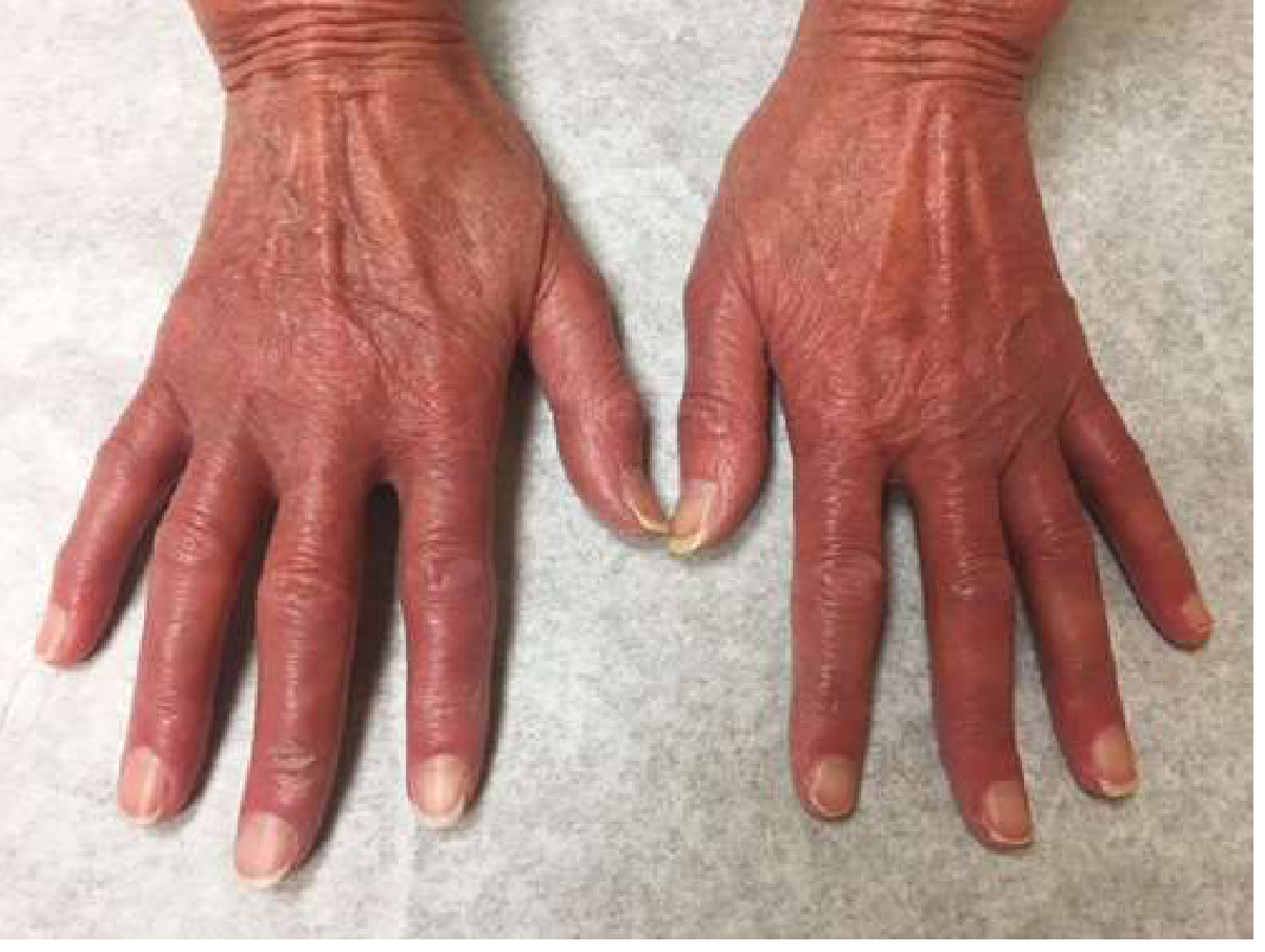
Gout / Hyperuricemia
- Symptomatic gout in 5-10% from high cell turnover and nucleic acid catabolism; uric acid stones
Splenomegaly
-
Present in two-thirds of patients at diagnosis
-
Harrison's, p. 862-863; Goldman-Cecil Medicine, p. 1750-1751; Henry's, p. 14
Laboratory Findings
| Parameter | Finding |
|---|---|
| Hemoglobin | >18.5 g/dL (men) / >16.5 g/dL (women) |
| Hematocrit | Often ≥60%; diagnostic threshold >49% men, >48% women |
| RBC count | 6-10 million/µL (up to 12 × 10¹²/L) |
| Serum EPO | Low (key diagnostic distinction from secondary polycythemia) |
| WBC | 10,000-50,000/µL; neutrophilia + basophilia |
| Platelets | >400,000/µL; often >1,000,000/µL; functionally abnormal |
| Neutrophil alkaline phosphatase (NAP) | Elevated in 80% |
| Uric acid | Elevated |
| ESR | Reduced (high viscosity) |
| Arterial O₂ saturation | Normal |
| Serum B12 | Often elevated (transcobalamin release from granulocytes) |
| JAK2 V617F mutation | Positive in >95% |
WHO Diagnostic Criteria (2016/2022)
Major criteria:
- Hemoglobin >16.5 g/dL (men) or >16.0 g/dL (women), OR hematocrit >49% (men) or >48% (women), OR increased red cell mass
- Bone marrow biopsy showing hypercellularity with panmyelosis (trilineage growth: erythroid, granulocytic, megakaryocytic proliferation) with pleomorphic mature megakaryocytes
- Presence of JAK2 V617F or JAK2 exon 12 mutation
Minor criterion:
- Subnormal serum EPO level
Diagnosis requires: All 3 major criteria, OR major criteria 1 + 2 with the minor criterion.
- Goldman-Cecil Medicine, p. 1750; Henry's, p. 14
Differential Diagnosis
PV must be distinguished from:
- Relative (spurious) polycythemia (Gaisböck's syndrome): plasma volume contraction from dehydration, diuretics, tobacco, androgens - no absolute increase in red cell mass
- Secondary absolute polycythemia: EPO levels are HIGH (opposite of PV):
- Hypoxia: COPD, high altitude, sleep apnea, high-affinity hemoglobin, carbon monoxide
- Ectopic EPO: hypernephroma, hepatocellular carcinoma, cerebellar hemangioblastoma, uterine fibroids
- Other MPNs: ET, PMF (can have same JAK2 V617F but different phenotype)
- CML (BCR-ABL1 positive - must be excluded)
Key distinguishing features of PV vs secondary polycythemia:
| Feature | PV | Secondary |
|---|---|---|
| Serum EPO | Low | High |
| Splenomegaly | Common | Absent |
| Leukocytosis/thrombocytosis | Yes | No |
| JAK2 mutation | >95% | No |
| Panmyelosis on BM | Yes | No |
Disease Course & Complications
Three phases:
- Proliferative phase: Controlled erythrocytosis, manageable with phlebotomy
- Stable phase: Iron deficiency limits red cell expansion; phlebotomy every 3 months
- Spent (post-PV MF) phase: Marrow fibrosis, cytopenias, massive splenomegaly, extramedullary hematopoiesis - occurs in a subset
Transformation:
- Post-PV myelofibrosis: variable frequency, higher with JAK2 V617F homozygosity
- AML transformation: ~1-2% without myelosuppressive therapy; higher (up to 10-15%) with alkylating agents or ³²P. Alkylating agents and radioactive phosphorus are leukemogenic and should be avoided
Survival: Without treatment, death from vascular complications within months. Median survival ~10 years with treatment (phlebotomy to maintain normal red cell mass).
- Robbins, Cotran & Kumar, p. 15; Harrison's, p. 864
Management
1. Phlebotomy (first-line for all patients)
- Goal: Hematocrit <45% in men, <42% in women (hematocrit ≥45% significantly increases thrombosis risk)
- Induces iron deficiency that limits re-expansion of red cell mass
- Once iron-deficient, phlebotomy usually needed only every 3 months
2. Low-dose Aspirin
- Reduces risk of thrombotic complications
- Caution: potentially harmful if red cell mass not controlled, and increases bleeding in patients >60 years
3. Cytoreductive Therapy (for high-risk patients)
Risk stratification:
- Low risk: Age <60, no thrombosis history
- High risk: Age ≥60 OR prior thrombosis
High-risk patients require cytoreduction in addition to phlebotomy:
| Drug | Notes |
|---|---|
| Hydroxyurea | Most widely used; effective but mutagenic (does not target the malignant HSC directly); leukemogenic with long use |
| Ruxolitinib (JAK1/2 inhibitor) | Targets involved HSCs; not mutagenic; reduces splenomegaly and pruritus; superior to best available therapy in trials |
| Pegylated IFN-α (ropeginterferon) | Targets malignant HSCs; achieves complete hematologic AND molecular remissions in ~20%; preferred in younger patients; biweekly dosing |
| Busulfan | Used in elderly; avoid alkylating agents for long-term use (leukemogenic) |
| ³²P (radioactive phosphorus) | Effective but leukemogenic; rarely used now |
4. Symptom Management
- Pruritus: Antihistamines, antidepressants (doxepin), ruxolitinib, pegIFN-α, hydroxyurea, PUVA
- Hyperuricemia/gout: Allopurinol (especially when starting chemotherapy)
- Erythromelalgia: Low-dose aspirin (usually very effective); platelet count reduction if needed
- Thrombosis: Anticoagulation; newer oral anticoagulants preferred over VKA (no monitoring required)
- Splenomegaly: Ruxolitinib or pegIFN-α; splenectomy rarely needed now with effective JAK2 inhibitors
5. Newer/Emerging Agents
- Ropeginterferon alfa-2b (Besremi): long-acting pegylated interferon; approved specifically for PV. A 2026 systematic review and meta-analysis confirms significant hematologic and molecular response rates.
- A 2024 meta-analysis confirmed ruxolitinib superior to best available therapy for hematocrit control, symptom burden, and spleen volume reduction in hydroxyurea-intolerant/resistant PV.
Key Points Summary
| Feature | Detail |
|---|---|
| Mutation | JAK2 V617F (>95%); exon 12 for most remainder |
| Mechanism | Constitutive JAK/STAT activation → EPO-independent erythropoiesis |
| EPO level | Low (key diagnostic finding) |
| Classic symptom | Aquagenic pruritus, erythromelalgia, plethora |
| Biggest complication | Thrombosis (cerebral, cardiac, Budd-Chiari) |
| Diagnosis | WHO criteria: panmyelosis + elevated Hgb/Hct + JAK2 mutation ± low EPO |
| First-line Rx | Phlebotomy to Hct <45% + low-dose aspirin |
| High-risk Rx | Add ruxolitinib or pegIFN-α (preferred) or hydroxyurea |
| Avoid | Alkylating agents, ³²P (leukemogenic) |
| Prognosis | Median survival ~10 years with treatment |
Sources: Harrison's Principles of Internal Medicine, 22E (2025); Robbins, Cotran & Kumar Pathologic Basis of Disease; Robbins & Kumar Basic Pathology; Goldman-Cecil Medicine; Henry's Clinical Diagnosis and Management by Laboratory Methods
This is a shared conversation. Sign in to Orris to start your own chat.