
Blood cancer and bone marrow cancer
blood cancer leukemia bone marrow smear histology
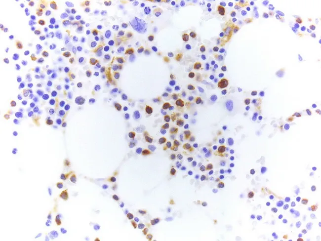
This histology image depicts a tartrate-resistant acid phosphatase (TRAP) stained smear from a bone marrow aspirate in hairy cell leukemia (HCL). The modality is bright-field microscopy. Hairy cell leukemia is a B-cell neoplasm characterized by bone marrow infiltration with mononuclear cells possessing delicate, cytoplasmic, hair-like projections. In TRAP cytochemistry, neoplastic cells exhibit red granular cytoplasmic staining due to tartrate-resistant acid phosphatase activity, a classic diagnostic feature; modern practice often relies on TRAP immunohistochemistry (IHC), which produces brown cytoplasmic positivity but is less specific than the cytochemical method. The image demonstrates scattered medium-to-large cells with abundant cytoplasm and fine cytoplasmic extensions, interspersed among normal hematopoietic elements and adipocytes; many cells show prominent TRAP positivity. Because TRAP activity is labile, slides must be air-dried and stained promptly after preparation. In this example, TRAP IHC has been applied, highlighting hairy cells with cytoplasmic labeling; however, positivity may be observed in other B-cell neoplasms, so interpretation should be integrated with morphology and immunophenotyping (e.g., CD11c, CD25, CD103). Clinically, HCL presents with cytopenias, splenomegaly, and marrow fibrosis; accurate histopathologic diagnosis informs targeted therapy with purine analogs and informs prognosis. This image is educational for hematopathology, cytochemistry, and diagnostic immunohistochemistry practice.
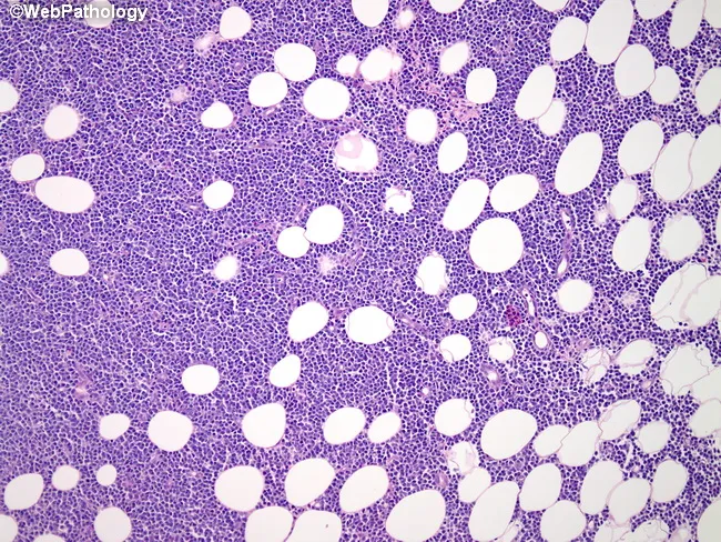
Bone marrow histology on this hematoxylin and eosin–stained section shows diffuse infiltration by mature B-lymphoid cells with prolymphocytic features, partially replacing normal adipocytic marrow. Infiltrating cells are medium to large with round to irregular nuclei, coarse chromatin, conspicuous nucleoli, and scant to moderate cytoplasm. Scattered prolymphocytes are present and background fat cells persist in residual islands; the overall architecture is variably effaced. This pattern is characteristic of bone marrow involvement by B-cell prolymphocytic leukemia (B-PLL). Although immunophenotype cannot be determined on this slide, typical profiles include bright surface immunoglobulin expression with CD19 and CD20 positivity, coexpression of CD5 and FMC7, and often absence of CD23. Clinically, B-PLL affects the elderly and presents with leukocytosis (often >100,000/μL), anemia, thrombocytopenia, and splenomegaly; B symptoms may be present. Relapsed disease can show central nervous system involvement, refractory hypercalcemia, or extramedullary masses. The histopathologic finding confirms marrow involvement and aids staging and management, while recognizing differential diagnoses such as chronic lymphocytic leukemia/small lymphocytic lymphoma, mantle cell lymphoma, hairy cell leukemia, and acute lymphoblastic leukemia. Integrate complete blood count, flow cytometry immunophenotyping, cytogenetics, and molecular studies for definitive classification and prognosis. This image depicts a bone marrow biopsy/aspirate specimen illustrating B-PLL–related marrow infiltration.
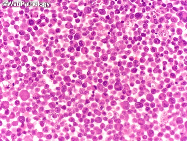
This brightfield light microscopy image depicts a dense bone marrow aspirate smear stained with hematoxylin and eosin. The cellularity is high with a uniform population of small to medium-sized lymphoid cells (lymphoblast-like cells) occupying much of the field. Nuclei are round to ovoid with finely dispersed, coarse chromatin, and scant basophilic cytoplasm; nucleoli are inconspicuous. The architecture is diffuse rather than follicular, with little evidence of erythroid or myeloid precursors; occasional mitotic figures may be present but are not prominent at this magnification. The histology shows a monotonous lymphoid infiltrate, a pattern that raises concern for a lymphoblastic process. Pathologic interpretation would emphasize lymphoid blasts consistent with acute lymphoblastic leukemia or lymphoblastic lymphoma; however, definitive classification requires ancillary studies such as flow cytometry immunophenotyping, immunohistochemistry, and genetic studies (cytogenetics/molecular). Clinically, such a pattern correlates with acute presentations: cytopenias, signs of marrow failure, fatigue, infections, or organomegaly. Potential uses include medical education for recognizing quantitative lymphoid blasts on bone marrow smears, training in differential diagnoses of monomorphic blue cell infiltrates, and optimizing search queries for pathology image datasets. Keywords include: lymphoblasts, pediatric ALL, bone marrow failure, marrow infiltration, hematopathology, immunophenotype, CD markers, prognosis guidance, diagnostic workflow, and educational value overall.
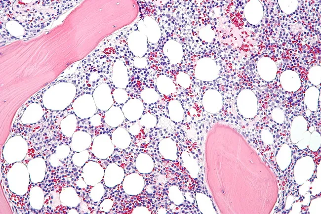
Light microscopy of a bone marrow biopsy from hairy cell leukemia (HCL) shows diffuse infiltration by small to medium lymphoid cells with abundant clear cytoplasm and oval, occasionally reniform nuclei; the cytoplasm creates a fried egg appearance. Hairy projections may be highlighted by immunostaining and by DBA.44 positivity, along with CD11c, CD25, and CD103 markers. The marrow is commonly hypercellular in established disease, whereas early involvement may be hypocellular and mimic aplastic anemia. Prominent reticulin fibrosis is typical and can hinder aspirate yield, making core biopsy essential for assessing infiltration and baseline disease burden. Normal hematopoietic elements, especially the myeloid lineage, are reduced and may display dysplastic changes resembling myelodysplastic syndrome. Plasma cells and mast cells may be mildly increased. In some cases, hairy cells assume a spindle-shaped morphology. The neoplastic cells are typically monotonous with minimal prominent nucleoli, and cytoplasm may exhibit cytoplasmic processes. Unlike several small B-cell lymphomas, discrete cellular aggregates are uncommon, with diffuse replacement predominating. Immunohistochemistry and flow cytometry confirm the diagnosis via characteristic immunophenotype. Clinically, marrow involvement informs staging, prognosis, and treatment planning, though peripheral blood smear and immunophenotypic profiling often suffice for diagnosis. This image, captured at high magnification on a hematoxylin and eosin stained section, showcases classic hairy cell features.
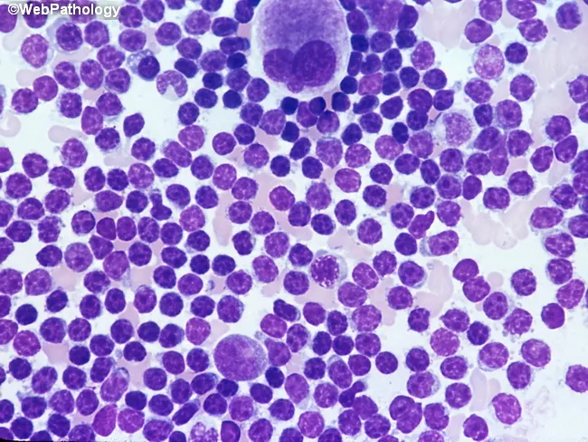
Bone marrow aspirate smear analyzed by light microscopy with high magnification reveals marrow involvement by chronic lymphocytic leukemia (CLL) cells. The predominant population consists of small, dark lymphocytes with scant cytoplasm and condensed, clumped nuclear chromatin, producing high nuclear-to-cytoplasmic ratios. Occasional larger forms and prolymphocytes may be present but are uncommon. A consistent finding is the relative scarcity of cytoplasm and uniform round to slightly irregular nuclei among the bulk of lymphoid cells. The smear may show scattered megakaryocytes, reflecting concurrent hematopoietic activity within the aspirate. Morphology mirrors circulating leukemic cells, consistent with systemic disease. Clinically, such marrow infiltration is associated with cytopenias and advanced disease stage, especially when diffuse, solid infiltration predominates in core biopsy patterns; these patterns correlate with adverse prognosis and with expression of prognostic markers such as ZAP-70. The image corresponds to a high-magnification field intended to document cellular detail rather than architectural marrow patterns. Diagnostic significance lies in confirming lymphoid marrow involvement by CLL, quantifying blast equivalents is not applicable, and differentiating from other small B-cell neoplasms relies on adjunct flow cytometry, cytogenetics, and immunophenotyping. This representation is typical for educational, diagnostic, and research contexts describing CLL marrow morphology.
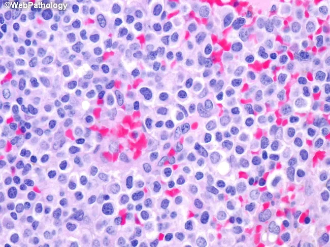
High-magnification bright-field light microscopy of a bone marrow hematopoietic smear stained with hematoxylin and eosin reveals a monotonous population of hairy cells adjacent to blood lakes. The neoplastic cells are uniform in size, with round to oval nuclei and inconspicuous nucleoli, and abundant clear cytoplasm creating the classic fried-egg appearance around the nucleus. Cytoplasmic projections (hair-like processes) may be subtle at this magnification but contribute to the characteristic 'hairy' cytology. The background shows scattered erythroid and myeloid cells with preserved but reduced overall marrow cellularity; focal vascular spaces appear as pink blood lakes infiltrated by neoplastic cells. The cells display minimal nuclear atypia, fine chromatin, and a low mitotic rate, consistent with a mature B-cell neoplasm. Immunophenotypic hallmarks, when tested, typically include CD11c, CD25, and CD103 positivity, with BRAF V600E mutation supporting diagnosis of hairy cell leukemia. Clinically, such marrow infiltration can cause cytopenias and splenomegaly; the morphological pattern is critical for prompt diagnosis and to guide targeted therapy (e.g., purine analogs, BRAF inhibitors). This image exemplifies the classic marrow involvement, providing a valuable reference for hematopathology education, differential diagnosis, and histomorphology training in leukemic lymphoid proliferations. Educationally, it reinforces recognition of distinct cytoplasmic clearing and nuclear features for learners.
multiple myeloma plasma cells bone marrow
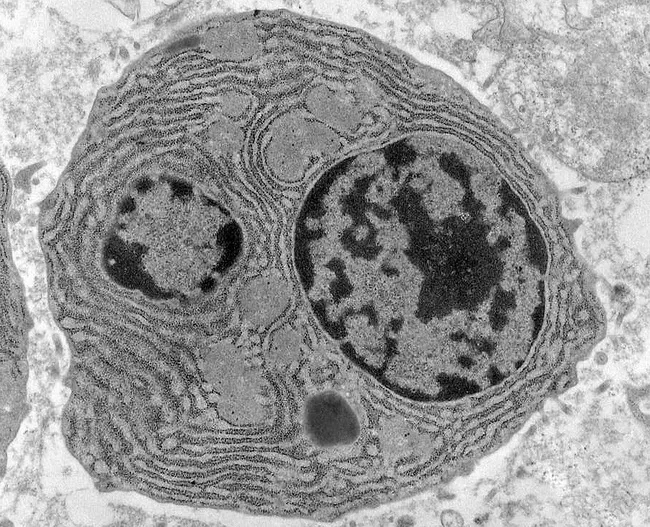
Transmission electron microscopy image of bone marrow plasma cells in multiple myeloma, highlighting ultrastructural features of malignant plasmacytosis. The specimen is a bone marrow biopsy/aspirate subjected to TEM to reveal subcellular architecture at high magnification. The primary subject is a plasma cell characterized by a prominent, eccentrically placed nucleus with chunky, irregular clumped chromatin and abundant cytoplasm. Bi- or multinucleation is frequently observed in myeloma cells. The cytoplasm shows extensive rough endoplasmic reticulum organized in parallel lamellae, reflecting heavy immunoglobulin synthesis. Light gray, round to ovoid cytoplasmic inclusions known as Russell bodies are evident within the cytoplasm, representing condensed immunoglobulin within intact endoplasmic reticulum. The cellular borders are well defined, and there is sometimes distension of the ER cisternae near the perinuclear region. The image underscores hallmark features of malignant plasma cells in multiple myeloma and demonstrates both proliferative activity and high secretory load. Clinically, these ultrastructural findings correlate with a high monoclonal immunoglobulin production and marrow infiltration, informing prognosis and therapeutic decisions when integrated with light-chain studies and routine histology. This EM view complements light microscopy by providing evidence of intracellular organelle expansion and intracellular inclusions that are not discernible with conventional staining. Potential educational use includes teaching plasmacytosis, plasma cell neoplasia morphology, and EM-based diagnostic histopathology.
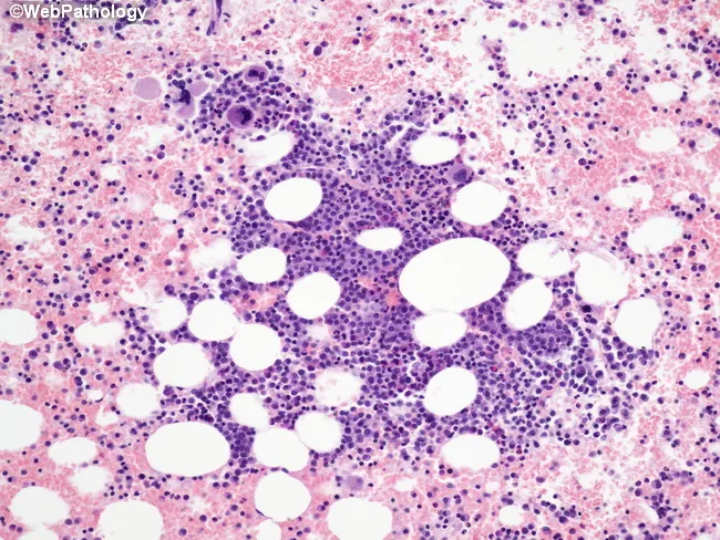
Bone marrow trephine biopsy prepared for light microscopy and stained with hematoxylin and eosin. The specimen shows bone trabeculae with islands of immature plasma cells dispersed within a background of normal hematopoiesis. In this case, multiple focal lesions are present, reflecting variable tumor burden and disease stage. The neoplastic plasma cells demonstrate a high nuclear-to-cytoplasmic ratio, round to oval nuclei, finely dispersed chromatin, and conspicuous nucleoli. Cytoplasm is scant, and some cells exhibit a plasmacytoid appearance consistent with early myeloma biology. The tumor islands are irregularly distributed and interposed between erythroid, myeloid, and megakaryocytic cells, indicating patchy infiltration rather than uniform replacement. Some large aggregates create focal crowding but do not completely efface the marrow architecture. Mitoses are infrequent in this field. The surrounding hematopoietic marrow appears variably preserved, with preserved megakaryopoiesis in residual areas. These histologic features are compatible with a plasma cell neoplasm and are typical of multiple myeloma in a relatively early focal stage, where trephine sampling may underrepresent overall tumor burden. Clinically, such findings correlate with laboratory monoclonal protein, skeletal surveys, and marrow plasma cell percentage for staging (ISS/RA). The described morphology supports diagnosis and informs prognosis, risk stratification, and treatment decisions in plasma cell myeloma.
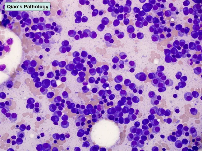
Bone marrow aspirate smear prepared with Wright-Giemsa stain was examined under light microscopy at 40X magnification. The smear demonstrates plasmacytosis with elevated plasma cells, comprising roughly 20% to 35% of nucleated cells in most cases of myeloma; in some preparations the marrow is nearly devoid of other hematopoietic elements due to extensive plasma cell replacement. Morphology spans the spectrum from mature, normal-appearing plasma cells to highly atypical plasmablasts. Mature cells show round to oval nuclei with clumped coarse chromatin and inconspicuous nucleoli; plasmablasts exhibit dispersed chromatin, prominent single nucleolus, and higher nuclear-to-cytoplasmic ratio. The cellular distribution may be diffuse or vaguely patterned with small clusters. This cytologic profile underpins the classification of myeloma variants into mature, intermediate, immature, and plasmablastic forms. Plasmablastic myelomas tend to be highly aggressive, with a poorer prognosis and median survival often less than one year, underscoring the clinical significance of morphologic assessment for risk stratification and treatment planning. The Wright-Giemsa preparation highlights cytoplasmic basophilia, nuclear pleomorphism, and mitotic activity in aggressive foci. This image provides a classic representation of plasmacytosis within marrow, illustrating the diagnostic importance of cellular morphology in multiple myeloma, plasmacytoma, and related plasma cell dyscrasias.
lymphoma Hodgkin Reed-Sternberg cells histology
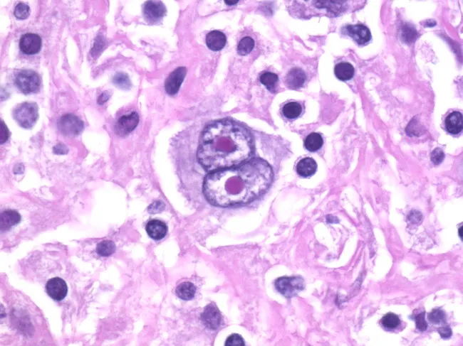
Histopathology: Light microscopy of an H&E-stained lymph node biopsy. This image highlights classic Reed-Sternberg (RS) cells within a mixed inflammatory background, a hallmark of classic Hodgkin lymphoma (cHL). The RS cells are large (up to ~100 microns) with abundant pale eosinophilic cytoplasm and prominent, bilobed to multinucleate nuclei. The nuclei show vesicular chromatin and thick nuclear membranes, with large central nucleoli creating an owl-eye appearance when two nuclei are juxtaposed. Variants, including giant RS cells and lacunar RS cells, may be seen in different HL subtypes. The surrounding milieu contains eosinophils, neutrophils, mature lymphocytes, plasma cells, and histiocytes, reflecting a characteristic reactive infiltrate. The nodal architecture is variably effaced, sometimes with formation of a rosette-like collar of T lymphocytes around RS cells. Immunophenotyping in practice typically demonstrates CD30 and CD15 positivity with weak PAX5 expression, supporting the HL diagnosis, and helps distinguish RS cells from RS-like cells in non-Hodgkin lymphomas or mimics such as anaplastic carcinoma. This histology provides essential diagnostic confirmatory evidence in suspected HL and informs prognosis and treatment planning (ABVD regimen, radiotherapy, or combined modality therapy). In some cases, classic RS cells may be absent, requiring ancillary testing and correlation with clinical findings, correlating with patient outcome.
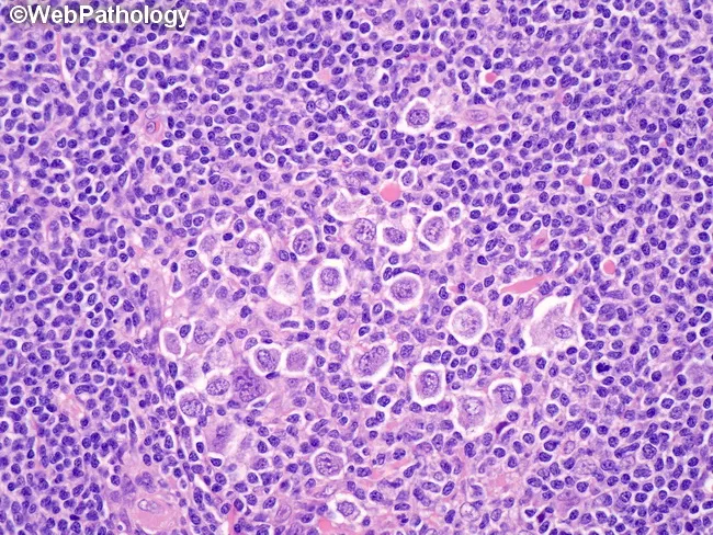
This histology image depicts lymph node tissue from a classic Hodgkin lymphoma with nodular sclerosis. The predominant cells are lacunar variants of Reed-Sternberg (RS) cells, characterized by multilobulated nuclei with coarse chromatin and small nucleoli, and a distinctive perinuclear cytoplasmic condensation that creates clear lacunar spaces in fixed tissues. The lacunae are artifacts of formalin fixation and are not present in B5-fixed specimens. Background comprises a mixed inflammatory milieu with small lymphocytes and scattered eosinophils embedded in fibrous nodules; the nodular architecture is evident with fibrous bands partitioning the lymph node into nodules, a hallmark of nodular sclerosis. RS cells may be solitary and scattered or form compact clusters and, in higher-grade nodular sclerosis (grade II), diffuse sheets. Clinical significance: The presence of lacunar RS cells in a fibrous nodal milieu supports a diagnosis of nodular sclerosis classic Hodgkin lymphoma. Differential considerations include other lymphadenopathies with RS-like cells (reactive conditions, other HL subtypes, or ALCL), but the combination of lacunar morphology, nodular sclerosis, and immunophenotype (CD30+, CD15+, PAX5 weak) is characteristic. This image is relevant for educational demonstrations of HL subtypes, fixation artifacts, and morphology-guided diagnosis, as well as educational references in pathology training and diagnostic review. Educational reference material.
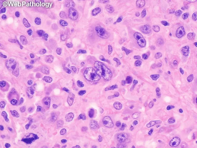
This histopathology image depicts a lymph node biopsy showing features diagnostic of lymphocyte-depleted classic Hodgkin lymphoma, reticular subtype. Using light microscopy on an H&E-stained paraffin section, the lymphoid architecture is markedly effaced by a cellular sheet of large, atypical mononuclear cells with prominent nucleoli. Occasional Reed-Sternberg cells are visible within the expansive background, including cells with multilobed or bilobed nuclei and prominent eosinophilic nucleoli, characteristic of Hodgkin lymphoma. The RS cells are scattered among numerous atypical mononuclear variants rather than forming a cohesive nodular structure. There is little-to-no fibrosis and only sparse non-neoplastic inflammatory cells, which is typical for the lymphocyte-depleted pattern. The background may show rimming by small lymphocytes and histiocytes in a reticular network, but overall cellularity is high. Immunophenotypic markers are not shown here, but in clinical practice RS cells typically express CD30 and CD15 with weaker PAX5 or B-cell markers. The diagnostic significance lies in recognizing the lymphocyte-depleted cHL morphology, which carries distinct clinical implications, often presenting with advanced stage disease and systemic symptoms. This image is useful for educational purposes, differential diagnosis conversation, and correlating histology with treatment planning (ABVD/BEACOPP regimens). Correlation with immunohistochemistry (CD30, CD15, PAX5) and EBV status further supports diagnosis in practice.
Blood Cancers and Bone Marrow Cancers: A Comprehensive Overview
1. Classification Overview
| Category | Cell of Origin | Examples |
|---|---|---|
| Acute Myeloid Leukemia (AML) | Myeloid progenitor | AML-M1 to M7 (FAB), core-binding factor AML |
| Chronic Myeloid Leukemia (CML) | Myeloid stem cell | BCR-ABL1+ CML |
| Myelodysplastic Neoplasms (MDS) | Myeloid progenitor | MDS with del(5q), MDS-EB |
| Acute Lymphoblastic Leukemia (ALL) | Lymphoid progenitor | B-ALL, T-ALL |
| Chronic Lymphocytic Leukemia (CLL) | Mature B-cell | CLL/SLL |
| Hodgkin Lymphoma | B-cell (Reed-Sternberg) | Nodular sclerosis, mixed cellularity |
| Non-Hodgkin Lymphoma (NHL) | B- or T-cell | Follicular, DLBCL, Burkitt, Mantle cell |
| Multiple Myeloma | Plasma cell | Myeloma, MGUS, Plasmacytoma |
| Myeloproliferative Neoplasms | Stem cell | Polycythemia vera, Essential thrombocythemia, Myelofibrosis |
- Robbins, Cotran & Kumar Pathologic Basis of Disease, Chapter 13
2. Leukemias
Acute Myeloid Leukemia (AML)
-
Pathogenesis: Two "hits" are typically required - a mutation that blocks differentiation (e.g., t(8;21) RUNX1-RUNX1T1; inv(16) CBFB-MYH11; t(15;17) PML-RARA) combined with one that promotes proliferation (e.g., FLT3-ITD, KIT mutations)
-
Diagnosis: >20% blasts in bone marrow aspirate (WHO criterion); some cytogenetic subtypes are diagnostic regardless of blast count
-
Presentation: Fever, pallor, fatigue, bleeding, petechiae
-
CBC: Anemia, thrombocytopenia, leukocytosis or leukopenia
-
Special case - APL (M3): t(15;17) PML-RARA - a hematologic emergency due to DIC risk; uniquely responsive to ATRA (all-trans retinoic acid) and arsenic trioxide
-
Treatment: Intensive induction chemotherapy (cytarabine + anthracycline "7+3"); targeted therapy for specific mutations (e.g., FLT3 inhibitors - midostaurin, gilteritinib; IDH inhibitors - enasidenib, ivosidenib); allogeneic stem cell transplant for high-risk disease
-
Prognosis: Overall cure rate ~70% in children; worse in older adults and high-risk cytogenetics
-
Tietz Textbook of Laboratory Medicine, 7th Ed; Swanson's Family Medicine Review
Acute Lymphoblastic Leukemia (ALL)
- Diagnosis: Dense infiltration of bone marrow by lymphoblasts (small cells with scant cytoplasm, fine chromatin, indistinct nucleoli)
- Genetics: Philadelphia chromosome (BCR-ABL1) occurs in ~20% of adult ALL - confers poor prognosis without TKI therapy; t(12;21) ETV6-RUNX1 is common in children and carries a favorable prognosis
- Treatment: Multi-agent chemotherapy (induction, consolidation, maintenance); CNS prophylaxis; TKIs (imatinib, dasatinib) for Ph+ ALL; CAR-T cell therapy (tisagenlecleucel) for relapsed/refractory B-ALL
- Prognosis: ~90% cure rate in children; ~40-50% in adults
Chronic Myeloid Leukemia (CML)
- Epidemiology: Median age ~60 years; ~15% of adult leukemias
- Phases: Chronic phase → Accelerated phase → Blast crisis (AML-like or ALL-like)
- Presentation: Splenomegaly, fatigue, night sweats; often asymptomatic (found on routine CBC)
- CBC: Markedly elevated WBC with all stages of granulocytes (left shift); basophilia is characteristic
- Treatment (revolution): Imatinib (Gleevec), the first BCR-ABL tyrosine kinase inhibitor (TKI), transformed CML from a fatal disease to one with near-normal life expectancy. Second-generation TKIs (dasatinib, nilotinib, bosutinib) and third-generation (ponatinib) address resistance
- Monitoring: Quantitative RT-PCR for BCR-ABL1 transcript levels; aim for deep molecular response (MR4.5)
- Goldman-Cecil Medicine; Braunwald's Heart Disease
Chronic Lymphocytic Leukemia (CLL)
- Immunophenotype: CD5+, CD19+, CD20 (dim), CD23+, surface Ig (dim) - this co-expression of CD5 with B-cell markers is pathognomonic
- Complications: Autoimmune hemolytic anemia, Richter transformation (to DLBCL), hypogammaglobulinemia, infections
- Staging: Rai (0-IV) or Binet (A-C) staging systems
- Treatment: Watch-and-wait for asymptomatic early disease; BTK inhibitors (ibrutinib, acalabrutinib, zanubrutinib) + BCL-2 inhibitor venetoclax are now standard of care for most patients
3. Bone Marrow-Primary Cancers: Myeloma and Related Disorders
Multiple Myeloma
Diagnostic Criteria - CRAB Features:
| Feature | Detail |
|---|---|
| C - hyperCalcemia | >1 mg/dL above normal or >11 mg/dL |
| R - Renal failure | Creatinine >2 mg/dL; light chain nephropathy (cast nephropathy) |
| A - Anemia | Hgb <10 g/dL or >2 g/dL below normal |
| B - Bone lesions | Osteolytic lesions on X-ray/CT/PET; punched-out skull lesions |
- Symptom to Diagnosis: An Evidence-Based Guide, 4th ed.; Robbins, Cotran & Kumar
Pathogenesis:
- Plasma cells home to bone marrow, produce osteoclast-activating factors (IL-1, RANKL), causing bone destruction
- Monoclonal immunoglobulin (usually IgG or IgA) or free light chains (Bence-Jones proteins) cause kidney injury, amyloidosis, and hyperviscosity
- Common genetic abnormalities: t(4;14), t(14;16) (poor prognosis); del(17p) TP53 (worst prognosis); t(11;14) (better prognosis)
Treatment:
- Transplant-eligible: VRd (bortezomib + lenalidomide + dexamethasone) induction → autologous stem cell transplant → lenalidomide maintenance
- Transplant-ineligible: VRd or DRd (daratumumab + lenalidomide + dexamethasone)
- Relapsed/refractory: Daratumumab (anti-CD38 monoclonal antibody), pomalidomide, carfilzomib, selinexor, belantamab mafodotin, CAR-T therapy (idecabtagene vicleucel, ciltacabtagene autoleucel)
- Bisphosphonates (zoledronic acid) or denosumab for bone disease
Spectrum of Plasma Cell Disorders:
- MGUS (Monoclonal Gammopathy of Undetermined Significance): Premalignant; ~1% per year risk of progression to myeloma
- Smoldering Multiple Myeloma: Intermediate; higher progression risk; no end-organ damage
- Plasma Cell Leukemia: >5% plasma cells in peripheral blood or >500/μL - aggressive variant
- Solitary Plasmacytoma: Single lesion; treated with radiotherapy; ~50% progress to myeloma
- Goldman-Cecil Medicine; Quick Compendium of Clinical Pathology, 5th ed.
4. Lymphomas
Hodgkin Lymphoma (HL)
- Immunophenotype: CD30+, CD15+, PAX5 (weak positive), CD45-
- Subtypes: Nodular Sclerosis (most common, ~70%); Mixed Cellularity; Lymphocyte-rich; Lymphocyte-depleted; Nodular Lymphocyte-Predominant HL (different biology)
- Association: Epstein-Barr virus (EBV) in a subset
- Bimodal age distribution: Young adults (15-35 yr) and older adults (>55 yr)
- Treatment: ABVD (doxorubicin, bleomycin, vinblastine, dacarbazine) ± radiotherapy; brentuximab vedotin (anti-CD30 ADC) for relapsed disease; checkpoint inhibitors (nivolumab, pembrolizumab) for relapsed/refractory HL
- Prognosis: >85% cure rate in early stage; ~65-70% in advanced stage
Non-Hodgkin Lymphomas (NHL)
| Lymphoma | Key Features | Treatment |
|---|---|---|
| Diffuse Large B-Cell Lymphoma (DLBCL) | Most common NHL; aggressive; CD20+ | R-CHOP (rituximab + chemo); CAR-T for relapse |
| Follicular Lymphoma | Indolent; t(14;18) BCL2; CD10+, BCL2+ | Watch-and-wait; R-CHOP; obinutuzumab; EZH2 inhibitors |
| Burkitt Lymphoma | Highly aggressive; c-MYC translocation; "starry sky" pattern; EBV-associated | Intensive short-duration chemo (CODOX-M/IVAC) |
| Mantle Cell Lymphoma | CD5+, cyclin D1+; t(11;14) | BTK inhibitors (ibrutinib, acalabrutinib); intensive chemo + ASCT |
| Marginal Zone Lymphoma | Indolent; associated with H. pylori (MALT), HCV, Sjogren's | Eradication therapy for H. pylori in gastric MALT |
- Robbins, Cotran & Kumar Pathologic Basis of Disease, Lymphoid Neoplasms chapter
5. Myeloproliferative Neoplasms (MPNs) and MDS
Myeloproliferative Neoplasms
- Polycythemia Vera (PV): JAK2 V617F mutation (>95%); excess red cells; risk of thrombosis and transformation to myelofibrosis/AML
- Essential Thrombocythemia (ET): JAK2/CALR/MPL mutations; excess platelets; thrombotic and hemorrhagic complications
- Primary Myelofibrosis (PMF): Marrow fibrosis, extramedullary hematopoiesis, splenomegaly; managed with ruxolitinib (JAK1/2 inhibitor) or stem cell transplant
Myelodysplastic Neoplasms (MDS)
- Common mutations: SF3B1 (favorable), TP53, ASXL1, EZH2 (adverse)
- del(5q) responds well to lenalidomide
- High-risk MDS treated with hypomethylating agents (azacitidine, decitabine) or allogeneic transplant
6. Common Presentations and Symptoms
| Symptom | Mechanism |
|---|---|
| Fatigue, pallor | Anemia (decreased RBCs) |
| Infections, fever | Neutropenia (decreased functional WBCs) |
| Bleeding, petechiae | Thrombocytopenia (decreased platelets) |
| Bone pain | Osteolysis (myeloma) or marrow expansion |
| Lymphadenopathy | Lymph node involvement |
| Splenomegaly/hepatomegaly | Extramedullary hematopoiesis or infiltration |
| Night sweats, weight loss | "B symptoms" - systemic inflammatory response |
7. Diagnostic Workup
- CBC with differential - first clue (cytopenias, blasts, atypical lymphocytes)
- Peripheral blood smear - morphology of abnormal cells
- Bone marrow aspirate + biopsy - essential for diagnosis, blast %, architecture
- Flow cytometry immunophenotyping - identifies cell lineage and aberrant markers
- Cytogenetics (karyotype + FISH) - detects chromosomal translocations
- Molecular/NGS panel - identifies actionable mutations (FLT3, IDH1/2, NPM1, TP53, etc.)
- Serum protein electrophoresis (SPEP) + immunofixation - detects M-protein in myeloma
- Imaging - CT/PET (lymphoma staging), skeletal survey/whole-body MRI (myeloma)
- LDH, uric acid, beta-2 microglobulin - markers of tumor burden and prognosis
8. General Treatment Modalities
| Treatment | Applications |
|---|---|
| Cytotoxic chemotherapy | ALL, AML, aggressive NHL |
| Targeted therapy (TKIs) | CML (imatinib, dasatinib), Ph+ ALL, FLT3-mutant AML |
| Monoclonal antibodies | Rituximab (CD20, NHL/CLL), daratumumab (CD38, myeloma), brentuximab (CD30, HL) |
| Immunomodulatory drugs (IMiDs) | Thalidomide, lenalidomide, pomalidomide (myeloma) |
| Proteasome inhibitors | Bortezomib, carfilzomib, ixazomib (myeloma) |
| BTK inhibitors | Ibrutinib, acalabrutinib, zanubrutinib (CLL, MCL) |
| BCL-2 inhibitors | Venetoclax (CLL, AML) |
| CAR-T cell therapy | Relapsed/refractory B-ALL, DLBCL, myeloma |
| Stem cell transplant | Autologous (myeloma); Allogeneic (AML, ALL, MDS, CML) |
| Hypomethylating agents | Azacitidine, decitabine (MDS, AML in elderly) |
| ATRA + arsenic trioxide | APL (AML-M3) |
| Radiation | Localized NHL, plasmacytoma, HL (involved field) |