
Polyuria
Reading File
Now let me search the medical library for polyuria content in parallel.
Finding Sources
Finding Sources
Excellent hits. Let me read the most comprehensive sources in parallel - Harrison's, Brenner's Kidney, Goldman-Cecil, and Comprehensive Clinical Nephrology.
Reading File
Reading File
Reading File
Reading File
Good content from Harrison's and Goldman-Cecil. Let me now get more from Harrison's (the diagnostic approach and management sections) and read the Comprehensive Clinical Nephrology polyuria section, plus fetch the diagnostic flowchart image.
Reading File
Reading File
Finding Sources
Loading Image
Excellent - I now have a comprehensive diagnostic flowchart from Harrison's plus detailed content from multiple authoritative textbooks. Let me get the DI treatment details and the Goldman-Cecil diagnostic flowchart image, plus a quick look at the Comprehensive Clinical Nephrology polyuria section.
Reading File
Finding Sources
Let me get the DI treatment section from Goldman-Cecil and the Goldman-Cecil diagnostic flowchart image.
Reading File
Loading Image
I now have comprehensive content. Let me get the treatment section from Goldman-Cecil block 36.
Reading File
I now have comprehensive content. Let me get the nephrogenic DI treatment continuation and also run a quick PubMed search for recent evidence.
Reading File
Searching PubMed
polyuria diabetes insipidus diagnosis treatment
I now have all the information needed for a comprehensive answer. Let me compose the full response.
Polyuria
Definition
Polyuria is conventionally defined as a urine output exceeding 3 L/day in adults (some sources use 2.5 L/day). It must be distinguished from urinary frequency, which involves frequent voiding of small volumes that do not total >3 L/day. A 24-hour urine collection may be needed to confirm true polyuria. - Goldman-Cecil Medicine, p. 1205
A physiology-based definition is more useful: polyuria exists when urine flow rate is higher than expected for the rate of solute excretion and vasopressin (AVP) activity in that clinical setting. - Brenner and Rector's The Kidney, p. 1024
Two Fundamental Categories
All polyuria falls into one of two mechanistic categories:
| Category | Mechanism | Urine Osmolality |
|---|---|---|
| Water diuresis | Failure of AVP secretion or renal AVP response | Low (<250 mOsm/kg) |
| Solute (osmotic) diuresis | Excessive non-reabsorbed solute in filtrate | High (>300 mOsm/kg) |
1. Water Diuresis
Water diuresis produces large volumes of dilute urine (<250 mOsm/kg) and occurs via three mechanisms:
A. Central Diabetes Insipidus (CDI)
Failure of AVP release from the posterior pituitary. Causes include:
- Idiopathic (selective destruction of AVP-secreting neurons, often autoimmune)
- Hypophysectomy, pituitary surgery, or trauma
- Neoplastic, inflammatory, vascular, or infectious hypothalamic/pituitary disease (trauma, tumor, sarcoidosis, histiocytosis, Sheehan's syndrome, Guillain-Barré, fat embolus, empty sella)
- Congenital lesions
Daily urine volume can reach up to 20 liters in complete CDI. - Goldman-Cecil Medicine, p. 1205
B. Nephrogenic Diabetes Insipidus (NDI)
Renal tubular resistance to AVP, preventing aquaporin-2 (AQP2) insertion into collecting duct cells.
- Congenital: Mutations in vasopressin V2 receptor gene (X-linked) or AQP2 gene
- Acquired tubular diseases: Pyelonephritis, analgesic nephropathy, multiple myeloma, amyloidosis, obstruction, sarcoidosis, hypercalcemia, hypokalemia, Sjögren's syndrome, sickle cell anemia
- Drugs/toxins: Lithium (most common drug cause - blocks AQP2 insertion), demeclocycline, methoxyflurane, ethanol, diphenylhydantoin, amphotericin B
C. Primary (Psychogenic) Polydipsia
Compulsive or habitual water drinking suppresses AVP and produces dilute urine. Causes include:
- Psychiatric disorders (most common - schizophrenia, bipolar disorder)
- Hypothalamic disease
- Drugs causing dry mouth (thioridazine, chlorpromazine, anticholinergics)
- Peripheral disorders causing elevated renin/angiotensin II
In primary polydipsia, extracellular volume is normal or expanded and plasma AVP is low because serum osmolality tends to be at the lower limits of normal. Urine osmolality may reach a minimum of ~50 mOsm/L. - Harrison's Principles of Internal Medicine 22E, p. 387
2. Solute (Osmotic) Diuresis
Occurs when large amounts of a poorly reabsorbed solute overwhelm proximal tubule reabsorption, dragging water along. Urine osmolality is typically >300 mOsm/kg.
Common causes:
- Glucosuria from uncontrolled diabetes mellitus - the most common cause of solute diuresis; causes hypertonic volume depletion
- Mannitol administration (iatrogenic)
- Radiocontrast media
- High-protein enteral/parenteral nutrition - increased urea production and excretion
- Post-obstructive diuresis - retained urea as primary osmotic agent
- Resolving ATN - natriuresis and polyuria from tubular damage
- Salt-wasting disorders (Bartter's syndrome, cystic kidney disease) - direct impairment of sodium reabsorption
- Renal failure - early sign is loss of concentrating ability (isosthenuria), presenting as nocturia and polyuria
Since urine sodium is less than blood sodium in glucosuric diuresis, more water than sodium is lost, leading to hypernatremia and hypertonicity. - Harrison's Principles of Internal Medicine 22E, p. 387
Diagnostic Approach
First step: Confirm polyuria with 24-hour urine collection.
Key test: Urine osmolality
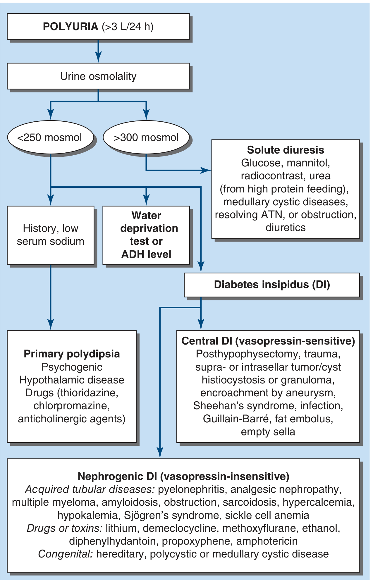
| Urine Osmolality | Interpretation | Next Step |
|---|---|---|
| <100 mOsm/kg | Water diuresis | History, serum sodium, AVP/copeptin level |
| 100-300 mOsm/kg | Indeterminate | Water deprivation test or copeptin stimulation |
| >300 mOsm/kg | Solute diuresis | Identify responsible solute (glucose, urea, sodium) |
| >800 mOsm/kg | Excludes diabetes insipidus | - |
Copeptin-Based Testing (Modern Approach)
Copeptin is cleaved from pre-pro-AVP during axonal transport - it is a reliable surrogate for AVP.
-
Copeptin ≥21.4 pmol/L: confirms partial or complete nephrogenic DI
-
Copeptin <2.6 pmol/L: identifies complete central DI
-
Intermediate levels: need water deprivation test OR hypertonic saline stimulation test
-
Goldman-Cecil Medicine, p. 2422
Water Deprivation Test
- Fluids withheld under controlled supervision; urine osmolality and body weight measured sequentially
- Endpoint: Two consecutive urine samples differing <10% in osmolality AND patient has lost ≥2% body weight
- 2 μg desmopressin (DDAVP) IV or SC is then given; urine osmolality checked at 2 hours
| Response Pattern | Urine Concentration (Dehydration) | Response to DDAVP |
|---|---|---|
| Normal | >800 mOsm/kg | <10% further rise |
| Complete CDI | Minimal (<300) | >50% rise |
| NDI | Minimal | No significant rise |
| Partial CDI / Primary Polydipsia | Moderate (300-800) | Partial rise (CDI) or minimal (PP) |
- Goldman-Cecil Medicine, p. 2422
Hypertonic saline infusion test: Infuse 3% NaCl to achieve serum Na ≥150 mmol/L, then measure plasma copeptin. Alternatively, arginine infusion with copeptin <3.5 pM at 60 min confirms DI.
Plasma AVP level is the recommended direct method for distinguishing CDI from NDI. - Harrison's 22E, p. 388
Treatment
Central DI
- Desmopressin (DDAVP) is the drug of choice - a synthetic AVP analogue with prolonged action and minimal vasopressor effects
- Oral tablets: start with half of a 0.1 mg tablet; typical schedule every 8-12 hours
- Intranasal spray: 10 μg fixed dose per spray
- IV/SC: 1-2 μg every 8-12 hours in hospitalized patients
- Monitor serum sodium regularly to prevent hyponatremia from over-treatment
- Post-traumatic/post-surgical CDI: may recover within the first year - "hold" one dose weekly to assess ongoing need
Nephrogenic DI
- Remove the offending drug if drug-induced (e.g., lithium)
- Amiloride 5-10 mg/day for lithium-induced NDI - blocks lithium entry into distal tubule cells
- Thiazide diuretics (e.g., hydrochlorothiazide): induce mild hypovolemia, increasing proximal tubular reabsorption of water and reducing distal flow
- NSAIDs occasionally used to reduce prostaglandin-mediated inhibition of AVP action
- Congenital NDI: high-dose DDAVP (10-20 μg SC or 100-200 μg intranasal) may work partially; thiazides + amiloride are the mainstay
Primary Polydipsia
- Treat the underlying psychiatric disorder
- Preliminary evidence: GLP-1 agonist dulaglutide 1.5 mg weekly may reduce thirst and fluid intake - Goldman-Cecil Medicine, p. 2424
Osmotic Diuresis
- Address the underlying cause (optimize glycemic control in DM, discontinue mannitol, manage post-obstructive state)
- Replace fluid and electrolyte losses
Gestational DI
- Desmopressin is the drug of choice - unaffected by placental vasopressinase (which degrades native AVP)
- Often resolves after delivery
Key Complications
- Hypernatremia and hyperosmolarity: develops if thirst mechanism is also impaired (dipsogenic failure + DI is particularly dangerous) - repeated episodes cause irreversible brain damage
- Bladder and upper tract dilatation: chronic massive polyuria from childhood can cause hydronephrosis and megabladder
- Hyponatremia: from over-treatment with desmopressin or primary polydipsia
- Volume depletion: particularly in solute diuresis (e.g., DM, post-obstructive)
Summary Table
| Condition | Urine Osm | Serum Osm | Serum Na | Response to DDAVP | Copeptin |
|---|---|---|---|---|---|
| Central DI (complete) | Low | High | High | >50% rise | Very low (<2.6) |
| Central DI (partial) | Low-moderate | High-normal | High-normal | Partial rise | Low-intermediate |
| NDI | Low | High | High | No rise | Very high (≥21.4) |
| Primary polydipsia | Very low (<50) | Low-normal | Low-normal | Minimal | Low |
| Solute diuresis | >300 | High-normal | High/variable | N/A | N/A |
Sources:
- Harrison's Principles of Internal Medicine 22E (2025), Chapter 55
- Goldman-Cecil Medicine International Edition, Chapters 102 & 206
- Brenner and Rector's The Kidney, Chapter 24
- Comprehensive Clinical Nephrology, 7th Edition
Recent PubMed evidence (2024-2026): A systematic review on gestational DI (PMID: 40440645) is available for that specific subtype. No major meta-analyses were found that would alter the above management framework.
Why does hypercalcemia cause Polyuria
Hypercalcemia causes nephrogenic DI through multiple mechanisms: high calcium directly impairs AVP (ADH) action on collecting duct cells, and also activates calcium-sensing receptors (CaSR) in the thick ascending limb that inhibit the NaCl cotransporter, washing out the medullary gradient needed for urine concentration.
Searching sources...
Finding Sources
Finding Sources
Good hits. Let me now read the detailed mechanistic sections from Brenner's Kidney, Medical Physiology, and NKF Primer.
Reading File
Reading File
Reading File
Reading File
Now let me get the CaSR mechanism in more detail from Medical Physiology, and the AQP2 downregulation detail.
Finding Sources
Finding Sources
Finding Sources
Reading File
Excellent - I found the key mechanistic quote from Comprehensive Clinical Nephrology and Harrison's. Let me read those sections in full detail.
Reading File
Reading File
I now have everything needed for a detailed mechanistic answer. Here it is:
Why Hypercalcemia Causes Polyuria
Hypercalcemia causes a form of acquired nephrogenic diabetes insipidus (NDI) - the kidneys produce large volumes of dilute urine not because AVP is absent, but because high calcium impairs the kidney's ability to concentrate urine through three distinct mechanisms.
Mechanism 1: CaSR Activation in the Thick Ascending Limb (TAL) - Destroys the Medullary Gradient
This is the primary mechanism.
The calcium-sensing receptor (CaSR) is expressed on the basolateral membrane of cells in the thick ascending limb of Henle (TALH) - the same receptor found in parathyroid glands that detects plasma calcium.
When serum calcium rises:
- High tubular fluid Ca²⁺ activates the CaSR on the basolateral surface of TAL cells
- This inhibits the NKCC2 cotransporter (Na⁺-K⁺-2Cl⁻), which is the principal NaCl reabsorber in the TAL
- It also inhibits ROMK channels (K⁺ recycling, needed to keep NKCC2 running) and directly inhibits paracellular NaCl permeability
The consequence: NaCl reabsorption in the TAL is reduced. Since the TAL is the "diluting segment" that builds the hyperosmotic medullary interstitium, this medullary concentration gradient collapses. Without a steep medullary gradient, even if AVP is present and AQP2 channels are open, water cannot be osmotically reabsorbed from the collecting duct into the interstitium - so the urine remains dilute and copious.
"Stimulation of the calcium receptor in the basolateral membrane of the thick ascending limb... reduces transcellular solute flux by inhibiting NKCC2 and ROMK channels and also by direct inhibition of paracellular permeability... Overall, this reduces the medullary osmotic gradient for water reabsorption." - Comprehensive Clinical Nephrology, 7th Ed.
"Calcium signals directly through the calcium-sensing receptor to downregulate Na⁺, K⁺, and Cl⁻ transport by the TALH and water transport in principal cells, thus reducing renal concentrating ability in hypercalcemia." - Harrison's Principles of Internal Medicine 22E, p. 388
Mechanism 2: CaSR Activation in the Collecting Duct Principal Cell - Blocks AQP2 Insertion
In the collecting duct, the CaSR is also present on the apical membrane of principal cells. When tubular Ca²⁺ is high:
- Apical CaSR activation prevents AVP-stimulated insertion of AQP2 water channels into the luminal membrane
- Without apical AQP2, the principal cell becomes water-impermeable regardless of AVP signaling
- Water cannot cross from the tubular lumen into the cell and onward into the interstitium
This is thought to be a physiologically protective mechanism - by reducing water reabsorption in the collecting duct when urine calcium is high, the kidney keeps urine calcium concentration relatively dilute, reducing the risk of calcium stone (nephrolithiasis) formation.
"With hypercalcemia, an elevated concentration of Ca²⁺ in tubular fluid activates the calcium-sensing receptor in the apical membrane of the principal cell, preventing insertion of AQP2. This is thought to be a protective mechanism to hinder kidney stone formation." - Comprehensive Clinical Nephrology, 7th Ed.
Mechanism 3: Downregulation of AQP2 Expression
Beyond acute AQP2 trafficking, chronic hypercalcemia also leads to:
- Downregulation of AQP2 gene expression in collecting duct cells (similar to what is seen in hypokalemia)
- This reduces the total cellular AQP2 pool available for membrane insertion
"Hypercalcemia has also been found to be associated with downregulation of AQP2." - Brenner and Rector's The Kidney
Summary Diagram of Mechanisms
HIGH SERUM Ca²⁺
       |
       ├──► CaSR activation (TALH basolateral)
       │         ↓
       │    ↓ NKCC2, ↓ ROMK, ↓ paracellular NaCl
       │         ↓
       │    ↓ Medullary osmotic gradient
       │         ↓
       │    Even with AQP2 open → ↓ water reabsorption
       │
       ├──► CaSR activation (Collecting duct apical)
       │         ↓
       │    Blocks AQP2 insertion into apical membrane
       │         ↓
       │    Collecting duct becomes water-impermeable
       │
       └──► Chronic: ↓ AQP2 gene expression
                 ↓
            Fewer AQP2 channels available

                ALL THREE → DILUTE, HIGH-VOLUME URINE (NDI)
The Vicious Cycle
The polyuria from NDI worsens the hypercalcemia:
- NDI → large volume diuresis → volume depletion
- Volume depletion → ↑ proximal tubule Na⁺ and Ca²⁺ reabsorption (solvent drag)
- Less Ca²⁺ reaches the urine → hypercalciuria falls → calcium not excreted
- Volume depletion → ↑ renin-angiotensin → further proximal Ca²⁺ retention
- Can also precipitate acute kidney injury → even less Ca²⁺ excretion
"The resulting diuresis worsens the hypercalcemia, because volume depletion limits the protective hypercalciuria and exacerbates the volume-dependent proximal tubule reabsorption of calcium." - NKF Primer on Kidney Diseases, 8th Ed.
This is why IV saline is the first-line treatment for symptomatic hypercalcemia - it breaks this cycle by restoring volume and promoting calciuresis.
Sources:
- Comprehensive Clinical Nephrology, 7th Ed. - detailed CaSR/AQP2 mechanism
- Harrison's Principles of Internal Medicine 22E (2025) - TALH CaSR signaling
- Brenner and Rector's The Kidney - AQP2 downregulation
- NKF Primer on Kidney Diseases, 8th Ed. - vicious cycle and clinical consequences
This is a shared conversation. Sign in to Orris to start your own chat.