
viral Pneumonia
Reading File
Now let me search the medical library and PubMed in parallel for viral pneumonia.
Finding Sources
Searching PubMed
"viral pneumonia"[MeSH Terms] AND management
Excellent - multiple authoritative sources found. Let me read the key sections in parallel.
Reading File
Reading File
Reading File
Excellent content retrieved. Let me now fetch more detail from Goldman-Cecil and Fishman's, plus grab the chest X-ray images and the pathology content.
Reading File
Reading File
Loading Image
Now I have comprehensive content from multiple authoritative textbooks. Let me also fetch the pathology image from Robbins and get the Goldman-Cecil inpatient treatment details.
Reading File
Loading Image
I have comprehensive information from multiple authoritative sources. Let me now compile a thorough response.
Viral Pneumonia
A comprehensive overview drawn from Murray & Nadel's Respiratory Medicine, Robbins Pathologic Basis of Disease, Rosen's Emergency Medicine, and Goldman-Cecil Medicine.
Definition & Overview
Viral pneumonia is an infection of the lower respiratory tract parenchyma caused by viruses, leading to inflammation of the alveoli and surrounding interstitium. It can range from mild self-limited disease to life-threatening illness requiring ICU-level care.
Common Causative Agents
The following viruses are the principal causes of community-acquired viral pneumonia:
| Virus | Notes |
|---|---|
| Influenza A & B | Leading cause in adults; pandemic potential via antigenic shift |
| Respiratory Syncytial Virus (RSV) | Most common in infants and young children |
| Human Metapneumovirus | Clinically similar to RSV |
| SARS-CoV-2 | COVID-19; major cause since 2020 |
| Adenovirus | Can cause severe pneumonia in military recruits |
| Rhinovirus | Usually mild, but can progress in immunocompromised |
| Varicella-Zoster Virus | Pneumonitis especially severe in pregnant women (35% mortality historically) |
| Rubeola (Measles) | Severe pneumonia in malnourished, immunosuppressed patients |
Risk factors for progression to severe lung disease include extremes of age, malnutrition, alcoholism, and underlying debilitating illnesses. - Robbins, Cotran & Kumar Pathologic Basis of Disease, p. 663
Pathogenesis
From Robbins Pathologic Basis of Disease, the general mechanisms of viral pneumonia are well understood:
- Viral attachment and entry: Viruses attach to and enter respiratory epithelial cells (e.g., influenza hemagglutinin binds sialic acid residues on cell-surface polysaccharides, followed by endosomal fusion).
- Cytopathic injury: Viral replication inhibits host cell mRNA translation and activates caspases, causing apoptosis of pneumocytes. Sodium channel inhibition causes fluid shifts leading to alveolar flooding.
- Inflammatory cascade: Dying cells release danger signals activating resident macrophages; inflammatory mediators (chemokines, cytokines) recruit neutrophils into the interstitium within 1-2 days.
- Impaired host defenses: Mucociliary clearance is disrupted, predisposing to bacterial superinfection (most commonly S. aureus, which can be life-threatening).
- Innate immune response: Type I interferon production upregulates MX1 gene (encodes a GTPase that interferes with viral replication); NK cells and cytotoxic T cells kill infected cells.
Antigenic concepts for influenza:
- Antigenic drift - spontaneous point mutations in hemagglutinin/neuraminidase genes; causes annual epidemics
- Antigenic shift - reassortment of entire gene segments from animal influenza strains (swine/avian); causes pandemics (e.g., 1918, 2009 H1N1)
Clinical Features
Adults
- Gradual onset with cough, fever, myalgia, headache, malaise
- May have rhinorrhea and pharyngitis
- Tachypnea, dyspnea, pleuritic chest pain in more severe disease
- Crackles on auscultation; wheezing possible
Children (Rosen's Emergency Medicine)
- More common in winter season
- Gradual onset with cough, congestion, and low-grade fever
- Tachypnea may be the only physical finding in mild cases
- Retractions, rales, and wheezing are common
- Severe: grunting, cyanosis, lethargy, dehydration, apnea (especially in infants < 3 months)
Radiology
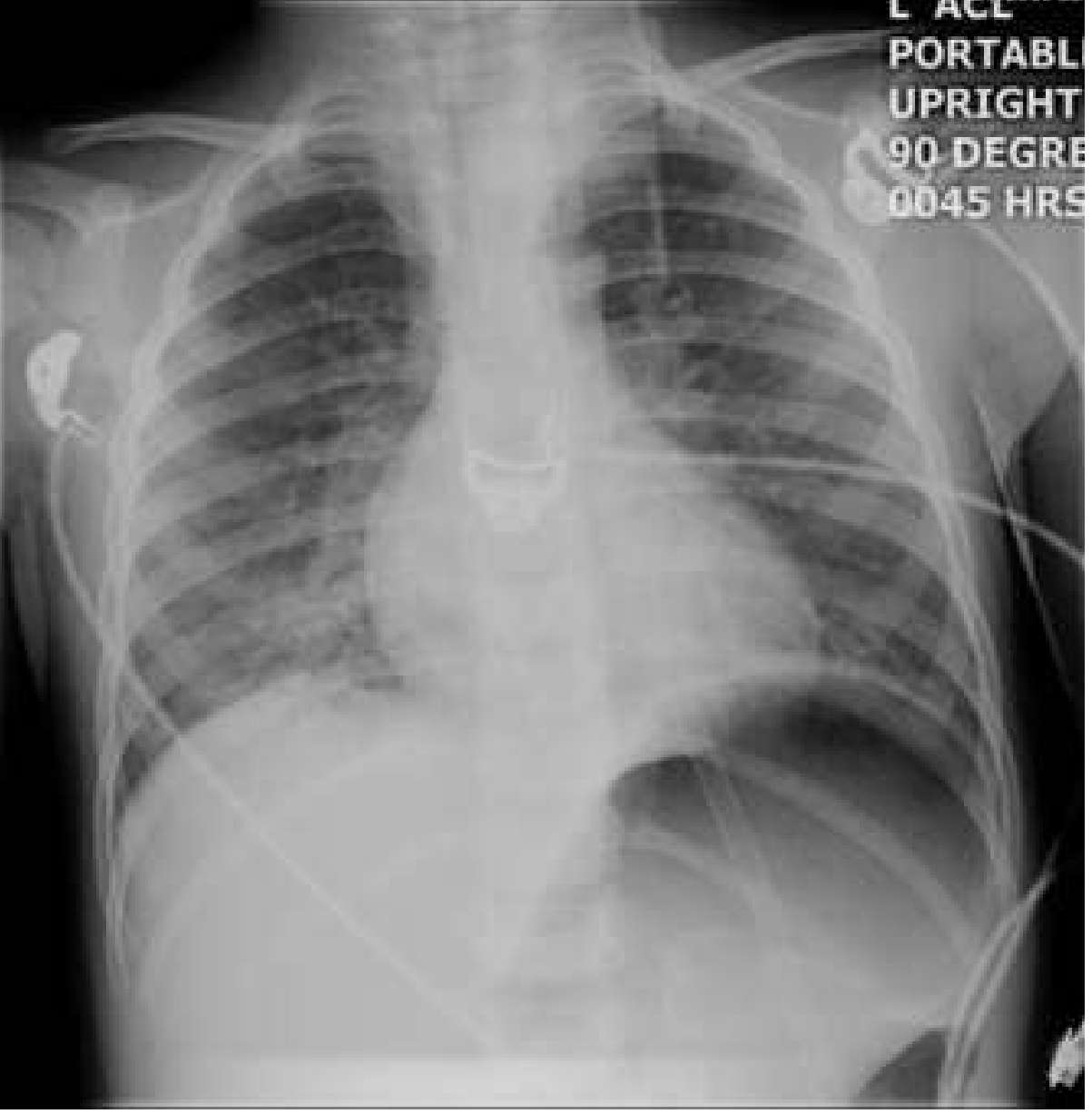
Chest X-ray in viral pneumonia typically shows:
- Hyperinflation
- Peribronchial thickening (cuffing)
- Diffuse bilateral interstitial infiltrates
- Patchy areas of consolidation (lobular atelectasis)
Lobar consolidation and pleural effusions can occur but are more characteristic of bacterial pneumonia.
Special Populations
Pregnancy (Murray & Nadel)
| Virus | Key Points |
|---|---|
| Influenza A/B | Significantly higher mortality than general population; 35% ICU admission and 7% death in 2009 H1N1 pandemic |
| Varicella | 35% mortality historically in pregnant women; treat with IV acyclovir |
| SARS-CoV-2 | Higher ICU/ventilation rates; risk factors include obesity, asthma, hypertension, age >35; use dexamethasone with caution (higher dose needed if preterm for fetal lung maturity) |
Vaccines: Inactivated influenza vaccine recommended in any trimester. Live attenuated vaccines (varicella, LAIV) are contraindicated in pregnancy.
Diagnosis
- Clinical: Winter presentation + fever + cough + interstitial infiltrates strongly suggests viral etiology
- Children generally do not require CXR or viral testing if clinical picture is clear
- Rapid antigen tests / PCR: for influenza, RSV, SARS-CoV-2
- Serology: Useful for retrospective diagnosis
- Lab findings: leukopenia or normal WBC (in contrast to leukocytosis in bacterial pneumonia); lymphopenia common with SARS-CoV-2
Management
Outpatient (Goldman-Cecil Medicine)
| Suspected Virus | Treatment |
|---|---|
| SARS-CoV-2 | Per COVID-19 protocols (nirmatrelvir/ritonavir if eligible) |
| Influenza | Oseltamivir 75 mg twice daily × 5 days |
| Other viral | Supportive care; treat for possible bacterial co-infection |
Inpatient (Goldman-Cecil Medicine)
- Influenza: Oseltamivir 75 mg twice daily × 5 days PLUS cover for bacterial co-infection (beta-lactam ± macrolide or respiratory fluoroquinolone)
- Other viral pneumonias: Treat as bacterial co-infection empirically (beta-lactam + macrolide or respiratory quinolone)
- SARS-CoV-2 severe: Dexamethasone, remdesivir; consider baricitinib or tocilizumab for cytokine storm
Supportive care
- Supplemental oxygen, escalating to high-flow nasal cannula or mechanical ventilation if needed
- Hydration
- Antipyretics/analgesics
- Bronchodilators if wheezing
Complications
- Bacterial superinfection (most common and serious - especially S. aureus, S. pneumoniae)
- ARDS (in severe influenza, COVID-19)
- Dehydration (especially in children)
- Bronchiolitis obliterans (rare, post-viral)
- Apnea (in infants)
- Secondary bacterial pneumonia (particularly S. aureus co-infection in influenza)
- Myocarditis (influenza, adenovirus)
Key Distinguishing Features: Viral vs. Bacterial Pneumonia
| Feature | Viral | Bacterial |
|---|---|---|
| Onset | Gradual | Abrupt |
| Fever | Low-grade | High-grade, rigors |
| WBC | Normal or low; lymphopenia | Leukocytosis with left shift |
| CXR | Bilateral interstitial infiltrates | Lobar consolidation |
| Sputum | Scant, watery | Purulent |
| Pleural effusion | Uncommon | More common |
| Procalcitonin | Low | Elevated |
Sources:
- Murray & Nadel's Textbook of Respiratory Medicine (Viral Pneumonia in Pregnancy chapter)
- Robbins, Cotran & Kumar Pathologic Basis of Disease, pp. 663-664
- Rosen's Emergency Medicine, p. 3192 (Pediatric Viral Pneumonia)
- Goldman-Cecil Medicine International Edition, Chapter 85 (Outpatient & Inpatient Management Tables)
Mode of Transmission
Viral pneumonia pathogens spread primarily via respiratory droplets, aerosols, and direct/indirect contact. Influenza and SARS-CoV-2 are classic examples of airborne/droplet transmission, while RSV spreads heavily via fomites. Let me pull detailed textbook content on this.
Searching sources...
Finding Sources
Finding Sources
Reading File
Reading File
Finding Sources
Reading File
Finding Sources
Reading File
Reading File
Loading Image
Excellent - rich, authoritative content from four major textbooks. Here is the complete answer:
Mode of Transmission - Viral Pneumonia
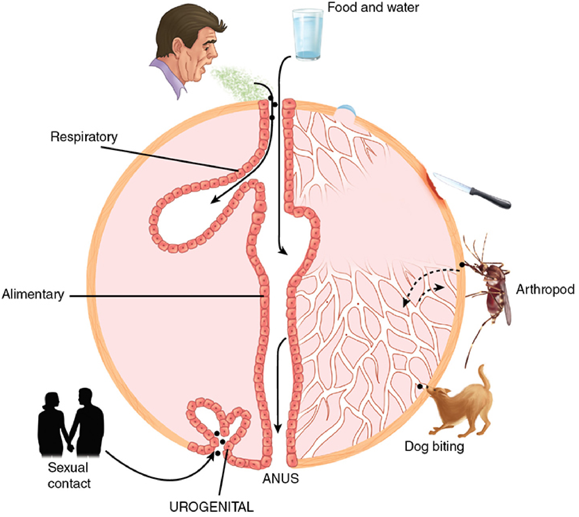
Primary Routes for Pneumonia-Causing Viruses
1. Large-Particle Respiratory Droplets (Most Common)
- Generated by coughing, sneezing, and talking
- Droplets are heavy (>5 µm) and travel only short distances (generally within 2 meters) before settling
- Require close proximity between source and recipient
- Principal route for: Influenza A & B, RSV, parainfluenza, coronavirus, SARS-CoV-2
"Influenza is spread person to person, primarily through large-particle respiratory droplet transmission (e.g., coughing or sneezing), which requires close proximity between the person who is the source and the person who is the recipient because droplets generally only travel short distances." - Red Book 2021, p. 724
2. Small-Particle Aerosols (Airborne Transmission)
- Particles <5 µm that remain suspended in air for extended periods
- Can travel beyond 2 meters and linger in enclosed spaces
- Longer-range airborne transmission may occur under unique conditions (e.g., aerosol-generating medical procedures like intubation, bronchoscopy)
- Particularly relevant for: Measles (highly airborne), Varicella-Zoster, SARS-CoV-2 (in poorly ventilated spaces)
"Large-droplet and small-particle aerosols traveling over a short range (generally within 2 meters) appear to be the predominant route; longer-range airborne transmission may also occur rarely under unique conditions." - Goldman-Cecil Medicine, p. 3520
3. Contact Transmission (Direct & Indirect)
Direct Contact
- Hand-to-mucosa self-inoculation: Virus picked up from contaminated surfaces or secretions transferred by touching the nose, eyes, or mouth
- Virus can survive on hands and surfaces for up to 24 hours (influenza)
- Important for: Influenza, RSV, rhinovirus, adenovirus
Indirect Contact (Fomites)
- Virus deposited on inanimate objects (doorknobs, toys, bedside rails) and transferred to mucous membranes
- RSV is classically transmitted this way - survives up to 6 hours on surfaces
- Route: Nose/mouth → hand or object → nose (rhinovirus, coronavirus, adenovirus)
"Another mode of transmission comes from contact with influenza virus from droplet-contaminated hands or surfaces, where it can remain for up to 24 hours, with transfer from hands to mucosal surfaces of the face." - Red Book 2021
4. Skin-to-Air-to-Respiratory Tract
- Skin vesicle discharge → air → respiratory tract
- Primary route for Varicella-Zoster virus (chickenpox/shingles)
- Also relevant for Smallpox virus
5. Zoonotic Transmission (Animal-to-Human)
| Animal Source | Mechanism | Virus |
|---|---|---|
| Chickens / Wild birds | Aerosol droplets from bird droppings/secretions; direct contact with infected birds or contaminated environments | Avian influenza H5N1, H7N9 |
| Swine | Aerosol droplets | Swine influenza H1N1 |
| Rodents | Inhalation of aerosolized excreta | Hantavirus pulmonary syndrome |
"Infection by avian viruses can occur after direct contact with infected birds or their excreta, exposure to contaminated environments and infectious aerosols, ingestion of inadequately cooked food, and sometimes by inoculation into the conjunctiva." - Goldman-Cecil Medicine
6. Vertical (Mother-to-Child) Transmission
- Less commonly causes pneumonia directly but relevant for congenital/perinatal disease
- In utero / transplacental: CMV, rubella, parvovirus B19
- Intrapartum (birth canal): HSV, hepatitis B/C, HIV
- Postpartum via breastfeeding: CMV, HTLV, HIV
Transmission Summary by Virus
| Virus | Primary Route | Secondary Route | Survival on Surfaces |
|---|---|---|---|
| Influenza A/B | Respiratory droplets | Aerosol, fomites | Up to 24 hours |
| RSV | Direct contact, fomites | Droplets | Up to 6 hours |
| SARS-CoV-2 | Droplets | Aerosol (enclosed spaces) | Hours to days |
| Measles | Airborne aerosol | Droplets | Up to 2 hours in air |
| Varicella-Zoster | Airborne (vesicle fluid) | Droplets, contact | Fragile; short-lived |
| Adenovirus | Droplets, fomites | Conjunctival | Weeks on surfaces |
| Hantavirus | Inhalation of rodent excreta | - | Variable |
| Avian influenza (H5N1) | Direct bird contact, aerosol | - | - |
| Rhinovirus | Fomites, hand-to-nose | Droplets | Up to 3 hours |
Key Epidemiological Points
- Incubation period of influenza: 1-4 days (mean 2 days); patients are infectious 24 hours before symptom onset - Red Book 2021
- Peak viral shedding in nasal secretions: first 3 days of illness; ceases within 7 days in most adults but can persist 10+ days in young children and immunocompromised patients
- The receptor binding site determines transmissibility: human influenza strains bind α-2,6-linked sialosaccharides (upper airway); avian strains prefer α-2,3-linked sialosaccharides (distal bronchioles/alveoli) - which is why avian flu is less efficiently transmitted human-to-human but causes more severe lower lung disease - Goldman-Cecil Medicine
Infection Control Implications
| Route | Precaution |
|---|---|
| Droplet | Surgical mask, limit to 2m distance |
| Airborne | N95 respirator, negative-pressure room |
| Contact | Gloves, gown, hand hygiene; avoid touching face |
| All routes | Hand washing (most effective single measure) |
Sources:
- Jawetz, Melnick & Adelberg's Medical Microbiology, 28th Ed. - Modes of Transmission
- Sherris & Ryan's Medical Microbiology, 8th Ed. - Table 7-1 Common Routes of Transmission
- Red Book 2021 (AAP Committee on Infectious Diseases) - Influenza Epidemiology
- Goldman-Cecil Medicine International Edition - Influenza Pathobiology, p. 3520
This is a shared conversation. Sign in to Orris to start your own chat.