
Osteomyelitis in 5 marks short note with digrmas pathology etiology flow charts where applicable
osteomyelitis pathology diagram bone infection
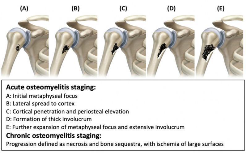
This medical illustration depicts the five-stage progression of osteomyelitis in the proximal humerus, categorized into acute and chronic phases. The series (A–E) visualizes the pathophysiology of bone infection: Stage A shows the initial metaphyseal focus of infection; Stage B demonstrates lateral spread toward the bone cortex; Stage C illustrates cortical penetration and subsequent periosteal elevation; Stage D identifies the formation of a thick involucrum (new bone growth surrounding infected tissue); and Stage E shows further expansion of the metaphyseal focus and extensive involucrum. The diagram highlights the anatomical relationship between the metaphysis, cortex, and periosteum during bacterial seeding and inflammatory response. Additionally, the content notes that chronic osteomyelitis is characterized by bone necrosis, formation of sequestra (detached dead bone), and large-surface ischemia. This anatomical diagram serves as an educational tool for medical students and clinicians to understand the radiological and pathological evolution of pediatric and adult bone infections.
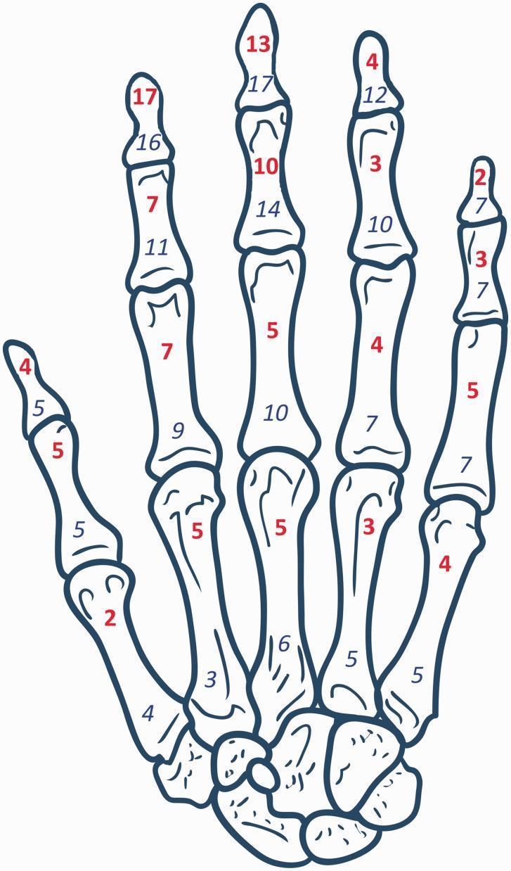
This medical illustration is an anatomical diagram of the human hand skeleton, used for clinical epidemiological mapping. It depicts the dorsal or palmar view of the metacarpals, carpal bones, and phalanges. The diagram functions as a data visualization tool for a cohort study on hand osteomyelitis in patients with digital arterial calcification. Each individual bone—proximal, middle, and distal phalanges, as well as the metacarpals—is overlaid with two sets of numerical data. Numbers in red, bold font represent the incidence of osteomyelitis at that specific anatomical site, while numbers in blue, italic font indicate the frequency of amputation for that bone. Visually, the data shows a higher incidence of pathology in the distal and middle phalanges, particularly of the index and middle fingers. This anatomical map is intended for advanced medical education and orthopedic research to illustrate the distribution of infection and surgical outcomes in high-risk populations, such as those with diabetes mellitus or end-stage renal disease.
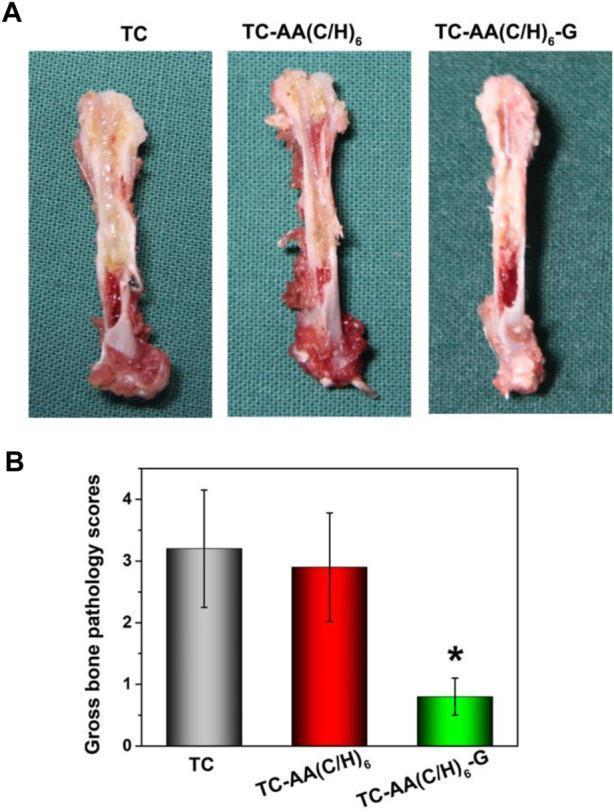
This composite educational image illustrates gross bone pathology in an experimental orthopedic model, focusing on implant-related infection. Panel A displays three longitudinal sections of animal femurs categorized by treatment group: TC (titanium coating), TC-AA(C/H)6 (multilayer coating), and TC-AA(C/H)6-G (gentamicin-loaded multilayer coating). The TC and TC-AA(C/H)6 specimens exhibit clear clinical signs of purulent osteomyelitis, characterized by visible intramedullary pus, severe structural degradation, and osteolytic lesions. In contrast, the TC-AA(C/H)6-G specimen shows preserved structural integrity with a healthy red marrow cavity and no visible signs of infection. Panel B provides a quantitative comparison through a bar graph of Gross Bone Pathology Scores. The TC group (gray bar) and TC-AA(C/H)6 group (red bar) show high pathology scores (approximately 3.0), whereas the TC-AA(C/H)6-G group (green bar) shows a significantly lower score (below 1.0). The asterisk (*) denotes statistical significance (p < 0.05). This visual demonstrates the efficacy of drug-loaded surface modifications in preventing implant-associated bone infections.
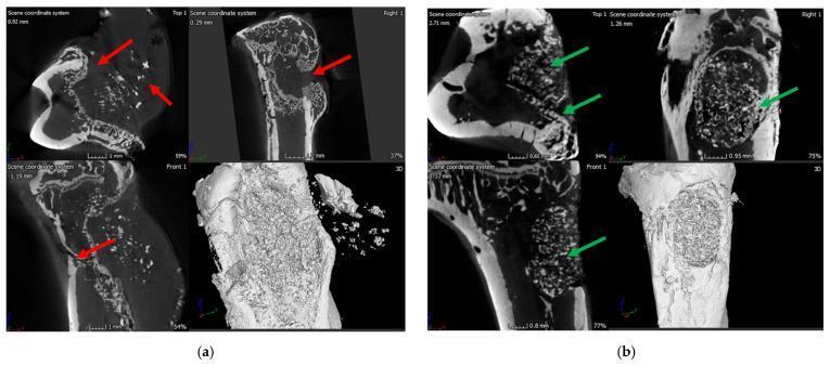
This diagnostic image set consists of Micro-CT (μ-CT) scans and 3D reconstructions of rat tibia used in a bone healing and infection study. Figure (a) illustrates an infection control group exhibiting characteristic signs of osteomyelitis. Red arrows highlight significant pathology, including a narrowing of the marrow space, deformed cortical bone, ectopic bone growth, and the presence of a sinus tract associated with a soft-tissue fibrous capsule. These features demonstrate severe osteolysis and disrupted bone architecture common in chronic infection. Figure (b) displays the treatment group using bioactive glass (BG). Green arrows indicate positive regenerative outcomes, specifically the formation of new immature cancellous and cortical bone within the previously drilled defect. The 3D and cross-sectional views show successful osseointegration and structural remodeling, contrasting the pathological destruction in the control group with the regenerative progression in the treatment group. This image is relevant for orthopedic research and the study of antibiotic-eluting bone graft substitutes.
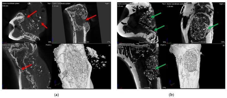
This comparative micro-computed tomography (μ-CT) study illustrates the pathophysiology of osteomyelitis and subsequent bone regeneration in a rat tibia model. Section (a) displays an untreated control group at 8 weeks post-infection with Staphylococcus aureus, showing significant structural pathology including ectopic bone growth (indicated by red arrows), narrowing of the medullary marrow space, and the formation of a sinus tract and pus-filled fibrous capsule. Section (b) demonstrates the therapeutic outcome 8 weeks after implantation of a vancomycin-loaded polymer/bioglass (BG) bone void-filling putty. This treated group exhibits successful infection eradication and advanced bone healing, characterized by the formation of new cortical and cancellous bone within the previous drilling site (indicated by green arrows). The images provide high-resolution cross-sectional and 3D reconstructions to demonstrate the efficacy of antibiotic-loaded scaffolds in treating orthopedic infections and promoting osteogenesis. This visual serves as an educational resource for medical microbiology, orthopedics, and biomaterials engineering.
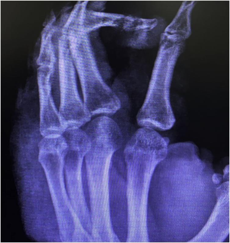
This diagnostic X-ray of the right hand demonstrates significant pathology localized to the third digit, consistent with chronic osteomyelitis. The imaging reveals extensive destruction of the cortical bone involving both the middle and distal phalanges. A hallmark finding of chronic infection, bone sequestration (sequestrum), is visible, characterized by fragments of necrotic, radiodense bone that have separated from the living bone tissue. The third finger shows marked anatomical deformity and soft tissue swelling. In contrast, the remaining phalanges, metacarpals, and visualized carpal bones maintain relatively normal bone density, cortical integrity, and joint alignment. This image serves as a clinical example of skeletal complications resulting from long-standing infections, such as those complicating cutaneous leishmaniasis with secondary bacterial superinfection. The primary educational focus is the radiological identification of cortical erosion and sequestrum formation in the context of chronic osteomyelitis.
acute osteomyelitis sequestrum involucrum histology microscopy
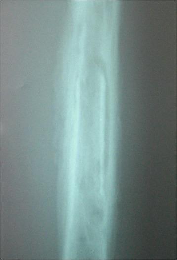
This diagnostic radiograph of a long bone demonstrates classic features of chronic haematogenous osteomyelitis, specifically classified as Beit CURE Type B2. The image reveals a central sequestrum, which is an isolated piece of necrotic, devascularized bone appearing as an area of increased radiopacity within the medullary canal. Surrounding the sequestrum is a well-defined involucrum, representing a layer of new periosteal bone formation. This involucrum appears structural and relatively normal in morphology, indicating that it is sufficiently stable to maintain the bone's integrity despite the underlying infection. The radiological presentation illustrates the characteristic pathological progression of chronic osteomyelitis where the body attempts to wall off infected necrotic tissue with a sheath of living bone. The image is an essential educational example for orthopedic surgery and radiology, highlighting the distinction between necrotic sequestrum and regenerative involucrum.
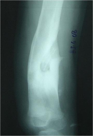
This diagnostic image is an anteroposterior x-ray radiograph of a long bone (tibia) demonstrating classic features of chronic haematogenous osteomyelitis. The radiograph reveals Type B3 classification findings characterized by the presence of a sequestrum—a dense, radiopaque segment of necrotic bone isolated from the surrounding living tissue. Encasing the sequestrum is a prominent involucrum, which appears as a thick layer of new periosteal bone formation. This involucrum is described as structural, sclerotic, and expanded, exhibiting significantly increased bone density and a widened diameter compared to normal bone morphology. The image illustrates the body's attempt to wall off infected necrotic tissue by creating a sheath of reactive bone. This visual is clinically significant for medical students and orthopedic specialists for identifying the radiographic hallmarks of pediatric chronic osteomyelitis and understanding the Beit CURE (BC) classification system for bone infections.
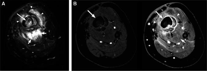
This diagnostic image set consists of axial MR images of the lower leg from a 4-year-old male, illustrating classic features of chronic osteomyelitis: involucrum and sequestrum. Image (A) is an axial T2-weighted fat-suppressed MRI demonstrating diffuse, high-signal intensity (edema) within the tibial medullary canal and the surrounding musculature. Image (B) shows side-by-side axial T1-weighted fat-suppressed images, pre-contrast (left) and post-contrast (right). The primary pathology shows a central, non-enhancing necrotic bone segment (sequestrum) indicated by a long white arrow. Surrounding this is a peripheral shell of new, enhancing bone formation (involucrum), highlighted by short black arrows. The surrounding soft tissues exhibit extensive enhancement and edema (arrowheads and short white arrows), consistent with myositis. These findings are pedagogically significant for distinguishing between viable inflammatory tissue (involucrum, myositis) and non-viable necrotic bone (sequestrum) in pediatric musculoskeletal infections. Target concepts include radiological identification of chronic infection markers and differentiate enhancement patterns in osteomyelitis.
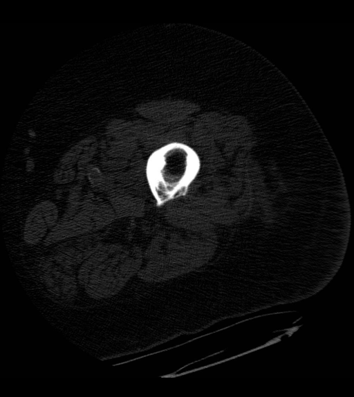
**Imaging Modality:** Axial Computed Tomography (CT) scan. **Anatomical Region:** Mid-shaft of a long bone (likely the femur or humerus) and surrounding soft tissues of the extremity. **Observed Pathology:** Chronic osteomyelitis with characteristic features of bone remodeling and sequestration. **Characteristic Visual Features:** * **Sequestrum:** A central, hyperdense fragment of necrotic bone is visible within the medullary cavity, detached from the surrounding living bone. * **Involucrum:** Marked circumferential cortical thickening and reactive sclerosis are present, representing the formation of new bone sheath around the infected site. * **Cortical Irregularity:** There is evidence of cortical erosion and architectural distortion. * **Soft Tissue Involvement:** Diffuse swelling and blurring of myofascial planes are noted in the adjacent soft tissues, suggesting associated inflammation or phlegmon. **Key Diagnostic Features:** The combination of a dense, sequestered bone fragment (sequestrum) surrounded by thickened reactive bone (involucrum) is pathognomonic for chronic osteomyelitis. This CT image effectively demonstrates the classic radiographic "bone within a bone" appearance used to differentiate chronic infectious processes from acute osteomyelitis or primary bone tumors.
OSTEOMYELITIS - Short Note (5 Marks)
ETIOLOGY
Routes of Infection
ROUTES OF INFECTION
         |
   ______|_______________________________
   |              |                     |
Hematogenous   Contiguous spread    Direct implantation
(most common   (from adjacent       (compound fractures,
in children)    soft tissue)         orthopedic surgery,
                                      diabetic foot)
Causative Organisms by Patient Group
| Patient Group | Most Likely Organism(s) |
|---|---|
| Children (most common) | Staphylococcus aureus |
| Neonates | Group B Streptococci, E. coli |
| Sickle cell anemia | Salmonella spp., gram-negative rods |
| Post-surgical / open fracture | Mixed organisms |
| Immunocompromised | Fungi, mycobacteria, gram-negatives |
| Diabetic foot | Polymicrobial |
Key point: S. aureus is the most common pathogen overall. Its cell wall proteins bind collagen in bone matrix, facilitating adherence. No organism is identified in ~50% of cases. (Robbins & Kumar Basic Pathology, p. 779)
PATHOGENESIS FLOWCHART
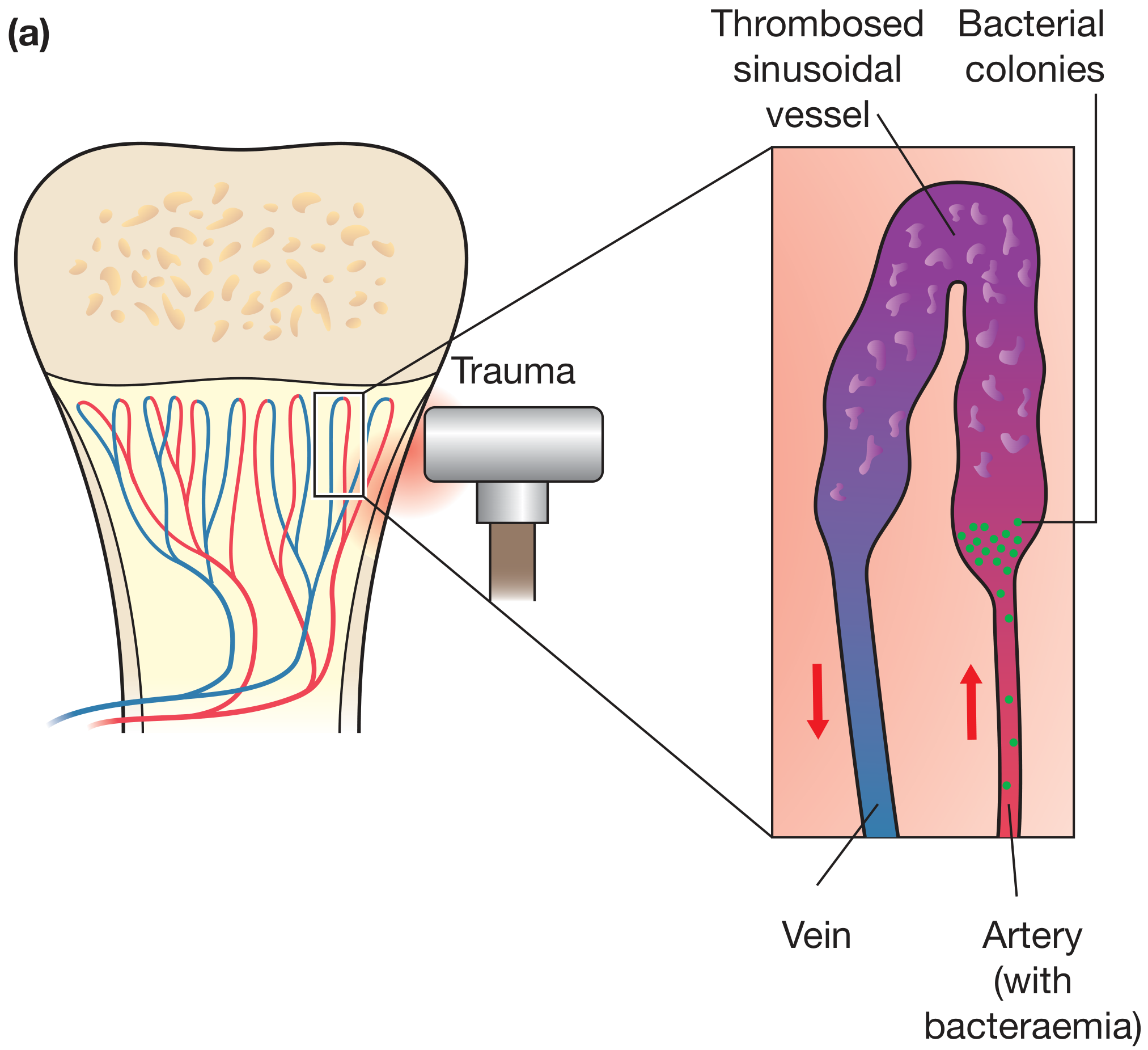
Pathogenesis Flow:
Bacteraemia
     |
     v
Seeding in metaphyseal sinusoids (slow flow + microtrauma)
     |
     v
Bacterial proliferation → Neutrophilic exudate (within 48h)
     |
     v
Increased intraosseous pressure → Vascular occlusion
     |
     v
BONE NECROSIS (osteocytes die within 48h)
     |
  ___|_______________________________________________
  |                                                 |
Spread through Haversian canals                  Epiphyseal spread
to PERIOSTEUM                                    → SEPTIC ARTHRITIS
  |
  v
Periosteal elevation (looser in children)
  |
  v
Subperiosteal abscess → Further ischaemia
  |
  v
Rupture → Soft tissue abscess → SINUS TRACT to skin
PATHOLOGY (Morphology)
Acute Phase (0-2 weeks)
- Bacteria proliferate; neutrophilic infiltrate is the hallmark
- Bone and marrow necrosis within 48 hours
- Pus tracks through Haversian systems to periosteum
- Subperiosteal abscess forms (periosteum easily lifted in children)
- Periosteal stripping worsens ischaemia
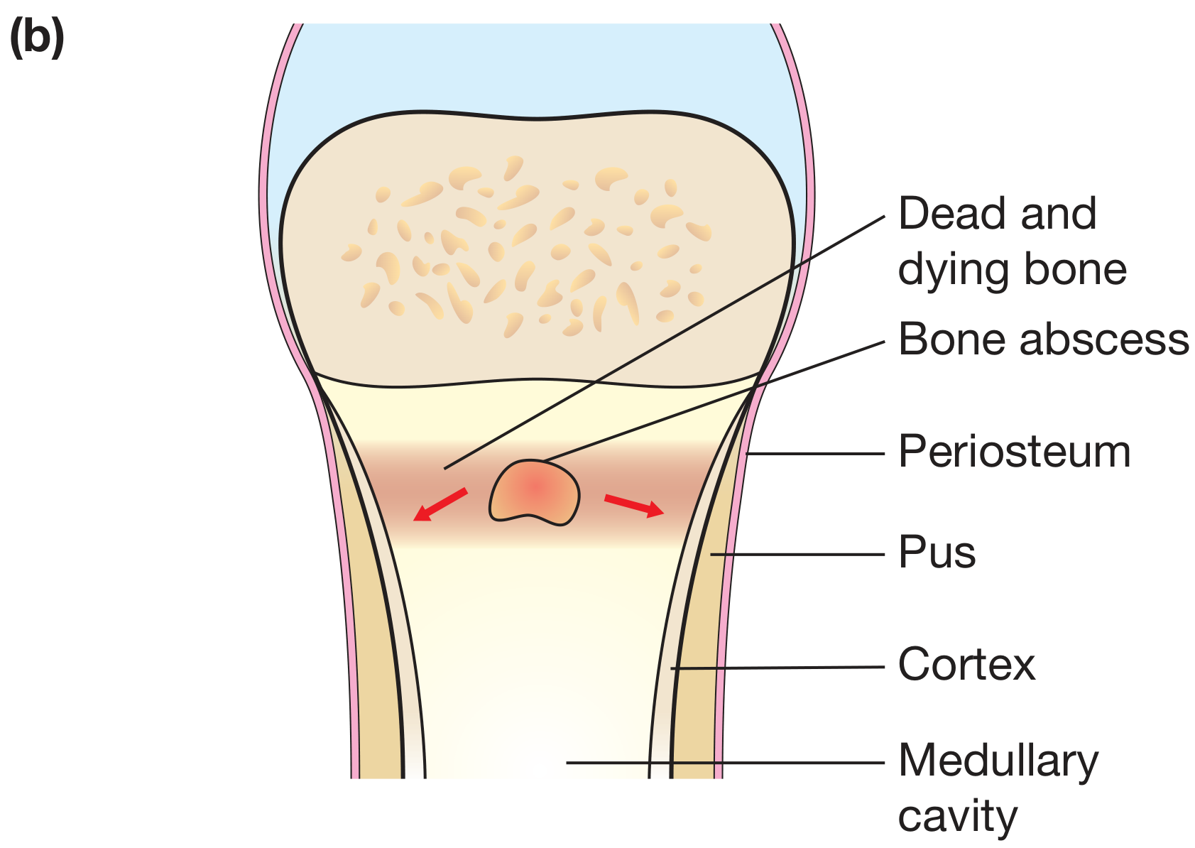
Chronic Phase (after 1 week)
- Chronic inflammatory cells release cytokines → osteoclastic resorption + fibrous tissue ingrowth
- Sequestrum - segment of dead, devitalized bone, separated from living tissue; appears as dense, radiodense fragment
- Involucrum - shell of new periosteal bone deposited around the dead bone; the periosteum is a potent stimulus for osteogenesis
- Results in "bone within a bone" appearance on imaging
CHRONIC OSTEOMYELITIS
        |
  Necrotic bone = SEQUESTRUM (dead, infected, avascular)
        |
  Surrounded by reactive new bone = INVOLUCRUM
        |
  Openings in involucrum = CLOACAE (pus discharge channels)
        |
  Tract to skin = SINUS TRACT (draining sinus)
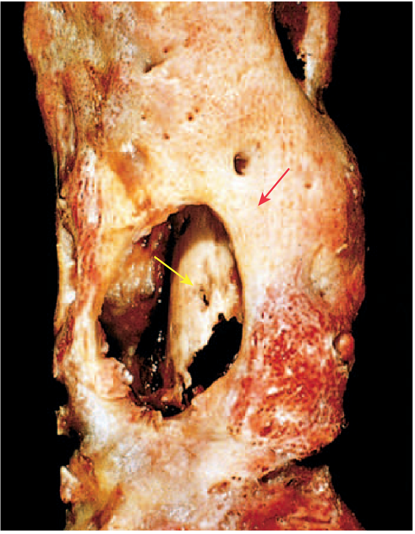
- Marrow fibrosis
- Sequestrum (necrotic bone trabeculae, empty lacunae)
- Lymphocytes and plasma cells (replaces the neutrophilic infiltrate of acute phase)
STAGES OF PROGRESSION (Imaging Diagram)
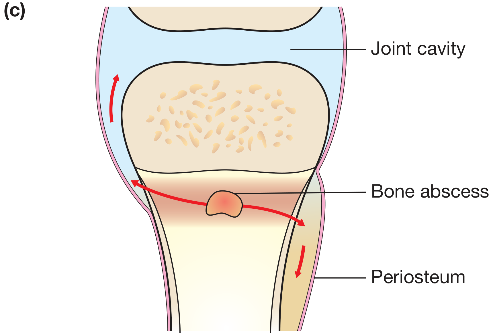
SPREAD TO JOINT (Important Special Case)
CLINICAL FEATURES
- Fever, malaise, chills, leukocytosis
- Throbbing pain and tenderness over bone
- Warmth, swelling, erythema over affected region
- In infants: unexplained fever, pseudoparalysis of limb
- X-ray: lytic focus + reactive bone (changes appear 10-14 days late)
- MRI: most sensitive and specific (early detection)
- Bone scan: useful when MRI unavailable
- Blood culture + bone biopsy/culture (needed to ID organism)
- ESR, CRP elevated; leukocytosis
COMPLICATIONS
COMPLICATIONS OF OSTEOMYELITIS
            |
    ________|_______________________________
    |           |           |              |
Pathologic   Septic      Squamous cell   Secondary
fracture    arthritis    carcinoma in    amyloidosis
                         sinus tract
            |
         Sepsis / endocarditis
TREATMENT (Principles)
- Antibiotics: IV initially; culture-guided; oral step-down once improving clinically and haematologically
- MRSA coverage where prevalence is high (e.g., vancomycin)
- Surgical drainage of pus (decompression of bone abscess / subperiosteal abscess)
- Rest/splintage of affected limb; analgesia
- Chronic osteomyelitis: surgical debridement of sequestrum (sequestrectomy) + saucerization; bone grafting if needed
MRI CORRELATION
Acc to harsh mohan for dental students
osteomyelitis jaw mandible dental X-ray panoramic
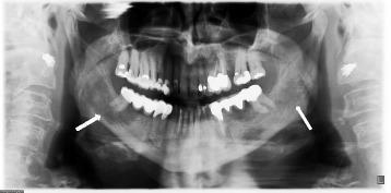
This diagnostic image is a panoramic X-ray (orthopantomogram) of the human maxillofacial region, showing the complete mandible, maxilla, and surrounding dental structures. Two white arrows indicate distinct pathological findings located bilaterally at the mandibular angles. These areas are characterized as osteolytic zones, presenting as radiolucent (darker) regions with ill-defined borders compared to the surrounding radiopaque cortical and cancellous bone. The osteolysis is observed on both the right and left sides of the lower jaw, with the right side showing more prominent involvement. Multiple dental restorations and evidence of previous dental procedures are visible across the dentition. The image serves as a clinical teaching tool for recognizing mandibular bone loss and differential diagnosis of systemic diseases manifesting in the jaw, such as sarcoidosis, osteomyelitis, or malignancy.
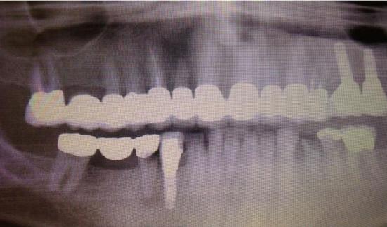
**Imaging Modality:** Orthopantomogram (Panoramic Dental X-ray). **Anatomical Region:** Maxilla and mandible with associated dentition. **Observed Pathology:** Medication-related osteonecrosis of the jaw (MRONJ). **Characteristic Visual Features:** The radiograph demonstrates ill-defined radiolucent and mixed radiopaque-radiolucent (moth-eaten) bone destruction. There is evidence of cortical bone erosion and sequestration, particularly noted in the alveolar regions. Findings include non-healing extraction sockets and widened periodontal ligament spaces. **Dental Status and Landmarks:** Extensive dental restorations are visible, including multiple radiopaque fixed prosthodontic bridges (maxillary and mandibular) and endosseous dental implants. There is significant generalized alveolar bone loss consistent with periodontitis. **Diagnostic Differentiating Features:** The combination of localized sequestrum formation, osteosclerosis, and persistent radiolucency in a patient with a history of bisphosphonate therapy is characteristic of drug-induced osteonecrosis. These features distinguish the condition from simple osteomyelitis or primary bone malignancy by the specific pattern of bone turnover suppression and necrotic bone involvement.
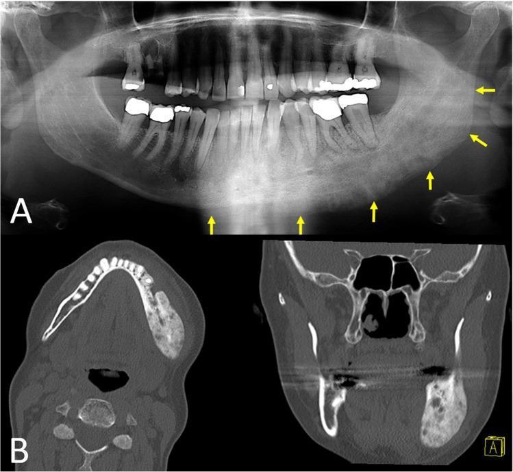
This composite educational image features a panoramic dental radiograph (A) and multiplanar CT scans (B) of a 50-year-old patient with chronic osteomyelitis of the left mandible. Panel A shows a panoramic X-ray demonstrating significant distension and deformity of the left hemimandible. Multiple yellow arrows indicate a mixed radiopaque and radiolucent appearance extending from the right parasymphyseal/premolar region to the posterior ramus, characterizing a combination of bone sclerosis and osteolytic destruction. Panel B contains axial and coronal CT slices that provide detailed visualization of the mandibular pathology. The left hemimandible exhibits marked cortical expansion and a heterogeneous internal structure. Notable features include increased bone marrow attenuation and irregular sclerotic masses interspersed with lucent areas, consistent with proliferative periostitis and reactive new bone formation. The right side of the mandible serves as a normal anatomical comparison, appearing non-expanded with clear cortical borders. This imaging illustrates the typical radiological features of secondary chronic osteomyelitis and proliferative periostitis in the maxillofacial region.
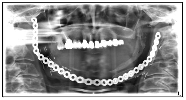
This panoramic dental X-ray (orthopantomogram) displays extensive surgical intervention and reconstruction of the mandible. A prominent, large-caliber metallic reconstruction plate with multiple circular screw holes spans the entire lower border of the mandible from the right to the left ramus. The image reveals significant continuity defects: there is a complete absence of native bone structure on the left side of the mandible and a marked discontinuity on the right, consistent with radical resection. The reconstruction plate is secured to the remaining mandibular segments with multiple screws; notably, some screws appear fractured or displaced. Visible radiopaque metallic clips are also scattered near the surgical site. The maxillary dentition remains relatively intact, showing normal alignment and restorations. The clinical significance of this imaging is to demonstrate post-surgical management of Medication-Related Osteonecrosis of the Jaw (MRONJ), highlighting the challenges of re-osteosynthesis following the failure of previous fibula grafts due to chronic inflammation and osteolysis.
OSTEOMYELITIS
(As per Harsh Mohan's Pathology - Dental Student Format | 5 Marks)
Note: Harsh Mohan's Textbook of Pathology is not available in our digital library. This note is compiled from Robbins & Kumar Basic Pathology, Robbins Pathologic Basis of Disease, and S Das's Manual on Clinical Surgery - all of which form the basis of Harsh Mohan's osteomyelitis chapter - with special emphasis on jaw osteomyelitis as relevant to dental students.
DEFINITION
ETIOLOGY & ROUTES OF INFECTION
ROUTES OF INFECTION IN OSTEOMYELITIS
              |
    __________|_____________________________
    |                 |                    |
HEMATOGENOUS    CONTIGUOUS SPREAD     DIRECT IMPLANTATION
(most common    (from adjacent        (tooth extraction,
in children;    soft tissue /         fractures, surgery,
via blood)      dental abscess)        implants)
                     ↑
          *** MOST IMPORTANT ROUTE
              FOR JAW OSTEOMYELITIS ***
Causative Organisms
| Setting | Organism |
|---|---|
| General (most common) | Staphylococcus aureus (80-90%) |
| Neonates | Group B Streptococcus, E. coli |
| Sickle cell anemia | Salmonella spp. |
| Jaw / dental (contiguous) | Mixed oral flora, anaerobes |
| Immunocompromised | Fungi, mycobacteria |
| Radiation/bisphosphonate | Sterile necrosis |
S. aureus cell wall proteins bind bone collagen, facilitating adherence. No organism found in ~50% of cases. (Robbins Pathologic Basis of Disease)
SPECIAL RELEVANCE TO DENTAL STUDENTS - OSTEOMYELITIS OF THE JAW
Why the Mandible is More Affected than the Maxilla
MANDIBLE more affected because:
→ Single blood supply (inferior alveolar artery)
→ Endarteritis → easy vascular occlusion → necrosis

MAXILLA less affected because:
→ Multiple anastomosing vertical arteries
→ Collateral blood supply maintained
THREE TYPES OF JAW OSTEOMYELITIS (S Das Classification)
(i) Acute Osteomyelitis of the Jaw
- Occasionally seen in infants as a complication of acute fevers (measles, scarlet fever)
- Either upper or lower jaw may be affected
- Features: swelling, redness, puffiness
- In maxilla: pressure may cause pus from nostril
- X-ray not helpful in early stages
(ii) Subacute Osteomyelitis (Commonest)
- Adults mainly affected
- Causes: Apical dental infection, alveolar abscess, fractured jaw, injudicious tooth extraction with poor general condition
- Mechanism: Endarteritis of the inferior alveolar artery → obstruction → bone necrosis
- Features: pain, swelling, tenderness, bone irregularity
- Vincent's symptom / Numbness of chin - increased tension in dental canal compresses the inferior dental nerve → numbness in distribution of mental nerve
- X-ray shows bone necrosis only after 3 weeks
(iii) Chronic Osteomyelitis
- Affects mandible more than maxilla
- Follows apical dental infection, alveolar abscess, fractures
- Also follows: radiation necrosis, phosphorus poisoning (phossy jaw), TB, syphilis, actinomycosis
- X-ray: localized osteitis, abscess (Brodie's abscess-like), sequestrum
PATHOGENESIS FLOWCHART
Dental infection / bacteraemia / trauma
              ↓
Bacterial seeding in bone (metaphysis in long bones;
dental apex / alveolus in jaw bones)
              ↓
Bacterial proliferation → NEUTROPHILIC EXUDATE
              ↓
Increased intraosseous pressure
              ↓
Vascular thrombosis → ISCHAEMIA
              ↓
BONE NECROSIS (within 48 hours)
              ↓
    __________|___________________________
    |                                    |
Pus spreads through Haversian           Spread to adjacent jaw →
canals → Periosteum elevated            Ludwig's angina / space infections
    ↓
Subperiosteal abscess
    ↓
Periosteum stripped → ↑↑ Ischaemia
    ↓
Rupture → Sinus tract (e.g., Median Mental Sinus)
    ↓ (if untreated, >1 week)
Chronic inflammatory cells / cytokines
    ↓
Osteoclastic resorption + Fibrous tissue ingrowth
    ↓
Dead bone = SEQUESTRUM
New periosteal bone = INVOLUCRUM
Opening in involucrum = CLOACA
PATHOLOGICAL STAGES
ACUTE PHASE (0-2 weeks)
- Neutrophilic infiltrate - hallmark
- Bone and marrow necrosis within 48 hours
- Subperiosteal abscess formation
- Periosteal elevation especially prominent in children (periosteum loosely attached)
CHRONIC PHASE (after 1-2 weeks)
- Lymphocytes and plasma cells replace neutrophils
- Marrow fibrosis
- Three key structures form:
┌─────────────────────────────────────────────────┐
│  SEQUESTRUM                                     │
│  = Fragment of dead, devitalized, avascular     │
│    bone; dense on X-ray (radiodense)            │
│  ↓ surrounded by ↓                             │
│  INVOLUCRUM                                     │
│  = Shell of new reactive periosteal bone        │
│    deposited around dead bone                   │
│  ↓ openings in involucrum ↓                    │
│  CLOACA                                         │
│  = Holes in involucrum through which pus        │
│    and fragments discharge → SINUS TRACT        │
└─────────────────────────────────────────────────┘
SPECIAL FORMS OF JAW OSTEOMYELITIS (Important for Dentistry)
1. Sclerosing Osteomyelitis of Garré
- Chronic form; commonly affects mandible in children
- A form of CRMO (Chronic Recurrent Multifocal Osteomyelitis) of the mandible
- Non-suppurative ossifying periostitis with subperiosteal new bone formation and sclerosis
- Features: pain and hard bony swelling of mandible
- Cultures usually negative; diagnosis by radiology + biopsy
- Biopsy: features of chronic osteomyelitis (Grainger & Allison's Diagnostic Radiology)
2. Median Mental Sinus
- A form of chronic osteomyelitis
- Apical abscess of lower incisor(s) → cortical penetration → pus accumulates under mentalis muscle → sinus discharges midline on the chin (S Das Manual on Clinical Surgery)
3. Alveolar Abscess → Osteomyelitis
- Progression pathway:
Acute pulpitis
     ↓
Root canal infection
     ↓
Localised osteitis (periapical)
     ↓
Alveolar abscess
     ↓  (if untreated)
OSTEOMYELITIS OF JAW
4. Radiation Osteomyelitis / Osteoradionecrosis
- Follows radiation therapy to jaw (e.g., for oral cancer)
- Radiation destroys vascularity → bone necrosis → secondary infection
- May present years after radiation
5. Bisphosphonate-Related Osteonecrosis of the Jaw (BRONJ / MRONJ)
- Associated with bisphosphonate therapy (used in osteoporosis, cancer)
- Triggered by dental extraction or trauma
- Non-healing necrotic bone, exposed through oral mucosa
RADIOLOGICAL FEATURES (X-Ray / OPG)
| Stage | Radiological Appearance |
|---|---|
| Early acute (<10 days) | Normal or subtle soft tissue swelling |
| Subacute (10-21 days) | Lytic areas; loss of lamina dura around roots |
| Chronic | "Moth-eaten" appearance; sequestrum (dense fragment); involucrum; sclerosis |
CLINICAL FEATURES
- Fever, toxaemia, malaise
- Severe pain over jaw, trismus (if masseter involved)
- Swelling, redness, oedema of gum
- Loose teeth in affected region
- Vincent's symptom - numbness of lower lip/chin (mental nerve compression)
- Regional lymphadenopathy
- Dull aching pain, persistent swelling
- Discharging sinus (may open on face or intraorally)
- Sequestrum palpable or visible
- Pathological fracture of mandible (in severe cases)
COMPLICATIONS
COMPLICATIONS
      |
   ___|_________________________________
   |          |           |            |
Pathologic  Septic    Ludwig's     Spread to
fracture   arthritis   angina      cavernous sinus
(mandible)            (lower jaw   (dental abscess →
                      infections)  cavernous sinus
                                   thrombosis)
   |              |
Squamous cell   Secondary
carcinoma in    amyloidosis
sinus tract     (rare, chronic)
TREATMENT
- Antibiotics: IV penicillin/clindamycin (covers oral flora); culture-guided; MRSA coverage where needed
- Surgical drainage of abscess (incision and drainage)
- Sequestrectomy - surgical removal of sequestrum
- Saucerization - removal of overhanging bone to allow drainage and healing
- Removal of offending tooth (the source of dental infection)
- Debridement of necrotic bone; bone grafting if needed
- Hyperbaric oxygen (HBO) - useful in radiation osteomyelitis