Current concepts of osteoarthritis md pathology 10 marks question give me beautiful answer that I can replicate in my exam
osteoarthritis joint pathology eburnation cartilage loss osteophyte
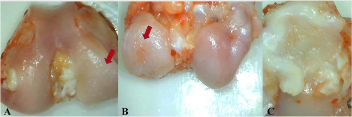
This set of gross pathological macrographs (labeled A, B, and C) illustrates severe osteoarthritic changes in a knee joint model. Image A and B focus on the femoral condyles, where red arrows highlight prominent eburnation, surface irregularities, and pitting in the load-bearing regions. Image B specifically demonstrates a localized full-thickness articular cartilage ulcer on the weight-bearing surface of the medial femoral condyle. Image C shows the medial tibial plateau, characterized by significant degenerative changes including peripheral whitish osteophyte formation and marginal erosions. These visual markers—cartilage loss, subchondral bone exposure (eburnation), and osteophytosis—are hallmark macroscopic features used to grade the severity of osteoarthritis and evaluate the impact of surgical interventions like meniscectomy on joint health. The images provide clinical context for orthopedic pathology and rheumatology education, emphasizing the progressive degradation of articular surfaces in chronic degenerative joint disease.
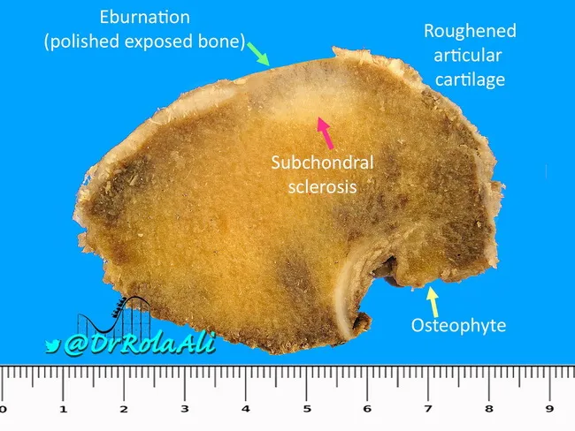
Imaging modality: Gross pathology photograph of a dissected femoral head demonstrating advanced degenerative joint disease consistent with osteoarthrosis. Specimen is the proximal femur with preserved cartilage surfaces (articular cartilage) inconsistent across regions; the dominant features include deformed, flattened joint surfaces and loss of articular cartilage bilaterally, exposing underlying subchondral bone. Subchondral sclerosis is evident as density increase beneath the cartilage, and full-thickness cartilage loss results in bone-on-bone contact at several points. Repeated articulation produces eburnation: polished, ivory-like exposed bone on opposing surfaces. Peripherally, marginal osteophytes project from the joint margin and localize along articular surfaces; these osteophytes may be covered by variable hyaline or fibrocartilage. The specimen shows deformity of the femoral head with contour irregularities and potential impingement of articular surfaces; roughened cartilage is present on exposed surfaces. The scale ruler at the base provides gross dimensions. Clinically, these findings correlate with chronic hip pain, reduced range of motion, and radiographic features of osteoarthritis such as joint space narrowing and osteophyte formation. This image is valuable for teaching gross pathology, orthopedic and rheumatology education, and cross-disciplinary differential diagnosis discussions, including osteoarthritis, secondary OA from prior trauma, and osteonecrosis with secondary degenerative changes.
Osteoarthritis (Degenerative Joint Disease) — 10 Mark Answer
1. Definition
2. Classification
| Type | Features |
|---|---|
| Primary (idiopathic) OA | Most common; insidious onset, no apparent cause; an "aging phenomenon"; oligoarticular, affects weight-bearing joints. Prevalence rises exponentially after age 50 — ~40% of people over 70 are affected. |
| Secondary OA | ~5% of cases; occurs in younger patients with a predisposing condition — joint deformity, prior joint injury/trauma, or systemic disease (diabetes, obesity). |
3. Pathogenesis (write as a flow/diagram — high yield)
- Chondrocyte injury in a genetically predisposed patient → alteration of the extracellular matrix (loss of proteoglycans, disruption of collagen network).
- Early OA: Chondrocytes attempt repair by proliferating and secreting matrix, but degradation (via matrix metalloproteinases, MMPs) exceeds repair.
- Mediators involved: IL-1, TNF, nitric oxide (NO), prostaglandin E2 (PGE2), TGF-β, BMP — these drive catabolic enzyme release and attempted (failed) repair.
- Late OA: Progressive loss of both matrix and chondrocytes, with damage extending into the subchondral bone.
- Biomechanical stress at the joint margins stimulates osteophyte (bony outgrowth) formation.
4. Morphology
- Chondrocyte loss and severe matrix degradation with full-thickness sloughing of cartilage
- Dislodged fragments of cartilage/bone float in the joint space as loose bodies ("joint mice")
- Exposed subchondral bone becomes the new articulating surface; friction polishes it to an ivory-like sheen — bone eburnation
- Small subchondral fractures allow synovial fluid to be forced into bone by a ball-valve mechanism, producing fibrous-walled subchondral cysts
- Osteophytes — bony outgrowths at the joint margins, capped by fibrocartilage/hyaline cartilage that gradually ossifies
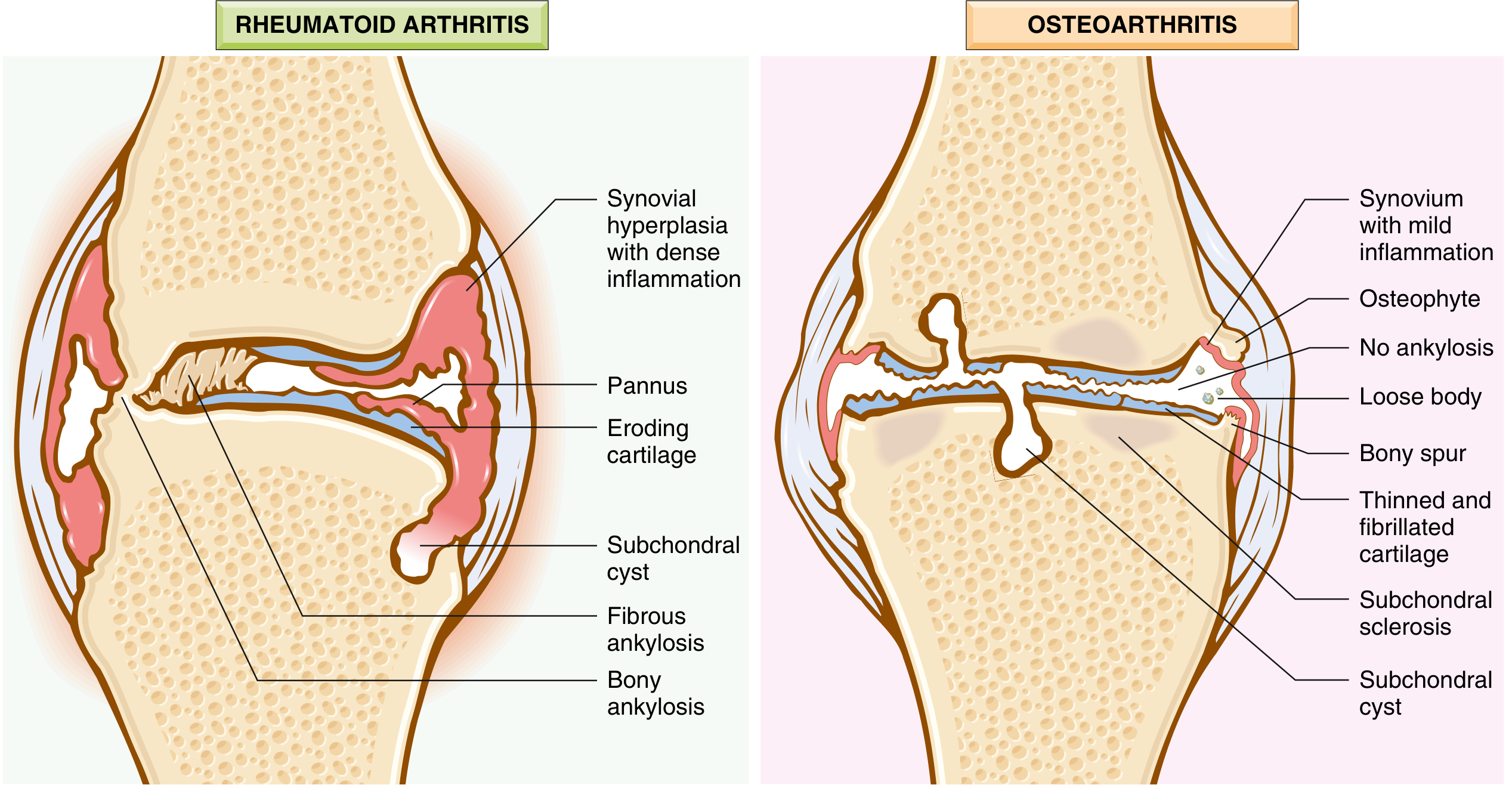
- Synovium: only mildly congested and fibrotic, with scattered chronic inflammatory cells (minimal inflammation, unlike RA)
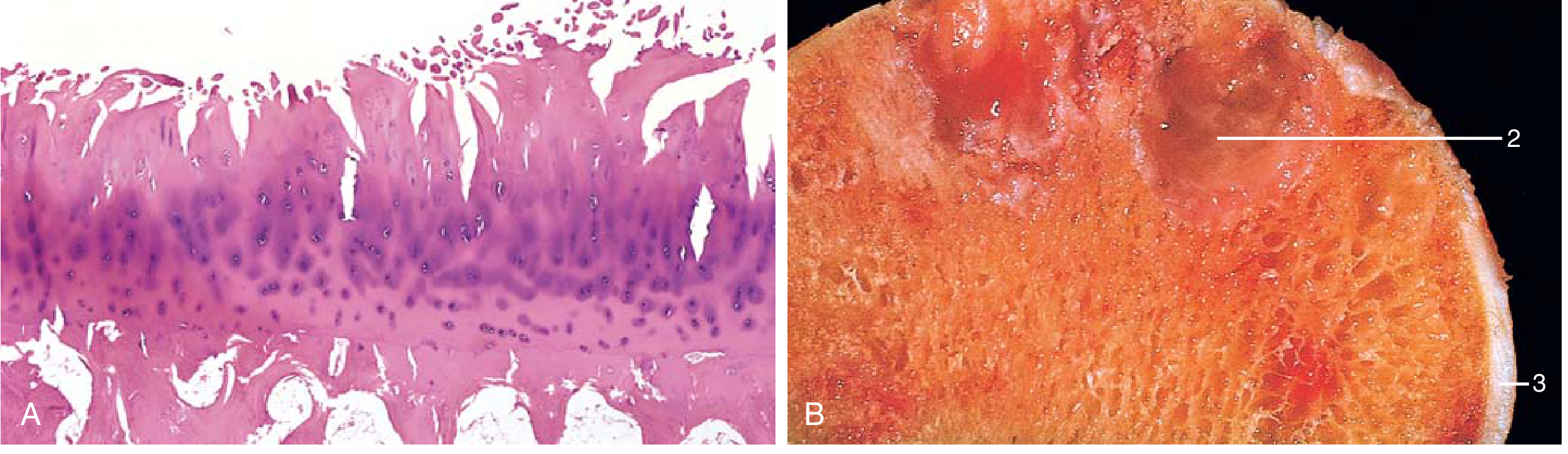
- Early: fibrillation (vertical clefts) of the articular cartilage surface
- Late: eburnated bone, subchondral cysts, residual fragmented cartilage
5. Clinical Features
- Onset typically in the 50s; if OA appears in a young person, look for an underlying secondary cause
- Joint pain worsening with use, morning stiffness, crepitus, and restricted range of motion
- Osteophyte impingement on spinal foramina → nerve root compression → radicular pain, muscle spasm/atrophy, neurologic deficits
- Joints commonly affected: hips, knees, lower cervical and lumbar vertebrae, PIP/DIP joints of fingers, first carpometacarpal joint, first tarsometatarsal joint
- Heberden nodes — prominent osteophytes at the DIP joints, more common in women
- Joint deformity may occur over time, but unlike RA, joint fusion (ankylosis) does NOT occur
- Radiographic severity correlates poorly with pain/disability
- No disease-modifying treatment exists: management is symptomatic — pain control, NSAIDs, intra-articular corticosteroids, activity modification, and joint replacement in severe disease
6. High-Yield Comparison Table: OA vs Rheumatoid Arthritis (write this — examiners love it)
| Feature | Osteoarthritis | Rheumatoid Arthritis |
|---|---|---|
| Primary pathogenic abnormality | Mechanical injury to articular cartilage | Autoimmunity |
| Role of inflammation | Secondary; exacerbates damage | Primary; T cells/antibodies destroy cartilage |
| Joints involved | Weight-bearing (knees, hips) | Small joints of hands first, then polyarticular |
| Pathology | Cartilage degeneration/fragmentation, osteophytes, subchondral cysts; minimal inflammation | Inflammatory pannus destroying cartilage; severe chronic inflammation; ankylosis |
| Serum antibodies | None | RF, ACPA (anti-citrullinated peptide antibody) |
| Other organ involvement | No | Yes (lung, heart, etc.) |