
Anatomy of external ear in simplified manner
Reading File
Now let me search the medical library for external ear anatomy content.
Finding Sources
Finding Sources
Good results. Let me read the most relevant sections from Gray's Anatomy and K.J. Lee's Otolaryngology in parallel.
Reading File
Reading File
Good - I have the Gray's Anatomy content. Let me now read more of it (the EAM section) and fetch the auricle diagram image in parallel.
Reading File
Loading Image
Loading Image
Now let me get the EAM (external acoustic meatus) section from Gray's Anatomy:
Reading File
Let me also get the detailed EAM content from the Cummings Otolaryngology section, and get the second anatomy diagram:
Reading File
Loading Image
I now have excellent content and images from Gray's Anatomy and Cummings Otolaryngology. Let me compile a clear, simplified answer.
Anatomy of the External Ear - Simplified
The external ear has two main parts: the Auricle (Pinna) and the External Acoustic Meatus (EAC). Together, they capture and channel sound toward the eardrum.
Part 1 - The Auricle (Pinna)
The visible part of the ear that sticks out from the side of your head. It is made of elastic cartilage covered by skin - except for the earlobe, which has no cartilage.
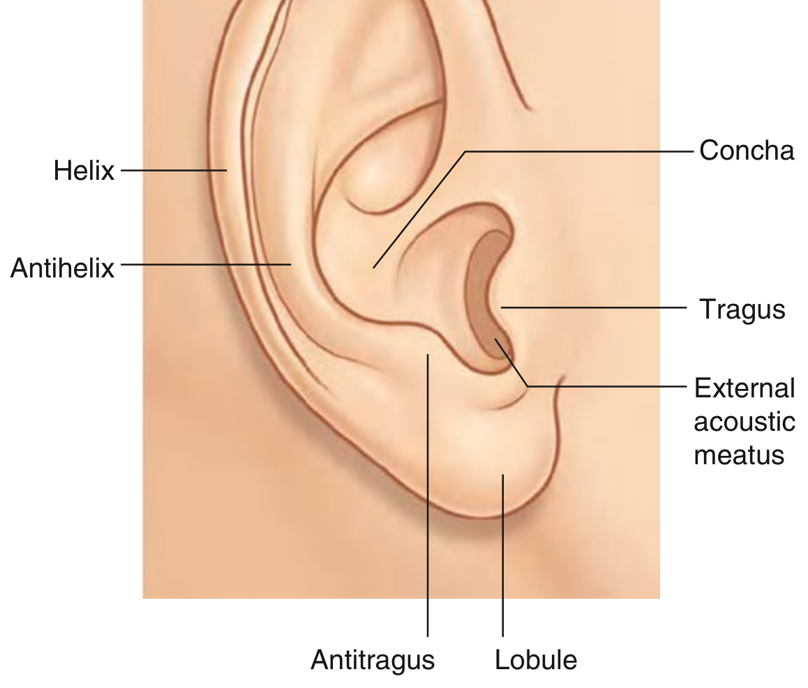
Here is a labeled diagram from Gray's Anatomy for Students:
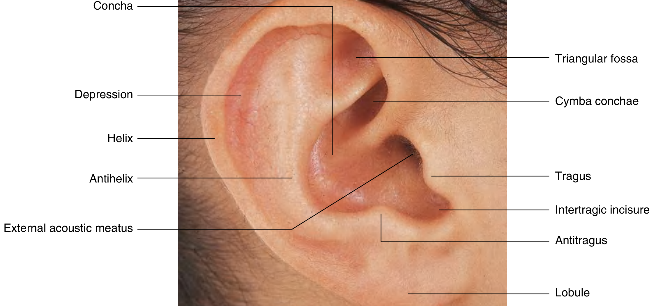
And a real photo of the external ear with all key landmarks labeled:
Key Landmarks of the Auricle
| Structure | What It Is |
|---|---|
| Helix | The large folded outer rim of the ear |
| Antihelix | A smaller curved ridge parallel and inside the helix |
| Scaphoid fossa | The depression (groove) between the helix and antihelix |
| Concha | The deep hollow bowl at the center of the ear - leads into the ear canal |
| Cymba conchae | The upper part of the concha (above the antihelix crus) |
| Triangular fossa | The depression in the fork of the antihelix (upper part) |
| Tragus | A small knob just in front of the ear canal opening |
| Antitragus | The small bump opposite the tragus, above the earlobe |
| Intertragic incisure | The notch (dip) between tragus and antitragus |
| Lobule (Earlobe) | The soft, fleshy lower tip - the only part with NO cartilage |
Simple way to remember: Helix = outer rim. Antihelix = inner ridge. Concha = the bowl. Tragus = the tab in front. Lobule = the earlobe.
Part 2 - The External Acoustic Meatus (EAC) / Ear Canal
The canal that runs from the concha inward to the eardrum (tympanic membrane). It is about 2.5 cm long and has two portions:
| Portion | Location | Lining |
|---|---|---|
| Cartilaginous (outer 1/3) | Lateral/outer part | Thick skin with hair follicles, sebaceous glands, and ceruminous (wax) glands |
| Bony (inner 2/3) | Medial/inner part | Thin, delicate skin without glands or hair; continuous with the eardrum |
- The narrowest point of the canal is the isthmus - the junction between the cartilaginous and bony parts.
- Cerumen (earwax) is produced only in the outer cartilaginous portion.
- The canal runs slightly anteroinferiorly - to straighten it for examination, pull the ear posterosuperiorly in adults.
Blood Supply
Comes from the external carotid artery via:
- Posterior auricular artery - supplies the posterior auricle
- Superficial temporal artery - supplies the anterior auricle and part of the canal
Nerve Supply (Sensory)
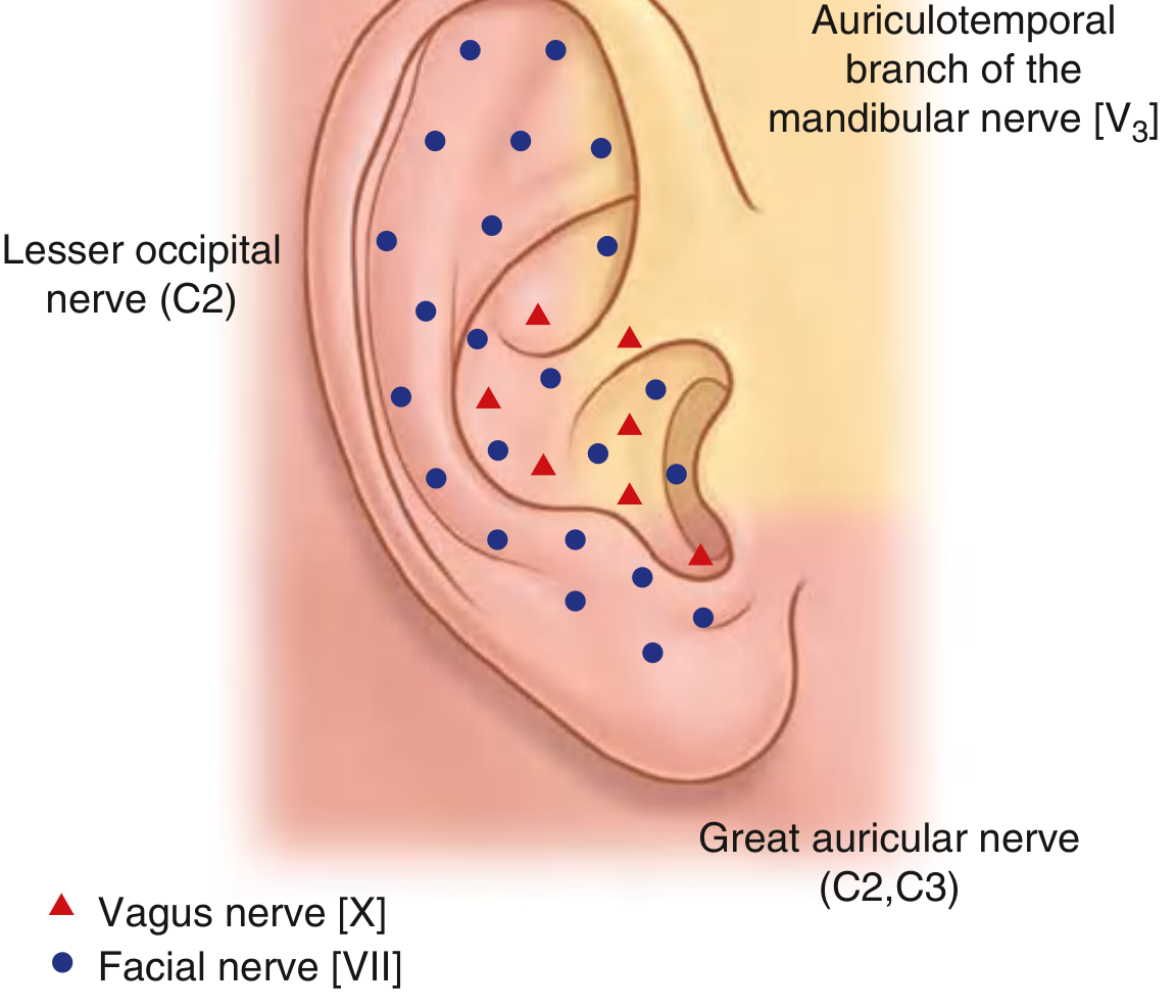
The auricle has a rich sensory nerve supply from multiple nerves - an important clinical reason why ear pain can be referred from many areas:
| Nerve | Area Supplied |
|---|---|
| Great auricular nerve (C2, C3) | Most of the auricle - anterior and posterior inferior surfaces |
| Lesser occipital nerve (C2) | Posterosuperior part |
| Auriculotemporal nerve (V3) | Anterior auricle and anterior wall of EAC |
| Vagus nerve (CN X) - Arnold's nerve | Posterior EAC wall and posterior auricle |
| Facial nerve (CN VII) | Small area of the auricle |
Clinical pearl: The vagus nerve (Arnold's nerve) supplies the EAC. Stimulating the posterior canal wall (e.g., during ear syringing) can trigger a cough reflex or even syncope (vasovagal response). This is called Arnold's reflex.
Muscles of the Auricle
Two groups:
- Intrinsic muscles - between cartilage parts; can slightly change shape
- Extrinsic muscles - anterior, superior, and posterior auricular muscles; connect to the scalp and can move the ear slightly
Both groups are supplied by the facial nerve (CN VII). These muscles are mostly vestigial in humans.
Development (Embryology - Quick Note)
The external ear develops from the 1st and 2nd branchial arches. Six tissue hillocks (hillocks of His) fuse to form the auricle. The ear canal (EAC) forms from the 1st branchial groove (cleft). Failure of development leads to conditions like aural atresia or preauricular sinuses.
Sources: Gray's Anatomy for Students, 4th ed., p. 1094-1097; Cummings Otolaryngology Head and Neck Surgery, Chapter 126.
This is a shared conversation. Sign in to Orris to start your own chat.