
Ingestional organophophorous compund poisioning
organophosphate poisoning management
organophosphate poisoning cholinergic syndrome clinical features
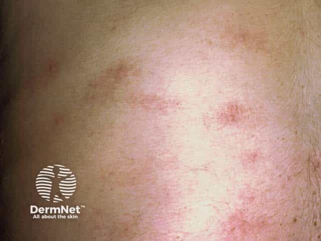
Clinical dermatology photograph of the skin displaying transient, pruritic wheals consistent with cholinergic urticaria. Modality: Dermatologic photography (clinical image); technique: high-resolution color photograph, close-up view of the cutaneous surface under standard ambient lighting; magnification not specified. Anatomical location: lower extremity skin, leg region, with clustered erythematous wheals visible on a lightly pigmented field. Visual features: well-circumscribed, raised erythematous plaques with blanching on pressure, variable size, often several millimeters to a centimeter, arranged scatteredly or in small groups; superficial dermal edema is suggested by diffuse redness and swelling, with surrounding fine border erythema. Clinical interpretation: wheals appear transient and pruritic, erupting in response to heat and exertion, a hallmark of cholinergic urticaria. Pathophysiology: mast cell degranulation and dermal edema in superficial plexuses; histology (if obtained) would show dermal edema with minimal epidermal change and mild perivascular infiltrates. Diagnostic significance: supports diagnosis when symptoms occur shortly after physical activity or temperature rise; aids differentiation from miliaria, contact dermatitis, or heat rash. Clinical relevance: helps guide management including antihistamines, trigger avoidance, and patient education; useful in educational and research settings for dermatology teaching, urticaria phenotyping, and exercise-induced hypersensitivity studies.
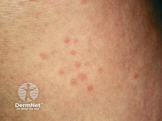
This is a dermatology clinical photograph of the skin surface demonstrating the typical small, transient wheals seen in cholinergic urticaria. The image shows numerous pinpoint to 3–4 mm erythematous, raised plaques scattered across the forearm in a crops-like distribution. Each wheal shows central blanching with a slightly edematous, ring-like border and a blanching halo consistent with superficial dermal edema. The lesions are pruritic and short-lived, usually resolving within 1–2 hours, often appearing after heat exposure, exercise, or emotional stress. In cholinergic urticaria, symptoms may be accompanied by warmth, sweating, or mild warmth-induced flushing. The focal photodermatographic appearance is not necessarily specific but the small size, rapid onset following provocative stimuli, and distribution pattern strongly support a physical urticaria rather than mast cell neoplasia or dermatitis. Clinically relevant features include the ability to trigger crops, readiness to respond to oral antihistamines, and potential improvement with pre-exercise cooling or preventive antihistamine therapy. The differential diagnosis includes other physical urticarias (dermographism, heat urticaria), ordinary urticaria, papular urticaria, and less commonly urticarial vasculitis. This image is most useful for educational illustration of cholinergic urticaria triggers: exercise, heat, stress; and for training in clinical recognition and patient counseling.
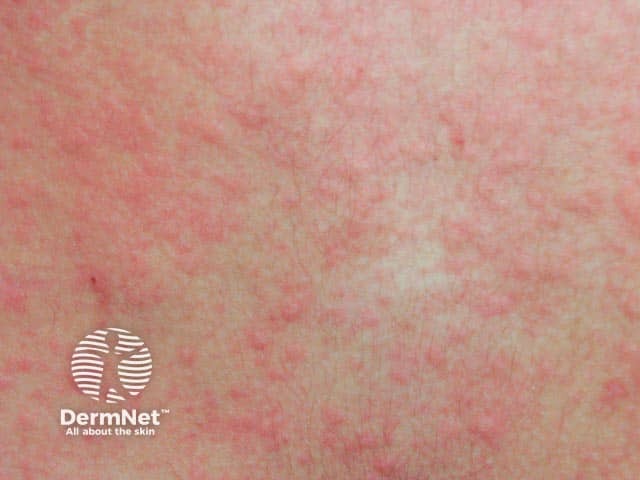
Imaging modality: Clinical photography of cutaneous surface using standard white-light illumination. Technique: frontal anterior view of the trunk with natural skin tone, without dermoscopy or special staining. Anatomical location: superficial skin of the torso with generalized distribution. Visual features: multiple small, erythematous wheals (1–5 mm) embedded in diffuse erythema; wheals are blanching under pressure and may coalesce into transient plaques; pruritus is often prominent. Pathophysiology: cholinergic urticaria is a heat- or sweat-triggered inducible urticaria resulting from mast cell degranulation. On clinical image, lesions appear as punctate or annular wheals in exposed or covered skin following heat exposure; lesions last minutes to an hour and may be accompanied by warmth, flushing, or burning sensation. Diagnostic significance: the presence of small, pruritic wheals after heat, exercise, or emotional stress supports the diagnosis; differential diagnoses include miliaria, ordinary urticaria, burns-related erythema, and dermographism. Clinical utility: this image illustrates characteristic appearance for educational reference, aids in recognition by clinicians and trainees, supports case-based teaching, and can be correlated with history and response to antihistamines. Potential use cases: medical education, dermatology exam prep, AI training for skin lesion classification, and decision support for heat-induced urticaria management.
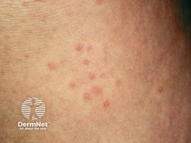
Clinical photography of a patient with cholinergic urticaria showing multiple small, erythematous, blanching wheals on the forearm. Imaging modality is clinical photography; technique involves a high-resolution, color-balanced image capturing superficial skin lesions in a macro-clinical view. The wheals are transient, pruritic, and vary in size from a few millimeters to up to several millimeters, with well-demarcated borders and pale centers. Triggered by heat, exercise, and emotional stress, the lesions can appear soon after sweating and typically resolve within minutes to hours. This presentation correlates with cholinergic urticaria, a physical urticaria subtype linked to cholinergic stimuli, requiring history of triggers and symptom timing. Differential diagnoses include other urticarias and heat-induced dermatoses; diagnostic significance lies in distinguishing exercise/sweat-induced wheals from dermatographism or drug reactions to guide management. Clinical utility includes educating patients on trigger mitigation, optimizing antihistamine therapy, and evaluating for underlying mast cell disorders if atypical features arise. Chronic management includes non-sedating H1 antihistamines at standard or increased dosing, with occasional H2 blockade. Patients are advised to avoid overheating, wear breathable clothing, and perform graded exercise; epinephrine is reserved for rare anaphylaxis. In ambiguous cases, exercise-provoked challenges or sweat tests may help; history differentiates from other physical urticarias.
Ingestional Organophosphorus Compound Poisoning
Overview
- Tintinalli's Emergency Medicine, p. 1342
- Rosen's Emergency Medicine, p. 3029
Pathophysiology
- Muscarinic receptors (parasympathetic postganglionic endings, sweat glands)
- Nicotinic receptors (autonomic ganglia, neuromuscular junctions)
- CNS cholinergic receptors
- Tintinalli's Emergency Medicine, p. 1342-1343
Clinical Features
1. Acute Cholinergic Crisis (Most Common)
| SLUDGE | DUMBELS |
|---|---|
| Salivation | Defecation |
| Lacrimation | Urination |
| Urination | Miosis |
| Defecation | Bradycardia, Bronchorrhea, Bronchospasm |
| GI cramps | Emesis |
| Emesis | Lacrimation |
| Salivation |
- Muscle fasciculations, cramps, weakness (including diaphragm)
- Mydriasis, pallor
- Tachycardia, hypertension (sympathetic ganglionic stimulation can override parasympathetic effects)
- Anxiety, restlessness, emotional lability
- Tremor, headache, dizziness
- Confusion, delirium, hallucinations
- Seizures, coma
Death is most commonly due to respiratory failure - from bronchorrhea + bronchospasm + respiratory muscle paralysis + CNS depression combined.
- Tintinalli's Emergency Medicine, p. 1343
2. Intermediate Syndrome
- Paralysis of neck flexor muscles
- Weakness of muscles innervated by cranial nerves
- Proximal limb muscle weakness
- Respiratory muscle paralysis (may require ventilatory support)
- Notably, no signs of cholinergic excess at this stage
- Symptoms typically resolve within 7 days
- Tintinalli's Emergency Medicine, p. 1343
- Adams and Victor's Principles of Neurology, p. 1224
3. Organophosphate-Induced Delayed Neuropathy (OPIDN)
- Symmetrical sensorimotor axonopathy (leg cramps → weakness → paralysis)
- Can mimic Guillain-Barré syndrome
- Cognitive dysfunction, impaired memory, mood changes
- Autonomic dysfunction, peripheral neuropathy, extrapyramidal signs
4. Chronic Toxicity
Diagnosis
| Test | Details |
|---|---|
| RBC acetylcholinesterase activity | More specific (reflects neuronal AChE); activity < 25% of normal = severe poisoning |
| Plasma pseudocholinesterase | More accessible; depressed in poisoning but less specific (also low in liver disease, pregnancy, malnutrition) |
| ECG | ST changes, peaked T-waves, AV block, prolonged QT |
| EMG | Identifies/quantifies AChE inhibition at neuromuscular junctions |
Management
Step 1 - Decontamination
- Skin/eye exposure: Remove all clothing (double-bag and discard), wash skin thoroughly with soap and water
- Ingestion: Gastric lavage may be considered if presentation is very early; activated charcoal has limited benefit due to rapid absorption and profuse vomiting/diarrhea
- Caregiver protection: Universal precautions (PPE), chemical-resistant gloves, eye protection - secondary contamination is a real risk
Step 2 - Stabilization & Supportive Care
- Airway is the priority - aggressive suctioning of secretions
- Intubation: Avoid succinylcholine if possible (it is metabolized by cholinesterases and may have prolonged effect of 4-6 hours in OP poisoning). Prefer rocuronium 1 mg/kg (non-depolarizing, not cholinesterase-dependent)
- Oxygen, mechanical ventilation as needed
- Benzodiazepines for seizures (diazepam/midazolam) - preferred over phenytoin
- Tachyarrhythmias generally resolve with atropinization; do not treat with beta-blockers
Step 3 - Antidote Therapy
A. Atropine (First-line - for muscarinic effects)
| Parameter | Detail |
|---|---|
| Initial dose (adult) | 1-3 mg IV (or 0.05 mg/kg in children) |
| Titration | Double the dose every 5 minutes |
| Endpoint ("atropinization") | Drying of respiratory secretions, easing respiratory effort, resolution of bronchospasm |
| Maintenance infusion | 10-20% of total cumulative loading dose per hour |
| Total doses needed | May require 200-500 mg in first hour in severe cases |
| NOT an endpoint | Mydriasis, tachycardia (these should not stop therapy) |
Tachycardia and mydriasis are NOT endpoints for stopping atropine - secretion control is the target.
B. Pralidoxime (2-PAM) - Oxime / Cholinesterase Reactivator
| Parameter | Detail |
|---|---|
| Adult dose | 1-2 g IV (30 mg/kg, max 2g) over 15-30 minutes |
| Pediatric dose | 25-50 mg/kg IV |
| Maintenance | 8-10 mg/kg/hr infusion |
| Continue until | Atropine no longer required |
| Early administration | Critical - efficacy decreases as aging progresses |
Soluble (fat-soluble) organophosphates may require prolonged treatment courses due to ongoing redistribution from tissue stores.
C. Benzodiazepines
- Diazepam 5-10 mg IV (or lorazepam/midazolam) for seizures and CNS agitation
- Should be given early in severe poisoning even without overt seizures (prophylactic)
Severity Classification
| Mild | Moderate | Severe |
|---|---|---|
| Miosis, excess secretions, GI symptoms | Bronchospasm, bradycardia, fasciculations | Seizures, coma, respiratory failure, severe bradycardia |
| RBC AChE > 50% | RBC AChE 20-50% | RBC AChE < 20% |
Key Differences: OP vs Carbamates
| Feature | Organophosphates | Carbamates |
|---|---|---|
| AChE binding | Irreversible (aging) | Reversible (spontaneous hydrolysis) |
| Duration of toxicity | Days-weeks | Hours (usually < 24h) |
| CNS penetration | Yes | Poor (adults) |
| Intermediate syndrome | Yes | Yes |
| Aging | Yes | No |
| Pralidoxime | Indicated (early) | Controversial, may be harmful (carbaryl) |
Disposition
- Mild cases: Observe for minimum 24 hours (symptoms can evolve)
- Moderate-severe: ICU admission with continuous monitoring
- Watch for intermediate syndrome (days 1-5) even after apparent recovery from acute phase
- Psychiatric evaluation mandatory in intentional ingestion
- Cholinesterase levels should be rechecked periodically
Recent Evidence (2024-2026)
- A 2025 systematic review (PMID 38797902) examined glycopyrrolate (a quaternary ammonium anticholinergic) as an alternative to atropine, finding it may offer benefits with less CNS side-effects in OP/carbamate poisoning.
- A 2026 umbrella review (PMID 42258859) of therapeutic interventions in OP poisoning synthesized evidence across multiple systematic reviews, providing updated guidance on treatment strategies.
Summary Algorithm
OP Ingestion
    ↓
Decontaminate + Secure Airway
    ↓
Atropine 1-3 mg IV → double q5min → target: dry secretions
    ↓
Pralidoxime 1-2g IV (early! before aging)
    ↓
Benzodiazepine for seizures
    ↓
ICU admission → watch for Intermediate Syndrome (day 1-5)
                          ↓
                     Respiratory monitoring + ventilatory support if needed