
Describe briefly about Vitamin A and vitamin d with information about synthesis , storage , uses, deficiency’s, symptoms, dosage , complications from Nelson and all the recent advances , guidelines from WHO,aap,iap, pubmed
"vitamin A" AND deficiency AND children
"vitamin D" AND deficiency AND children
WHO AAP IAP vitamin A vitamin D guidelines 2024 2025 children supplementation
"vitamin D" AND supplementation AND guidelines
vitamin A deficiency xerophthalmia Bitot spots night blindness
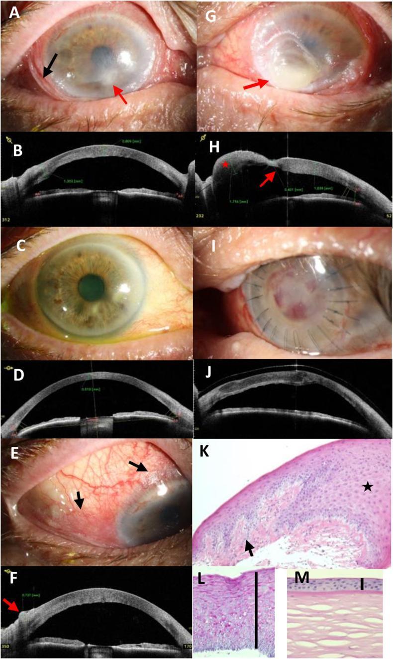
This composite figure illustrates the clinical and diagnostic progression of severe xerophthalmia and keratomalacia secondary to vitamin A deficiency in a 70-year-old male. Panels A, E, and G show clinical photographs highlighting characteristic Bitot's spots (foamy keratinized conjunctival plaques), corneal stromal infiltrates, and advanced keratomalacia with focal bulging (staphyloma formation). Panels B, D, F, H, and J utilize Anterior Segment Optical Coherence Tomography (AS-OCT) to quantify pathology, revealing conjunctival thickening (up to 737 μm), corneal stromal edema (1039 μm), and focal thinning (401 μm). Post-treatment panels (C, D, I, J) demonstrate clinical resolution after vitamin A supplementation and tectonic lamellar keratoplasty, with AS-OCT confirming successful graft adaptation. Histopathological sections (K, L) using HE and PAS stains contrast the diseased cornea against a normal control (M), showing massive epithelial hyperplasia (approximately 20 cell layers), focal keratinization, loss of Bowman's layer, and granular PAS-positive deposits, pathognomonic for squamous metaplasia in the setting of nutritional deficiency.
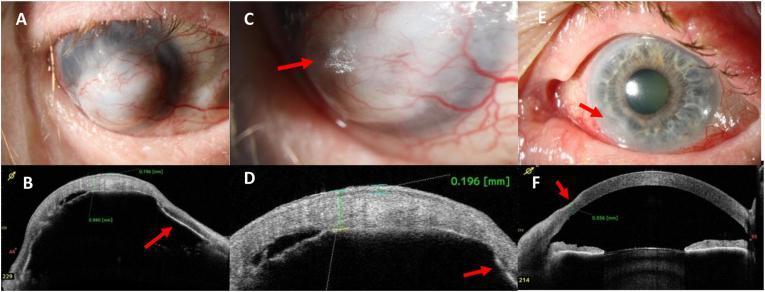
This composite image presents clinical photographs and Anterior Segment Optical Coherence Tomography (AS-OCT) findings in a case of severe keratomalacia associated with Vitamin A deficiency. Panels A-D focus on the right eye (OD). Panel A shows advanced corneal melting (keratomalacia) characterized by a bulging, whitish corneal surface completely overgrown by vascularized conjunctival tissue (conjunctivalization). Panel C highlights keratinization on the corneal surface, resembling Bitot's spots (arrow). Panels B and D provide AS-OCT cross-sections showing massive corneal thickening (940 μm), epithelial thickening (196 μm), and complete iris-to-cornea attachment (athalamia/anterior synechiae, arrow). Panels E and F focus on the left eye (OS). Panel E demonstrates bulbar conjunctival hyperemia and a hazy peripheral lesion at the 7-8 o'clock position (arrow). Panel F (AS-OCT) confirms slight peripheral corneal thinning to 556 μm in that same region (arrow). This series illustrates the range of ophthalmic manifestations of systemic malnutrition, from early peripheral thinning to end-stage keratectasia and corneal scarring.
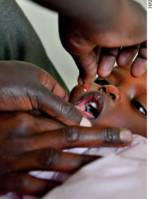
This clinical photograph demonstrates the oral administration of high-dose vitamin A supplementation to a pediatric patient. The image shows an adult health worker or caregiver using a manual technique to ensure safe delivery: one hand gently stabilizes the child's jaw and depresses the lower lip to keep the mouth open, while the other hand squeezes a small capsule or dispenser. A single, translucent red droplet of the liquid supplement is captured in mid-air, directed toward the child's open mouth. The child, appearing to be an infant or toddler, has their mouth open wide, revealing the tongue and primary dentition. This visual illustrates a common public health intervention used in nutritional programs to prevent vitamin A deficiency (VAD) and its associated complications, such as xerophthalmia and increased susceptibility to infections. The focus is on the practical application and administration technique within a community health or pediatric clinical setting.
rickets vitamin D deficiency bowing legs bone changes children
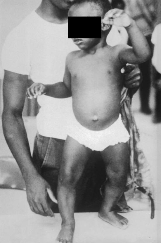
This clinical photograph displays the classic musculoskeletal manifestations of vitamin D deficiency rickets in a young child. The most prominent feature is significant genu varum (bowing of the legs), where the weight-bearing long bones show lateral curvature due to impaired mineralization of the growth plate and osteoid matrix. Additionally, the child exhibits a distended abdomen (potbelly), which is a common clinical sign associated with rickets due to hypotonia of the abdominal muscles. The upper extremities appear thin, suggesting poor muscle development or nutritional deficiency. This archival image serves as a pedagogical tool for identifying systemic skeletal deformities resulting from metabolic bone disease. Key educational concepts include the relationship between vitamin D deficiency, calcium homeostasis, and the softening of bones (osteomalacia) during childhood development. The clinical relevance extends to pediatrics, endocrinology, and public health nutrition.
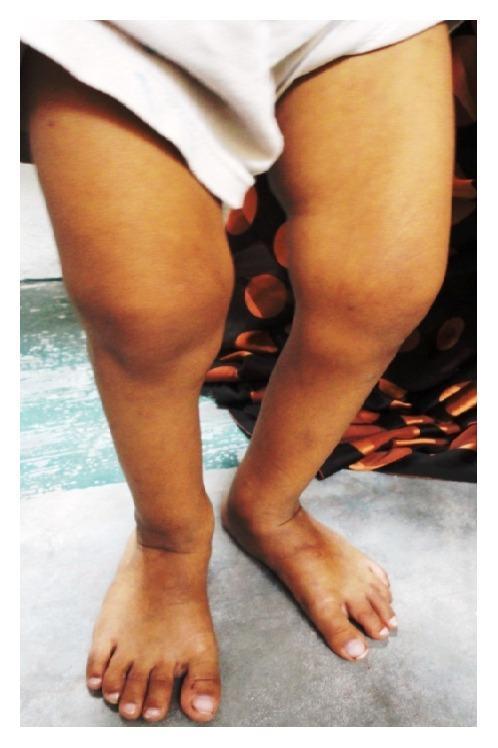
A clinical photograph of the lower extremities of a pediatric patient exhibiting signs of metabolic bone disease, likely rickets. The image demonstrates a prominent bilateral bowing of the legs (genu varum), characterized by a distinct outward curvature of the femoral and tibial shafts. There is marked enlargement and widening of the ankle joints, consistent with metaphyseal flaring. The skin over the affected regions appears intact without discoloration or acute inflammatory changes. This presentation is a classic clinical manifestation of vitamin D deficiency or hereditary rickets, illustrating the skeletal deformities that arise from impaired mineralization of the growth plates and osteoid matrix during development. The image serves as an educational tool for identifying physical signs of systemic pediatric bone pathology and understanding the orthopedic consequences of chronic mineral deficiencies.
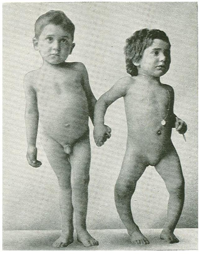
This clinical photograph displays a 6.5-year-old boy (left) and his 4-year-old sister (right) presenting with characteristic skeletal manifestations of rickets, likely secondary to severe vitamin D deficiency. The boy exhibits genu valgum (knock-knees), characterized by inward angling of the knees, while the girl demonstrates significant genu varum (bow legs), showing a lateral outward curvature of the femurs and tibiae. Both children show evidence of growth retardation (short stature for age) and additional skeletal deformities, including potentially widened epiphyses at the wrists and ankles. The image serves as a classic educational example of how nutritional or metabolic bone diseases interfere with proper mineralization of the growth plate, leading to progressive softening and bowing of weight-bearing long bones during childhood development.
Vitamin A and Vitamin D - A Comprehensive Pediatric Review
VITAMIN A (Retinol)
Structure and Forms
- Retinol - the transport and storage form
- Retinal - the aldehyde form (used in vision)
- Retinoic acid - the active transcriptional form
- Retinyl esters - the storage form in the liver
- Provitamin A carotenoids - plant precursors (most important: β-carotene, which contributes ~30% of dietary vitamin A)
Synthesis and Metabolism
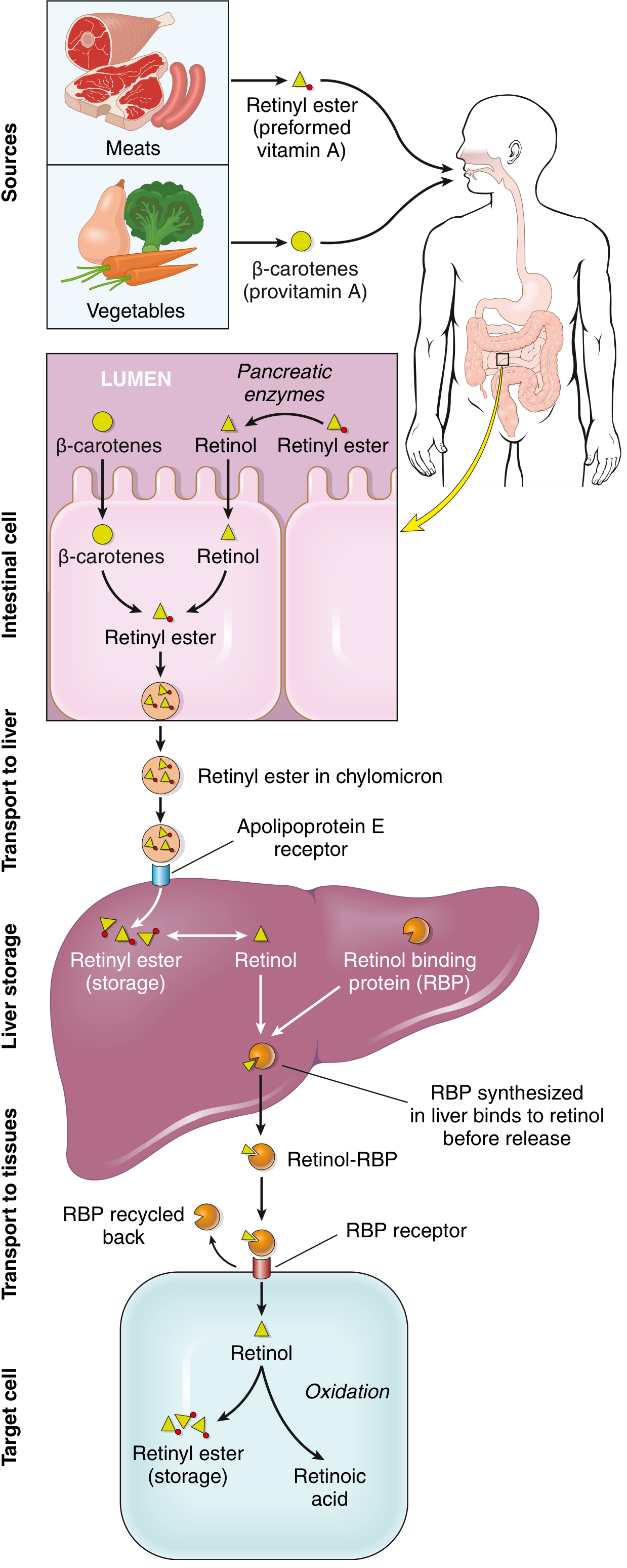
- Animal sources (preformed vitamin A): liver, fish, eggs, milk, butter
- Plant sources (provitamin A): yellow/leafy green vegetables - carrots, squash, spinach, broccoli
- Requires bile, pancreatic enzymes, and antioxidant activity in food
- Retinol (as retinyl ester) and β-carotene absorbed through the intestinal wall
- β-carotene is converted to retinol within intestinal cells
- Retinol is packaged into chylomicrons and transported to the liver
- Liver takes up retinol via the apolipoprotein E receptor
- >90% of body's vitamin A reserves stored in the liver, predominantly as retinyl ester in perisinusoidal (Ito/stellate) cells
- Hepatic reserves are sufficient for at least 6 months in a well-nourished person
- For transport from liver to periphery: retinol binds to Retinol-Binding Protein (RBP), which is synthesized in the liver
- RBP is recycled after retinol is delivered to target cells via cell-surface RBP receptors
- In target cells: retinol may be stored as retinyl ester or oxidized to retinoic acid
Functions
-
Vision (rhodopsin synthesis): Vitamin A is a component of rhodopsin (rod cells) and three iodopsins (cone cells). Synthesis of all four visual pigments is reduced in deficiency. Retinal combines with opsin protein to form these pigments.
-
Epithelial differentiation: Retinoic acid binds to Retinoic Acid Receptors (RARs), forming heterodimers with Retinoic X Receptors (RXRs). These RAR/RXR heterodimers bind to Retinoic Acid Response Elements (RAREs) in regulatory DNA, controlling genes encoding growth factor receptors, tumor suppressor proteins, and secreted factors. Without vitamin A, mucus-secreting columnar epithelium undergoes squamous metaplasia → keratinizing epithelium.
-
Metabolic effects: Retinoids inhibit adipogenesis and stimulate lipid breakdown. RXR can heterodimerize with PPARs and Vitamin D receptors.
-
Immune function: Vitamin A promotes regeneration of damaged epithelia and is required for optimal immune function. Supplementation reduces diarrhea morbidity by ~15% and mortality by ~30%. Reduces overall child mortality by 20-30% in deficient populations.
Deficiency
- Poor nutrition (most common globally)
- Fat malabsorption (celiac disease, Crohn's, ulcerative colitis)
- Bariatric surgery
- Continuous use of mineral oil laxatives
- Liver disease (reduces RBP synthesis)
- Protein-energy malnutrition (reduces RBP synthesis even with adequate hepatic stores)
Clinical Signs and Symptoms
| WHO Grade | Finding |
|---|---|
| XN | Night blindness (nyctalopia) - earliest sign |
| X1A | Conjunctival xerosis (dryness) |
| X1B | Bitot's spots (foamy, keratinized plaques on conjunctiva) |
| X2 | Corneal xerosis |
| X3A | Corneal ulceration/keratomalacia <1/3 cornea |
| X3B | Keratomalacia ≥1/3 cornea → blindness |
| XS | Corneal scar |
| XF | Xerophthalmic fundus |
Dosage - Treatment and Supplementation
| Age | Dose |
|---|---|
| <6 months | 50,000 IU/day PO × 2 days |
| 6 months to <1 year | 100,000 IU/day PO × 2 days |
| 1-5 years | 200,000 IU/day PO × 2 days |
- Infant: 1,500 IU/day
- Child 1-3 yr: 5,000 IU/day
- Child 4-8 yr: 5,000-10,000 IU/day
- Child ≥9 yr/adolescent: 10,000 IU/day
- RDA expressed in retinol equivalents (reflecting both preformed vitamin A and β-carotene)
- 900 mcg RAE/day for adult males; lower for children
Toxicity / Complications of Excess
- Headache, dizziness, vomiting, stupor, blurred vision
- May mimic brain tumor (pseudotumor cerebri/raised ICP, papilledema)
- Irritability, GI disturbance, rash
- Weight loss, anorexia, nausea, vomiting
- Bone and joint pain
- Increased osteoclast activity → bone resorption → fracture risk
- Hepatotoxicity (fibrosis, cirrhosis with very high doses)
- Skin changes (peeling, alopecia)
VITAMIN D (Calciferol)
Structure and Forms
- Vitamin D3 (Cholecalciferol): Endogenously synthesized in skin from 7-dehydrocholesterol under UV-B; also from animal dietary sources
- Vitamin D2 (Ergocalciferol): From plant sources (ergosterol); less potent than D3
- 25-hydroxyvitamin D [25(OH)D]: Circulating storage form; used for assessment of vitamin D status
- 1,25-dihydroxyvitamin D [1,25(OH)₂D = Calcitriol]: The biologically active form
Synthesis and Metabolism
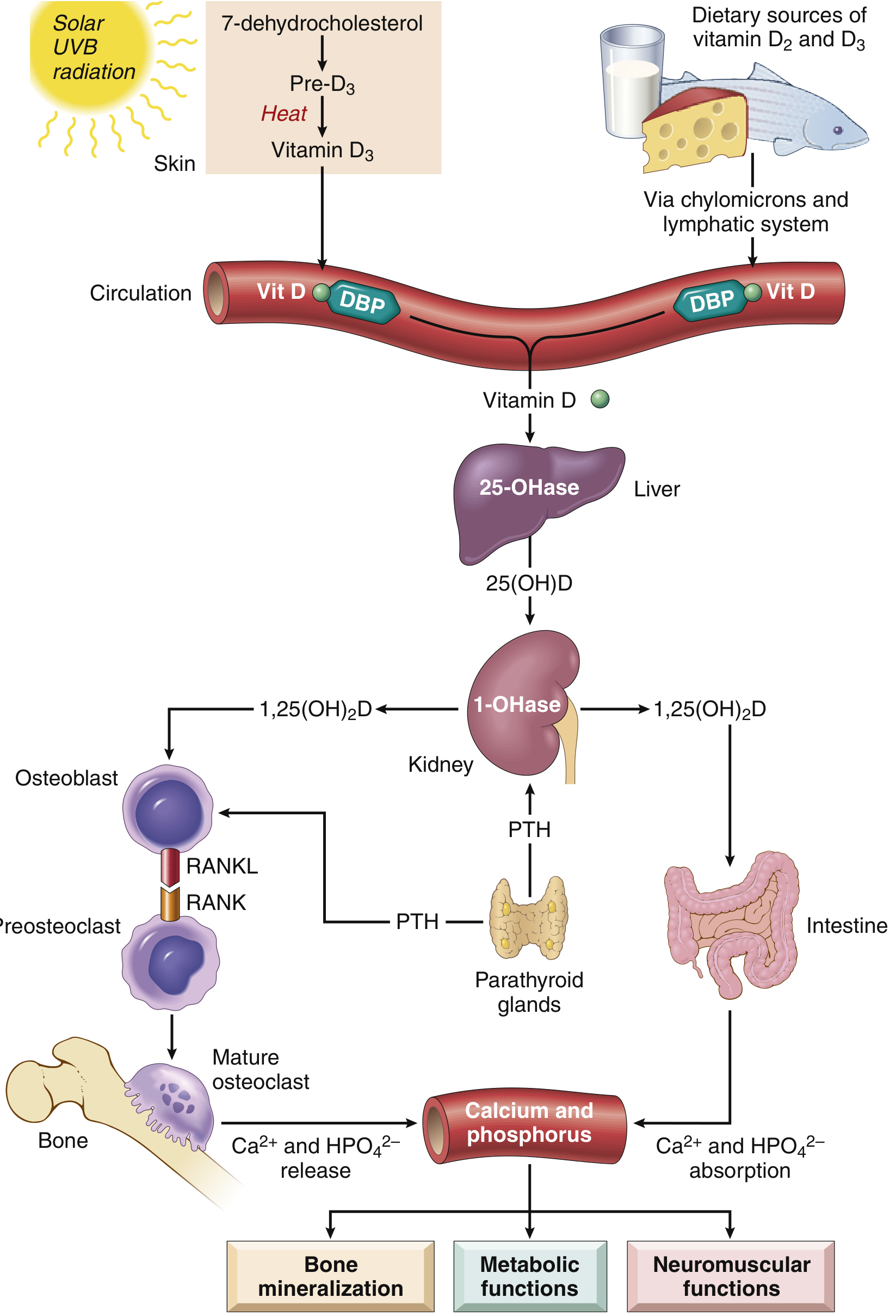
- Solar UV-B irradiation converts 7-dehydrocholesterol in skin → Pre-vitamin D3 → (heat) → Cholecalciferol (D3)
- Melanin competes for UV-B; darker skin = reduced D3 production
- Deep-sea fish (salmon, tuna, mackerel), fish liver oils
- Egg yolks, fortified milk/dairy, mushrooms (UV-exposed)
- Plant sources contain ergosterol → vitamin D2
- Vitamin D (from skin or gut) binds to vitamin D-binding protein (DBP/α1-globulin) in plasma → transported to liver
- Liver 25-hydroxylase converts → 25(OH)D (the circulating storage form; half-life ~2-3 weeks)
- Kidney 1α-hydroxylase converts → 1,25(OH)₂D (Calcitriol) (the active form)
- Hypocalcemia → ↑PTH → activates 1α-hydroxylase → ↑1,25(OH)₂D
- Hypophosphatemia → directly activates 1α-hydroxylase
- High 1,25(OH)₂D → feedback inhibition of 1α-hydroxylase
Functions
- Intestine: Stimulates Ca²⁺ and HPO₄²⁻ absorption by upregulating calcium transport proteins in enterocytes
- Kidney: Stimulates Ca²⁺ reabsorption in distal tubules via calbindin, plasma membrane calcium pump, epithelial calcium channel
- Bone: Required for mineralization of osteoid matrix and epiphyseal cartilage; upregulates RANKL on osteoblasts → osteoclast activation → calcium/phosphate mobilization
- Parathyroid: High 1,25(OH)₂D decreases PTH gene transcription (negative feedback)
Deficiency
- Insufficient sunlight exposure (high latitudes, indoor lifestyle, dark skin, cultural clothing, sunscreen use)
- Dietary insufficiency
- Fat malabsorption (celiac disease, cholestatic liver disease, Crohn's disease, cystic fibrosis)
- Prematurity (vitamin D crosses placenta in last trimester only)
- Chronic kidney disease (impaired 1α-hydroxylation)
- Anticonvulsant drugs (phenytoin, phenobarbitone - interfere with gut absorption and alter metabolism)
- Vitamin D-dependent rickets Type I (inherited 1α-hydroxylase deficiency)
- Vitamin D-resistant rickets (renal tubular phosphate leak)
- Maternal vitamin D deficiency during pregnancy
| Status | 25(OH)D Level |
|---|---|
| Deficiency | <12 ng/mL (<30 nmol/L) |
| Insufficiency | 12-20 ng/mL (30-50 nmol/L) |
| Sufficiency | >20 ng/mL (>50 nmol/L) |
Clinical Signs and Symptoms
- Skeletal deformities from impaired bone mineralization:
- Craniotabes (ping-pong ball skull in infants)
- Frontal bossing, delayed fontanelle closure
- Rachitic rosary (beading at costochondral junctions)
- Harrison's sulcus (groove along lower thorax)
- Genu varum (bowing of legs) or genu valgum (knock-knees) in toddlers
- Widened wrists and ankles (metaphyseal flaring)
- Pathological fractures
- Hypocalcemia: Tetany, convulsions, laryngospasm, prolonged QTc
- Myopathy: Hypotonia, proximal muscle weakness, delayed walking
- Growth retardation
- Dental abnormalities: Enamel defects, delayed eruption
- Increased respiratory infections (hypotonia of respiratory muscles)
- Low/normal serum calcium
- Low serum phosphate
- Elevated alkaline phosphatase (hallmark)
- Low 25(OH)D
- Elevated PTH (secondary hyperparathyroidism)
- Widening and cupping/fraying of metaphyses
- Decreased bone density
- Bowing of long bones
- Bone pain (especially axial skeleton, lower limbs)
- Proximal myopathy, waddling gait
- Looser's zones (pseudofractures) on imaging
- Increased fracture risk (vertebral bodies, femoral necks)
- Histology: thickened layers of eosinophilic unmineralized osteoid around basophilic mineralized trabeculae
Dosage
- All breastfed infants: 400 IU/day starting within the first 2 months of life, continuing throughout childhood/adolescence
- Formula-fed infants consuming <500 mL/day fortified formula: also supplement 400 IU/day
| Population | Dose |
|---|---|
| Pregnant/lactating women | 600 IU/day |
| Breastfed infants (first year) | 400 IU/day from first few days of life |
| Children 1-3 years | 600 IU/day |
| Children 4-10 years | 600-1,000 IU/day |
| Adolescents | 600 IU/day |
- Children <1 year: 2,000 IU/day × 12 weeks
- Children >1 year: 3,000 IU/day × 12 weeks
- Children >3 years with confirmed deficiency: 60,000 IU once weekly × 6 weeks (stoss therapy)
- Children with malabsorption: higher doses as needed
| Age | Insufficiency (12-20 ng/mL) | Deficiency (<12 ng/mL) |
|---|---|---|
| <1 year | 1,000 IU once daily | 2,000-4,000 IU once daily |
| ≥1 year | 2,000 IU once daily | 5,000-6,000 IU once daily OR 50,000 IU once weekly |
- Age <10 yr: Insufficiency: 2,000 IU/day; Deficiency: 5,000 IU/day
- Age ≥10 yr: Insufficiency: 4,000-6,000 IU/day; Deficiency: 10,000 IU/day or 50,000 IU/week
Toxicity / Complications of Excess
- Hypercalcemia: nausea, vomiting, anorexia, weakness, constipation, polyuria, polydipsia
- Hypercalciuria → nephrolithiasis, nephrocalcinosis
- Metastatic calcification (soft tissues, blood vessels, kidneys, cornea, lungs)
- Raised serum 25(OH)D (typically >150 ng/mL or 375 nmol/L)
- Prolonged excess → renal failure
- Infants <6 months: 1,000 IU/day
- Infants 6-12 months: 1,500 IU/day
- Children 1-3 years: 2,500 IU/day
- Children 4-8 years: 3,000 IU/day
- Children >8 years/adults: 4,000 IU/day
RECENT ADVANCES AND GUIDELINE UPDATES (2023-2026)
Vitamin A - Recent Evidence
-
Measles management (2026): A systematic review (Kaur et al., 2026) confirmed that high-dose vitamin A supplementation remains a key therapeutic strategy in acute measles management, reducing severity, ocular complications, and mortality - especially in vitamin A-deficient populations. The WHO 2-dose regimen is strongly supported.
-
Neonatal respiratory outcomes (2024): A meta-analysis by Li et al. (2024) showed significant association between vitamin A status and neonatal respiratory diseases, suggesting supplementation may have a protective role in premature infants.
-
Pregnancy supplementation (2023): A meta-analysis (Ma et al., 2023) found that vitamin A supplementation during pregnancy improved birth weight and infant growth outcomes, particularly in deficient populations.
-
Biofortification: WHO and UNICEF continue to push crop biofortification (β-carotene-rich orange sweet potato, golden rice) as a sustainable strategy to address VAD in low-income countries.
Vitamin D - 2024 Endocrine Society Clinical Practice Guideline
- Does not recommend routine vitamin D screening in the general population
- Does not support supplementation for prevention of cardiovascular disease, diabetes, cancer, or autoimmune diseases (insufficient evidence)
- Does recommend supplementation to prevent rickets in infants and young children, and to prevent upper respiratory tract infections in children
- Supports empirical supplementation (without screening) in:
- Infants (400-600 IU/day)
- Children and adolescents (600-1,000 IU/day)
- Pregnant women
- Dark-skinned individuals at higher latitudes
- The guideline is controversial - a rebuttal (Nwosu, 2025) criticized the lack of specific guidance for preterm infants and downplaying of vitamin D's role in immune and metabolic health in children.
QUICK-REFERENCE COMPARISON TABLE
| Feature | Vitamin A | Vitamin D |
|---|---|---|
| Type | Fat-soluble, retinoid | Fat-soluble, secosteroid |
| Main form | Retinol (transport), Retinyl ester (storage) | Cholecalciferol (D3) → Calcitriol (active) |
| Endogenous synthesis | No | Yes (skin, UV-B) |
| Primary storage | Liver (Ito cells), >90% | Liver and adipose tissue |
| Activation | Oxidation to retinoic acid | Liver (25-OH) → Kidney (1,25-OH₂) |
| Key receptor | RAR/RXR nuclear receptors | VDR (nuclear receptor) |
| Classic deficiency disease | Xerophthalmia, night blindness | Rickets (children), Osteomalacia (adults) |
| First sign of deficiency | Night blindness | Hypocalcemia, craniotabes |
| Key lab marker | Serum retinol | Serum 25(OH)D |
| Deficiency treatment (child) | 200,000 IU × 2 days (measles) | 3,000 IU/day × 12 weeks (IAP) |
| Prophylaxis dose | Not routinely supplemented universally | 400 IU/day (infants, AAP/IAP) |
| Toxicity | Pseudotumor cerebri, teratogenicity | Hypercalcemia, nephrocalcinosis |
| Teratogenic | Yes (high dose - Category X) | No (at recommended doses) |
Key References
- Robbins & Kumar Basic Pathology, pp. 292-296 - primary source for metabolism, functions, deficiency pathology
- Harriet Lane Handbook (23rd ed., Johns Hopkins) - dosing tables
- Tietz Textbook of Laboratory Medicine (7th ed.) - lab assessment
- IAP Consensus on Vitamin D (PMC11964374) - Indian pediatric guidelines
- 2024 Endocrine Society CPG on Vitamin D (Pilz et al., 2026)
- Measles management systematic review (Kaur et al., 2026)
- Vitamin A in neonatal respiratory disease (Li et al., 2024)
Describe briefly about trace elements with information about synthesis storage uses deficiency symptoms dosage complications from Nelson and all the recent advances guidelines from WHO aap iap pubmed
"trace element" deficiency children
WHO AAP IAP guidelines zinc iodine copper selenium supplementation children 2024 2025
IAP zinc supplementation dosing diarrhea children guideline mg
zinc supplementation children diarrhea
WHO iodine deficiency disorders universal salt iodization guideline update
Trace Elements - Comprehensive Pediatric Review
1. ZINC
- Periorificial and acral dermatitis (dry, scaly, eczematous plaques around mouth, buttocks, hands - often secondarily infected with Candida)
- Growth retardation, hypogonadism in males
- Dysgeusia, poor appetite, alopecia
- Poor wound healing, abnormal dark adaptation
- Impaired immune function, increased infection susceptibility, diarrhea
- Infants <6 months: 10 mg/day for 10-14 days
- Children ≥6 months: 20 mg/day for 10-14 days
- IAP 2006 guideline: uniform 20 mg elemental zinc during diarrhea + 7 days post-cessation for children >3 months
- Preventive dosing: 10-15 mg/day shows modest reduction in diarrhea incidence
2. COPPER
- Myeloneuropathy - lower limb paresthesias, spasticity, gait difficulty, brisk reflexes, extensor plantar responses
- Hematologic: microcytic anemia, neutropenia, occasional pancytopenia
- Menkes disease (X-linked, infancy onset by 3 months): poor growth, "kinky hair" (pili torti), severe neurodevelopmental regression, connective tissue abnormalities
- Skin/hair hypopigmentation, bone abnormalities
3. IODINE
- Congenital iodine deficiency (cretinism): severe intellectual disability, short stature, coarse facial features, protruding tongue, umbilical hernia. Severity depends on timing - deficiency before fetal thyroid function is established (maternal T3/T4 dependent) causes the most severe intellectual impairment.
- Goiter (TSH-driven thyroid enlargement) - most common cause of nontoxic goiter worldwide
- Older children/adults: hypothyroidism - fatigue, cold intolerance, constipation, growth delay, cognitive slowing
- Universal salt iodization: 15-40 ppm (mg/kg) iodine in salt at production/retail level
- Global coverage: ~89% of population uses iodized salt (2020 data); ~1 billion people still without adequate access
- WHO recommends monitoring urinary iodine excretion in population surveys as the key adequacy indicator
4. SELENIUM
- Keshan disease: multifocal myocarditis → fatal cardiomyopathy, primarily in women and young children; cardiomegaly, arrhythmias, muscle pain/weakness, white nail beds, hypopigmentation of skin/hair
- Kashin-Beck disease: osteoarthropathy affecting epiphyseal/articular cartilage → enlarged joints, shortened digits
5. OTHER TRACE ELEMENTS (Brief)
| Element | Deficiency Effect | Excess/Toxicity |
|---|---|---|
| Manganese | No well-defined human deficiency syndrome; role in antioxidant defense and macronutrient metabolism | Occupational/parenteral nutrition exposure → neurotoxicity (parkinsonism-like) |
| Chromium | Impaired glucose tolerance (potential link to insulin resistance) | Occupational exposure → renal failure, dermatitis, pulmonary cancer risk |
| Fluoride | No defined deficiency state; important for dental/bone structure | Dental fluorosis (mottled, pitted enamel - occurs during enamel calcification in first 7 years of life); Skeletal fluorosis with chronic intake >3-6 mg/L (osteosclerosis on imaging); endemic in parts of India |
| Molybdenum | Extremely rare; cofactor deficiency reported only in genetic sulfite oxidase deficiency | Rare; occupational exposure only |
RECENT ADVANCES AND GUIDELINE UPDATES (2023-2026)
- A 2024 systematic review and meta-analysis (Ali et al., J Glob Health) reconfirmed efficacy of zinc supplementation for acute and persistent watery diarrhea in children, supporting current WHO/UNICEF dosing.
- A 2025 systematic review (Pradhan et al.) highlighted poor adherence to oral zinc supplementation regimens in real-world settings as a major limiting factor in effectiveness - an implementation gap rather than efficacy gap.
- The 2024 WHO update on childhood pneumonia and diarrhea now includes a conditional recommendation for reduced zinc dosing (5 mg/day for up to 14 days) in select settings, reflecting evidence that lower doses achieve similar efficacy with less vomiting risk - a shift from the traditional 10-20 mg/day regimen.
- A 2025 systematic review (Zuo et al., Occup Environ Med) examined maternal trace element and toxic metal exposure in relation to fetal and early childhood growth - reinforcing the importance of maternal trace element status for offspring outcomes.
- A 2024 meta-analysis (Abbasi et al.) found altered serum trace element concentrations (copper, zinc, selenium, magnesium) in children with febrile seizures compared to controls, suggesting a possible role in seizure susceptibility - though causality remains unproven.
- Preterm neonates on parenteral nutrition may require higher iodine, selenium, and copper provision than previously recommended, per updated pediatric parenteral nutrition trace element guidance.
QUICK REFERENCE TABLE
| Element | Deficiency Disease | Hallmark Sign | Standard Pediatric Dose |
|---|---|---|---|
| Zinc | Acrodermatitis enteropathica | Periorificial/acral dermatitis | 10-20 mg/day (diarrhea) |
| Copper | Copper deficiency myeloneuropathy, Menkes disease | Gait disturbance, kinky hair | 2 mg oral, 1-3×/day |
| Iodine | Cretinism, goiter | Intellectual disability, goiter | Iodized salt (15-40 ppm) |
| Selenium | Keshan disease, Kashin-Beck disease | Cardiomyopathy, joint disease | Individualized (PN: ~2-3 mcg/kg/day) |
| Fluoride | None defined | - | Community water fluoridation |
Give this in a pdf format You can also add information from Elizebeth nutrition from paediatrics and give a detailed pdf for all the above information
mkdir -p /tmp/workspace/nutrition-pdf && ls -la /tmp/workspace/
mkdir -p /tmp/workspace/nutrition-pdf/assets && cd /tmp/workspace/nutrition-pdf/assets && \ curl -s -o vitA_metabolism.png "https://cdn.orris.care/cdss_images/4fae2b23df63655fa88e068f141bbbd4ba5113f362aca6daf8c5418732591ad1.png" && \ curl -s -o vitD_metabolism.png "https://cdn.orris.care/cdss_images/d95a7fc67a2846589f7abbf021f1756247d1d25d48292588b7328eb5e9a67a45.png" && \ curl -s -o xerophthalmia.jpg "https://cdn.orris.care/cdss_images/pmc_clinical_VQA_e9f4bc4267b4e79d145285a3f5fd7547ecac48c4ae082cba6babbe2567cc713d.jpg" && \ curl -s -o rickets.jpg "https://cdn.orris.care/cdss_images/pmc_clinical_VQA_1cca75faf4740f41e18a1e7ba5947034550c16fa04f9f1036024dae4179eedda.jpg" && \ curl -s -o zinc_deficiency.png "https://cdn.orris.care/cdss_images/04c52796dd70c4798a0cf527e26172243744950b44da8c7bc84cd05f22eeeace.png" && \ ls -la /tmp/workspace/nutrition-pdf/assets/
~/nutrition-pdf/build_pdf.py
#!/usr/bin/env python3
"""
Build a comprehensive PDF on Vitamin A, Vitamin D and Trace Elements
for pediatric nutrition, compiled from Nelson-equivalent textbooks
(Robbins & Kumar Basic Pathology, Harriet Lane Handbook, Tietz Lab Medicine,
Harrison's Principles of Internal Medicine, Fitzpatrick's Dermatology, etc.),
WHO / AAP / IAP guidelines, and recent PubMed evidence (2023-2026).
"""
from reportlab.lib.pagesizes import A4
from reportlab.lib.units import cm
from reportlab.lib import colors
from reportlab.platypus import (
SimpleDocTemplate, Paragraph, Spacer, Table, TableStyle, Image,
PageBreak, ListFlowable, ListItem, HRFlowable
)
from reportlab.lib.styles import getSampleStyleSheet, ParagraphStyle
from reportlab.lib.enums import TA_CENTER, TA_LEFT, TA_JUSTIFY
from reportlab.pdfgen import canvas
import os
ASSETS = "/tmp/workspace/nutrition-pdf/assets"
OUT = "/tmp/workspace/nutrition-pdf/Vitamins_A_D_and_Trace_Elements_Pediatric_Nutrition.pdf"
styles = getSampleStyleSheet()
styles.add(ParagraphStyle(name="MainTitle", fontSize=24, leading=28, alignment=TA_CENTER,
textColor=colors.HexColor("#1a3c6e"), fontName="Helvetica-Bold", spaceAfter=10))
styles.add(ParagraphStyle(name="SubTitle", fontSize=13, leading=17, alignment=TA_CENTER,
textColor=colors.HexColor("#555555"), fontName="Helvetica", spaceAfter=6))
styles.add(ParagraphStyle(name="H1", fontSize=17, leading=21, textColor=colors.HexColor("#1a3c6e"),
fontName="Helvetica-Bold", spaceBefore=14, spaceAfter=8,
borderWidth=0, borderColor=colors.HexColor("#1a3c6e")))
styles.add(ParagraphStyle(name="H2", fontSize=13.5, leading=17, textColor=colors.HexColor("#b5451b"),
fontName="Helvetica-Bold", spaceBefore=10, spaceAfter=6))
styles.add(ParagraphStyle(name="H3", fontSize=11.5, leading=15, textColor=colors.HexColor("#2e6e3e"),
fontName="Helvetica-Bold", spaceBefore=6, spaceAfter=4))
styles.add(ParagraphStyle(name="Body", fontSize=10, leading=14.5, alignment=TA_JUSTIFY, spaceAfter=6))
styles.add(ParagraphStyle(name="BodyBold", parent=styles["Body"], fontName="Helvetica-Bold"))
styles.add(ParagraphStyle(name="Caption", fontSize=8.5, leading=11, alignment=TA_CENTER,
textColor=colors.HexColor("#555555"), fontName="Helvetica-Oblique", spaceAfter=10))
styles.add(ParagraphStyle(name="Citation", fontSize=8.5, leading=12, alignment=TA_LEFT,
textColor=colors.HexColor("#444444"), fontName="Helvetica-Oblique"))
styles.add(ParagraphStyle(name="Bullet", parent=styles["Body"], leftIndent=10, spaceAfter=3))
TABLE_HEADER_STYLE = TableStyle([
("BACKGROUND", (0, 0), (-1, 0), colors.HexColor("#1a3c6e")),
("TEXTCOLOR", (0, 0), (-1, 0), colors.white),
("FONTNAME", (0, 0), (-1, 0), "Helvetica-Bold"),
("FONTNAME", (0, 1), (-1, -1), "Helvetica"),
("FONTSIZE", (0, 0), (-1, -1), 8.5),
("GRID", (0, 0), (-1, -1), 0.5, colors.grey),
("ROWBACKGROUNDS", (0, 1), (-1, -1), [colors.white, colors.HexColor("#f2f2f2")]),
("VALIGN", (0, 0), (-1, -1), "TOP"),
("TOPPADDING", (0, 0), (-1, -1), 4),
("BOTTOMPADDING", (0, 0), (-1, -1), 4),
("LEFTPADDING", (0, 0), (-1, -1), 5),
("RIGHTPADDING", (0, 0), (-1, -1), 5),
])
story = []
# ---------------------------------------------------------------- TITLE PAGE
story.append(Spacer(1, 4*cm))
story.append(Paragraph("Vitamin A, Vitamin D & Trace Elements", styles["MainTitle"]))
story.append(Paragraph("A Comprehensive Pediatric Nutrition Reference", styles["SubTitle"]))
story.append(Spacer(1, 0.5*cm))
story.append(HRFlowable(width="60%", thickness=1.2, color=colors.HexColor("#b5451b"), hAlign="CENTER"))
story.append(Spacer(1, 1*cm))
story.append(Paragraph(
"Synthesis & Metabolism · Storage · Functions & Uses · Deficiency & Symptoms · "
"Dosage & Supplementation · Toxicity & Complications · Recent Advances & Guidelines "
"(WHO / AAP / IAP / PubMed)", styles["SubTitle"]))
story.append(Spacer(1, 3*cm))
story.append(Paragraph(
"<b>Compiled from:</b> Robbins & Kumar Basic Pathology; Robbins, Cotran & Kumar Pathologic Basis of Disease; "
"The Harriet Lane Handbook (Johns Hopkins, 23rd ed.); Tietz Textbook of Laboratory Medicine (7th ed.); "
"Harrison's Principles of Internal Medicine (22E, 2025); Fitzpatrick's Dermatology; Andrews' Diseases of the Skin; "
"Yamada's Textbook of Gastroenterology; Goldman-Cecil Medicine; Bradley & Daroff's Neurology; "
"Ganong's Review of Medical Physiology; Harper's Illustrated Biochemistry; Kanski's Clinical Ophthalmology; "
"Park's Textbook of Preventive and Social Medicine — supplemented with WHO, AAP, and IAP guidelines and "
"PubMed literature (2023-2026).", styles["Citation"]))
story.append(Spacer(1, 1*cm))
story.append(Paragraph(
"<i>Note: A dedicated title by \"Elizabeth\" on pediatric nutrition/child health was not found in the indexed "
"medical textbook library used for this compilation. All content below is drawn from the authoritative "
"textbooks listed above, which cover equivalent Nelson Textbook of Pediatrics-level material on these topics, "
"plus current international guidelines.</i>", styles["Citation"]))
story.append(PageBreak())
# ---------------------------------------------------------------- TOC (manual)
story.append(Paragraph("Contents", styles["H1"]))
toc_items = [
"1. Vitamin A (Retinol)",
"2. Vitamin D (Calciferol)",
"3. Trace Elements Overview",
"4. Zinc",
"5. Copper",
"6. Iodine",
"7. Selenium",
"8. Manganese, Chromium, Fluoride & Molybdenum",
"9. Comparative Summary Tables",
"10. Recent Advances & Guideline Updates (WHO / AAP / IAP / PubMed, 2023-2026)",
"11. References",
]
story.append(ListFlowable([ListItem(Paragraph(t, styles["Body"]), leftIndent=6) for t in toc_items],
bulletType="bullet"))
story.append(PageBreak())
def h1(text):
story.append(Paragraph(text, styles["H1"]))
story.append(HRFlowable(width="100%", thickness=0.8, color=colors.HexColor("#1a3c6e")))
story.append(Spacer(1, 6))
def h2(text):
story.append(Paragraph(text, styles["H2"]))
def h3(text):
story.append(Paragraph(text, styles["H3"]))
def body(text):
story.append(Paragraph(text, styles["Body"]))
def bullets(items):
story.append(ListFlowable(
[ListItem(Paragraph(i, styles["Bullet"]), leftIndent=8, bulletFontSize=8) for i in items],
bulletType="bullet", start="circle"))
story.append(Spacer(1, 4))
def img(path, width=11*cm, caption=None):
try:
im = Image(path, width=width, height=width*0.75)
im.hAlign = "CENTER"
story.append(Spacer(1, 4))
story.append(im)
if caption:
story.append(Paragraph(caption, styles["Caption"]))
except Exception as e:
pass
def cite(text):
story.append(Paragraph(text, styles["Citation"]))
story.append(Spacer(1, 4))
def make_table(data, col_widths=None):
t = Table(data, colWidths=col_widths, repeatRows=1)
t.setStyle(TABLE_HEADER_STYLE)
story.append(t)
story.append(Spacer(1, 8))
# ============================================================ 1. VITAMIN A
h1("1. Vitamin A (Retinol)")
h2("Structure and Forms")
body("Vitamin A is a generic term for a group of fat-soluble compounds: <b>retinol</b> (transport/storage form), "
"<b>retinal</b> (aldehyde, used in vision), <b>retinoic acid</b> (active transcriptional form), and "
"<b>retinyl esters</b> (hepatic storage form). Provitamin A carotenoids (mainly beta-carotene) are plant-derived "
"precursors, contributing about 30% of dietary vitamin A. Retinoids refer to both natural and synthetic "
"vitamin A-related chemicals.")
h2("Synthesis and Metabolism")
body("<b>Dietary sources:</b> Preformed vitamin A comes from animal foods — liver, fish, eggs, milk, butter. "
"Provitamin A (beta-carotene) comes from yellow/leafy green vegetables such as carrots, squash, and spinach.")
body("<b>Absorption:</b> Requires bile, pancreatic enzymes, and dietary antioxidant activity. Retinyl esters and "
"beta-carotene are absorbed in the intestine; beta-carotene is converted to retinol within intestinal cells "
"and packaged into chylomicrons for transport to the liver.")
body("<b>Storage:</b> The liver takes up retinol via the apolipoprotein E receptor. <b>More than 90% of the body's "
"vitamin A reserves are stored in the liver</b>, predominantly as retinyl ester in perisinusoidal stellate "
"(Ito) cells. In a well-nourished person, hepatic reserves suffice for at least <b>6 months</b>.")
body("<b>Transport:</b> Retinol is released from the liver bound to <b>Retinol-Binding Protein (RBP)</b>, "
"synthesized in the liver. Peripheral tissues take up retinol via cell-surface RBP receptors; RBP is then "
"recycled to the blood. In target cells, retinol is stored as retinyl ester or oxidized to retinoic acid.")
img(f"{ASSETS}/vitA_metabolism.png", width=10.5*cm,
caption="Fig 1. Vitamin A metabolism – dietary sources, intestinal absorption, hepatic storage, and "
"peripheral transport. (Robbins & Kumar Basic Pathology)")
h2("Functions / Uses")
bullets([
"<b>Vision:</b> Component of rhodopsin (rod cells) and iodopsins (cone cells); essential for vision in reduced light. Synthesis of all four visual pigments falls in deficiency.",
"<b>Epithelial differentiation:</b> Retinoic acid binds Retinoic Acid Receptors (RAR), forming RAR/RXR heterodimers that bind Retinoic Acid Response Elements, regulating genes for growth factor receptors and tumor suppressors. Deficiency causes squamous metaplasia of normal columnar epithelium.",
"<b>Metabolic effects:</b> Retinoids inhibit adipogenesis and stimulate lipid breakdown via RXR heterodimerization with PPARs and Vitamin D receptors.",
"<b>Immune function:</b> Vitamin A promotes epithelial regeneration and optimal immune function. Supplementation reduces diarrhea-related morbidity by ~15% and mortality by ~30%; reduces overall child mortality by 20-30% in deficient populations.",
])
h2("Deficiency")
body("<b>Causes:</b> Poor nutrition (globally most common), fat malabsorption (celiac disease, Crohn's disease, "
"ulcerative colitis), bariatric surgery, chronic mineral oil laxative use, liver disease (reduced RBP "
"synthesis), protein-energy malnutrition.")
body("<b>Epidemiology:</b> Leading cause of preventable childhood blindness worldwide; affects an estimated 250 "
"million preschool-age children (WHO).")
h3("Clinical Signs and Symptoms — WHO Grading of Xerophthalmia")
make_table([
["WHO Grade", "Finding"],
["XN", "Night blindness (nyctalopia) – earliest sign"],
["X1A", "Conjunctival xerosis (dryness)"],
["X1B", "Bitot's spots (foamy keratinized conjunctival plaques)"],
["X2", "Corneal xerosis"],
["X3A", "Corneal ulceration / keratomalacia, <1/3 cornea"],
["X3B", "Keratomalacia, \u22651/3 cornea \u2192 blindness"],
["XS", "Corneal scar"],
["XF", "Xerophthalmic fundus"],
], col_widths=[3*cm, 12*cm])
img(f"{ASSETS}/xerophthalmia.jpg", width=10*cm,
caption="Fig 2. Clinical xerophthalmia and keratomalacia due to vitamin A deficiency, showing Bitot's spots, "
"corneal infiltrates, and keratomalacia with AS-OCT correlation.")
bullets([
"<b>Respiratory tract:</b> Squamous metaplasia of mucociliary epithelium \u2192 recurrent pulmonary infections.",
"<b>Urinary tract:</b> Keratin debris \u2192 renal and bladder stones.",
"<b>Skin:</b> Follicular hyperkeratosis / papular dermatosis, plugged adnexal ducts.",
"<b>Systemic:</b> Increased susceptibility to infections (measles, malaria, diarrhea), impaired wound healing.",
])
cite("Source: Robbins & Kumar Basic Pathology, pp. 292-293; Kanski's Clinical Ophthalmology (WHO grading table); Harper's Illustrated Biochemistry.")
h2("Dosage — Treatment and Supplementation")
h3("Measles Supplementation (Harriet Lane Handbook, 23rd ed. / WHO)")
make_table([
["Age", "Dose"],
["<6 months", "50,000 IU/day PO \u00d7 2 days"],
["6 months to <1 year", "100,000 IU/day PO \u00d7 2 days"],
["1-5 years", "200,000 IU/day PO \u00d7 2 days"],
], col_widths=[6*cm, 9*cm])
body("A third dose may be given 2-4 weeks later if the patient has ocular signs of deficiency or is severely "
"malnourished. <b>WHO-recommended indications:</b> children 6 months-2 years who are hospitalized, or with "
"immunodeficiency, ophthalmic evidence of deficiency, impaired GI absorption, moderate-severe malnutrition, "
"or recent immigration from a high measles-mortality area.")
h3("Cystic Fibrosis Dosing (Harriet Lane Handbook)")
make_table([
["Age", "Dose"],
["Infant", "1,500 IU/day"],
["1-3 years", "5,000 IU/day"],
["4-8 years", "5,000-10,000 IU/day"],
["\u22659 years / adolescent", "10,000 IU/day"],
], col_widths=[6*cm, 9*cm])
body("<b>Malabsorption syndrome prophylaxis</b> (child >8 yr / adult): 10,000-50,000 IU/day of a water-miscible "
"product. RDA is expressed in retinol activity equivalents (RAE), reflecting contributions of both preformed "
"vitamin A and beta-carotene.")
h2("Toxicity / Complications of Excess")
h3("Acute hypervitaminosis A")
bullets(["Headache, dizziness, vomiting, stupor, blurred vision",
"May mimic a brain tumor (pseudotumor cerebri, raised ICP, papilledema)",
"Irritability, GI disturbance, rash"])
h3("Chronic hypervitaminosis A")
bullets(["Weight loss, anorexia, nausea, vomiting",
"Bone and joint pain; increased osteoclast activity \u2192 fracture risk",
"Hepatotoxicity (fibrosis, cirrhosis with very high doses)",
"<b>Teratogenicity (Category X):</b> high-dose retinol/retinyl esters and synthetic retinoids (isotretinoin, acitretin) carry high risk of craniofacial, CNS, cardiovascular and thymic malformations."])
cite("Source: Robbins & Kumar Basic Pathology, p. 293; Harriet Lane Handbook.")
story.append(PageBreak())
# ============================================================ 2. VITAMIN D
h1("2. Vitamin D (Calciferol)")
h2("Structure and Forms")
bullets([
"<b>Vitamin D3 (Cholecalciferol):</b> synthesized endogenously in skin from 7-dehydrocholesterol under UV-B; also from animal dietary sources.",
"<b>Vitamin D2 (Ergocalciferol):</b> from plant sources (ergosterol); less potent than D3.",
"<b>25-hydroxyvitamin D [25(OH)D]:</b> circulating storage form used to assess vitamin D status (half-life ~2-3 weeks).",
"<b>1,25-dihydroxyvitamin D [1,25(OH)\u2082D = Calcitriol]:</b> the biologically active form.",
])
h2("Synthesis and Metabolism")
body("<b>Endogenous synthesis (~90% of needs):</b> Solar UV-B converts 7-dehydrocholesterol in skin \u2192 "
"pre-vitamin D3 \u2192 (heat) \u2192 cholecalciferol (D3). Melanin competes for UV-B, so darker skin reduces D3 "
"production.")
body("<b>Dietary sources (~10%):</b> deep-sea fish (salmon, tuna, mackerel), fish liver oils, egg yolks, fortified "
"milk/dairy, UV-exposed mushrooms.")
h3("Metabolic Pathway")
bullets([
"Vitamin D binds <b>vitamin D-binding protein (DBP, an alpha1-globulin)</b> in plasma \u2192 transported to liver.",
"<b>Liver 25-hydroxylase</b> converts vitamin D \u2192 <b>25(OH)D</b> (storage form).",
"<b>Kidney 1alpha-hydroxylase</b> converts 25(OH)D \u2192 <b>1,25(OH)\u2082D (calcitriol)</b> (active form).",
])
h3("Regulation of Renal 1alpha-hydroxylase")
bullets([
"Hypocalcemia \u2192 \u2191PTH \u2192 activates 1alpha-hydroxylase \u2192 \u2191 1,25(OH)\u2082D",
"Hypophosphatemia \u2192 directly activates 1alpha-hydroxylase",
"High 1,25(OH)\u2082D \u2192 feedback inhibition of 1alpha-hydroxylase",
])
img(f"{ASSETS}/vitD_metabolism.png", width=10*cm,
caption="Fig 3. Vitamin D metabolism: cutaneous/dietary synthesis, hepatic 25-hydroxylation, renal "
"1-hydroxylation to calcitriol, and downstream actions on bone, intestine and kidney.")
h2("Functions / Uses")
body("Calcitriol acts like a steroid hormone, binding the nuclear <b>Vitamin D Receptor (VDR)</b> present in most "
"nucleated cells, inducing transcription of target genes.")
bullets([
"<b>Intestine:</b> Stimulates calcium and phosphate absorption via enterocyte calcium transport proteins.",
"<b>Kidney:</b> Stimulates calcium reabsorption in distal tubules (calbindin, calcium pump, epithelial calcium channel).",
"<b>Bone:</b> Required for mineralization of osteoid matrix and epiphyseal cartilage; upregulates RANKL on osteoblasts \u2192 osteoclast activation \u2192 calcium/phosphate mobilization.",
"<b>Parathyroid:</b> High 1,25(OH)\u2082D decreases PTH gene transcription (negative feedback).",
"<b>Other:</b> Immune modulation, neuromuscular function. Observational (non-causal) links to cardiovascular disease, diabetes, autoimmune disease, and cancer.",
])
h2("Deficiency")
h3("Causes")
bullets([
"Insufficient sunlight (high latitude, indoor lifestyle, dark skin, cultural clothing, sunscreen)",
"Dietary insufficiency; fat malabsorption (celiac disease, cholestatic liver disease, Crohn's disease, cystic fibrosis)",
"Prematurity (vitamin D crosses the placenta mainly in the last trimester)",
"Chronic kidney disease (impaired 1alpha-hydroxylation)",
"Anticonvulsants (phenytoin, phenobarbitone)",
"Inherited: Vitamin D-dependent rickets type I (1alpha-hydroxylase deficiency); Vitamin D-resistant rickets (renal phosphate leak)",
"Maternal vitamin D deficiency during pregnancy",
])
h3("Diagnostic Thresholds (IAP / Endocrine Society)")
make_table([
["Status", "25(OH)D Level"],
["Deficiency", "<12 ng/mL (<30 nmol/L)"],
["Insufficiency", "12-20 ng/mL (30-50 nmol/L)"],
["Sufficiency", ">20 ng/mL (>50 nmol/L)"],
], col_widths=[6*cm, 9*cm])
h3("Clinical Features — Rickets (Children)")
bullets([
"Craniotabes (ping-pong ball skull in infants); frontal bossing; delayed fontanelle closure",
"Rachitic rosary (costochondral beading); Harrison's sulcus",
"Genu varum (bow legs) or genu valgum (knock-knees); widened wrists/ankles (metaphyseal flaring)",
"Pathological fractures; growth retardation; dental enamel defects, delayed eruption",
"Hypocalcemia: tetany, convulsions, laryngospasm, prolonged QTc",
"Myopathy: hypotonia, proximal weakness, delayed walking; increased respiratory infections",
])
img(f"{ASSETS}/rickets.jpg", width=8.5*cm,
caption="Fig 4. Classic rickets in a young child — genu varum (bowing of legs) and distended abdomen "
"from impaired bone mineralization due to vitamin D deficiency.")
h3("Biochemical and Radiological Findings")
bullets([
"Low/normal serum calcium; low serum phosphate; <b>elevated alkaline phosphatase</b> (hallmark)",
"Low 25(OH)D; elevated PTH (secondary hyperparathyroidism)",
"Radiographs: metaphyseal widening/cupping/fraying, decreased bone density, long bone bowing",
])
h3("Osteomalacia (Adults)")
bullets(["Bone pain (axial skeleton, lower limbs); proximal myopathy, waddling gait",
"Looser's zones (pseudofractures); increased fracture risk (vertebral bodies, femoral necks)",
"Histology: thickened unmineralized osteoid layers around normally mineralized trabeculae"])
cite("Source: Robbins & Kumar Basic Pathology, pp. 294-296; Tietz Textbook of Laboratory Medicine, p. 1683.")
h2("Dosage")
h3("AAP Recommendations")
bullets(["All breastfed infants: <b>400 IU/day</b> starting within the first 2 months of life, continued through childhood/adolescence.",
"Formula-fed infants consuming <500 mL/day fortified formula: also supplement 400 IU/day."])
h3("IAP Recommendations (India)")
make_table([
["Population", "Dose"],
["Pregnant / lactating women", "600 IU/day"],
["Breastfed infants (first year)", "400 IU/day from first few days of life"],
["Children 1-3 years", "600 IU/day"],
["Children 4-10 years", "600-1,000 IU/day"],
["Adolescents", "600 IU/day"],
], col_widths=[7*cm, 8*cm])
h3("IAP Treatment of Confirmed Deficiency")
bullets(["Children <1 year: 2,000 IU/day \u00d7 12 weeks",
"Children >1 year: 3,000 IU/day \u00d7 12 weeks",
"Children >3 years with confirmed deficiency: 60,000 IU once weekly \u00d7 6 weeks (stoss therapy)",
"Malabsorption: higher doses as required"])
h3("Harriet Lane Handbook — Cholecalciferol Dosing by 25(OH)D Level")
make_table([
["Age", "Insufficiency (12-20 ng/mL)", "Deficiency (<12 ng/mL)"],
["<1 year", "1,000 IU once daily", "2,000-4,000 IU once daily"],
["\u22651 year", "2,000 IU once daily", "5,000-6,000 IU once daily OR 50,000 IU once weekly"],
], col_widths=[4*cm, 5.5*cm, 5.5*cm])
body("<b>Malabsorptive conditions (non-CF):</b> Age <10 yr – insufficiency 2,000 IU/day, deficiency "
"5,000 IU/day. Age \u226510 yr – insufficiency 4,000-6,000 IU/day, deficiency 10,000 IU/day or 50,000 "
"IU/week.")
h2("Toxicity / Complications of Excess")
body("Hypervitaminosis D is rare and almost always from supplementation, not sunlight.")
bullets(["Hypercalcemia: nausea, vomiting, anorexia, weakness, constipation, polyuria, polydipsia",
"Hypercalciuria \u2192 nephrolithiasis, nephrocalcinosis",
"Metastatic calcification (soft tissue, vessels, kidney, cornea, lung)",
"Elevated serum 25(OH)D, typically >150 ng/mL (375 nmol/L); prolonged excess \u2192 renal failure"])
h3("Safe Upper Limits")
make_table([
["Age Group", "Upper Limit"],
["Infants <6 months", "1,000 IU/day"],
["Infants 6-12 months", "1,500 IU/day"],
["Children 1-3 years", "2,500 IU/day"],
["Children 4-8 years", "3,000 IU/day"],
["Children >8 years / adults", "4,000 IU/day"],
], col_widths=[7*cm, 8*cm])
cite("Source: Tietz Textbook of Laboratory Medicine; Harriet Lane Handbook.")
story.append(PageBreak())
# ============================================================ 3. TRACE ELEMENTS OVERVIEW
h1("3. Trace Elements — Overview")
body("Trace elements are inorganic micronutrients required in very small amounts (\u00b5g/L in body fluids, mg/kg "
"in tissue) but essential for metalloenzyme function, growth, and immunity. Homeostasis depends on regulated "
"intestinal absorption, plasma transport proteins, tissue storage/uptake mechanisms, and excretion (mainly "
"fecal for most trace metals; urinary for some). Deficiency can arise from poor dietary intake, malabsorption, "
"or increased losses due to disease, injury, infection, or catabolic states (burns, short bowel syndrome, "
"prolonged parenteral/enteral nutrition without adequate trace element supplementation).")
body("The clinically important trace elements in pediatrics are <b>Zinc, Copper, Iodine, Selenium, Manganese, "
"Chromium, Fluoride, and Molybdenum.</b> Documented clinical trace element deficiency syndromes exist for "
"Cu, Zn, Se, and Cr.")
cite("Source: Tietz Textbook of Laboratory Medicine, Chapter on Trace Elements, pp. 1311-1314.")
# ============================================================ 4. ZINC
h1("4. Zinc")
h2("Synthesis / Absorption")
body("Zinc is not synthesized endogenously and is entirely dietary. About 15-35% of ingested zinc is absorbed, "
"mainly in the proximal small intestine, via a carrier-mediated process and a non-carrier diffusion process "
"involving <b>metallothionein</b>. Synthesis of metallothionein increases when zinc intake is high, limiting "
"further absorption — a key homeostatic control mechanism.")
h2("Storage / Function")
body("There is no major dedicated storage organ; zinc is distributed across muscle, bone, liver, and skin. It is "
"an essential cofactor for more than 300 metalloenzymes, and is required for DNA/RNA synthesis, immune "
"function, wound healing, taste perception (gustin), and retinol-binding protein synthesis (linking zinc and "
"vitamin A status).")
h2("Deficiency")
h3("Causes")
bullets(["Intestinal malabsorption syndromes (inflammatory bowel disease, cystic fibrosis)",
"Liver disease; nephrotic syndrome (urinary loss); alcoholism (renal loss)",
"Anorexia nervosa, food faddism, extensive cutaneous burns",
"Prolonged parenteral or enteral nutrition with inadequate zinc",
"High-phytate diets (rural populations in parts of Iran, Turkey, former Yugoslavia)",
"Penicillamine therapy (e.g., in Wilson disease); bariatric surgery",
"<b>Acrodermatitis Enteropathica (AE):</b> inherited autosomal recessive defect in intestinal zinc transporter (ZIP4 protein, SLC39A4 gene); classically presents at weaning from breast milk to formula/cereal (lower zinc bioavailability)."])
h3("Symptoms")
bullets(["<b>Dermatitis:</b> dry, scaly, eczematous plaques around the mouth, buttocks and acral areas; often secondarily infected with <i>Candida albicans</i>; paronychia and bright erythema of fingers",
"Growth retardation in children/adolescents; hypogonadism in males",
"Dysgeusia, poor appetite, alopecia",
"Poor wound healing; abnormal dark adaptation; impaired mentation",
"Chronic/subacute deficiency (mild, 40-60 mcg/dL): psoriasiform dermatitis of hands, feet, knees"])
img(f"{ASSETS}/zinc_deficiency.png", width=9*cm,
caption="Fig 5. Zinc deficiency – dry, scaly eczematous plaques around the buttocks with secondary "
"Candida infection, and acral changes with paronychia and erythema of the fingers.")
h2("Dosage (WHO / UNICEF / IAP — Acute Diarrhea Management)")
make_table([
["Age", "Dose", "Duration"],
["Infants <6 months", "10 mg/day", "10-14 days"],
["Children \u22656 months", "20 mg/day", "10-14 days"],
], col_widths=[5*cm, 5*cm, 5*cm])
body("IAP (2006) guideline: uniform 20 mg elemental zinc during diarrhea plus 7 days post-cessation for children "
">3 months. Preventive dosing of 10-15 mg/day shows a modest reduction in diarrhea incidence.")
h2("Complications of Excess")
bullets(["Vomiting (most common, dose-related side effect)",
"<b>Copper deficiency</b> (excess zinc induces enterocyte metallothionein, which blocks copper absorption)",
"Immune suppression; reduced HDL cholesterol",
"FDA safe upper limit for supplemental use: ~40 mg/day"])
cite("Source: Fitzpatrick's Dermatology; Tietz Textbook of Laboratory Medicine; Yamada's Gastroenterology; WHO/UNICEF guidance; IAP 2006 Guidelines on Acute Diarrhea Management.")
story.append(PageBreak())
# ============================================================ 5. COPPER
h1("5. Copper")
h2("Synthesis / Absorption")
body("Copper is not synthesized; it is absorbed in the stomach and proximal jejunum. Bile contributes ~5 mg/day "
"to total copper turnover. Absorption is reduced by excess dietary zinc or iron, and by ascorbate (which "
"reduces Cu\u00b2\u207a to Cu\u207a).")
h2("Storage / Transport")
body("Newly absorbed copper is transported bound to albumin/transcuprein to the liver, where it is incorporated "
"into <b>ceruloplasmin</b> (which carries 60-95% of circulating copper). Ceruloplasmin is an acute-phase "
"reactant, rising in pregnancy, liver disease, and infection.")
h2("Deficiency")
h3("Causes")
bullets(["Prior gastric surgery (bypass, gastrectomy) – copper is absorbed in the stomach/proximal jejunum",
"Excess dietary zinc (upregulates enterocyte metallothionein, decreasing copper absorption) – including denture creams containing zinc",
"Malnutrition, prematurity, total parenteral nutrition, copper-chelating agents"])
h3("Symptoms")
bullets(["<b>Myeloneuropathy:</b> lower limb paresthesias, weakness, spasticity, gait difficulty; brisk reflexes; extensor plantar responses; sensorimotor axonal polyneuropathy",
"<b>Hematologic:</b> microcytic anemia, neutropenia, occasionally pancytopenia",
"<b>Menkes disease</b> (X-linked recessive, congenital, onset by 3 months): poor growth, \"kinky hair\" (pili torti / trichopoliodystrophy), severe neurodevelopmental regression, connective tissue abnormalities",
"Skin/hair hypopigmentation; bone abnormalities"])
h2("Dosage (Treatment)")
body("Oral copper sulfate or gluconate, <b>2 mg, one to three times daily.</b> If oral replacement is ineffective, "
"elemental copper (as copper sulfate/chloride) 2 mg IV daily for 3-5 days, then weekly for 1-2 months until "
"levels normalize, followed by resumption of oral daily therapy.")
h2("Complications")
body("Neurologic improvement with replacement may take many months or may not occur at all; hematologic indices "
"typically normalize more readily. (Note: copper <i>excess</i>/impaired excretion, as in Wilson disease, "
"causes hepatic and neurologic copper accumulation with low ceruloplasmin, and is treated by chelation with "
"penicillamine or with zinc, not relevant to primary dietary deficiency states.)")
cite("Source: Bradley & Daroff's Neurology in Clinical Practice; Harrison's Principles of Internal Medicine (22E); Yamada's Textbook of Gastroenterology; Goldman-Cecil Medicine; Henry's Clinical Diagnosis and Management by Laboratory Methods.")
# ============================================================ 6. IODINE
h1("6. Iodine")
h2("Function / Synthesis")
body("Iodine is not synthesized in the body; it is obtained from iodized salt, seafood, and dairy. It is the "
"essential substrate for thyroid hormone synthesis (T3/T4). Deiodinase enzymes (selenoproteins) convert T4 "
"to the biologically active T3, linking iodine and selenium metabolism.")
h2("Deficiency")
body("Endemic in mountainous/inland regions with iodine-poor soil (Himalayas, inland China, parts of Africa). "
"WHO estimates that <b>54 countries remain iodine deficient</b>.")
h3("Symptoms")
bullets(["<b>Congenital iodine deficiency (cretinism):</b> severe intellectual disability, short stature, coarse facial features, protruding tongue, umbilical hernia. Severity depends on timing – deficiency occurring before the fetal thyroid gland is functional (when the fetus depends on maternal T3/T4 crossing the placenta) causes the most severe intellectual impairment.",
"<b>Goiter</b> – TSH-driven thyroid enlargement; the most common cause of nontoxic goiter worldwide",
"Older children/adults: hypothyroidism – fatigue, cold intolerance, constipation, growth delay, cognitive slowing"])
h2("Dosage / Prevention (WHO / UNICEF)")
bullets(["Universal salt iodization: <b>15-40 ppm (mg/kg)</b> iodine in salt at the point of production/retail",
"Global coverage: ~89% of the population uses iodized salt (2020 data); ~1 billion people still lack adequate access",
"WHO recommends monitoring urinary iodine excretion in population surveys as the key indicator of adequacy"])
h2("Complications")
body("Untreated congenital deficiency causes irreversible neurodevelopmental damage — screening and early "
"correction are time-critical. Excess iodine (rare, from over-supplementation) can precipitate thyrotoxicosis "
"or, paradoxically, worsen hypothyroidism in susceptible individuals.")
cite("Source: Robbins, Cotran & Kumar Pathologic Basis of Disease; Sabiston Textbook of Surgery; Katzung's Basic and Clinical Pharmacology; WHO/UNICEF/ICCIDD Guidelines on Salt Iodization.")
# ============================================================ 7. SELENIUM
h1("7. Selenium")
h2("Function")
body("Selenium is a component of glutathione peroxidase (antioxidant defense) and deiodinase enzymes (thyroid "
"hormone metabolism), and is metabolically interdependent with iodine and vitamin E.")
h2("Deficiency")
body("<b>Causes:</b> low soil-selenium regions (endemic areas of China), restricted protein diets, unsupplemented "
"parenteral nutrition, malabsorption.")
h3("Symptoms — Two Classic Syndromes")
bullets(["<b>Keshan disease:</b> multifocal myocarditis \u2192 fatal cardiomyopathy, primarily affecting women and young children in endemic areas; cardiomegaly, arrhythmias, muscle pain/weakness, white nail beds, hypopigmentation of skin/hair (pseudoalbinism). Findings resolve with selenium supplementation.",
"<b>Kashin-Beck disease:</b> osteoarthropathy affecting the epiphyseal and articular cartilage and growth plates \u2192 enlarged joints, shortened fingers and toes."])
h2("Diagnosis and Dosage")
body("Diagnosis: plasma selenium level and glutathione peroxidase activity. Treatment: selenium supplementation "
"for both acute correction and long-term maintenance (individualized dosing; parenteral nutrition trace "
"element mixes for infants typically supply ~2-3 mcg/kg/day).")
h2("Complications of Excess (Toxicity — can be acutely fatal)")
bullets(["Dry, brittle hair with exfoliative scalp dermatitis and alopecia",
"Brittle nails with white horizontal streaking, eventual nail loss",
"Garlic/sour-milk breath odor, hypersalivation, nausea, vomiting, diarrhea",
"Corrosive hemorrhagic gastritis, potentially progressing to deep gastric ulcer",
"Peripheral anesthesia, hyperreflexia, convulsions, paralysis",
"Acute tubular necrosis \u2192 potential acute renal failure requiring dialysis"])
cite("Source: Fitzpatrick's Dermatology; Andrews' Diseases of the Skin; Fuster and Hurst's The Heart; Tietz Textbook of Laboratory Medicine.")
# ============================================================ 8. OTHER TRACE ELEMENTS
h1("8. Manganese, Chromium, Fluoride & Molybdenum")
make_table([
["Element", "Deficiency Effect", "Excess / Toxicity"],
["Manganese", "No well-defined human deficiency syndrome; role in antioxidant defense and macronutrient metabolism", "Occupational/parenteral nutrition exposure \u2192 neurotoxicity (parkinsonism-like features)"],
["Chromium", "Impaired glucose tolerance (possible link to insulin resistance)", "Occupational exposure \u2192 renal failure, dermatitis, pulmonary cancer risk"],
["Fluoride", "No defined deficiency state; important for dental/bone structure", "Dental fluorosis (mottled, pitted enamel during years of tooth calcification, first 7 years of life); Skeletal fluorosis with chronic intake >3-6 mg/L (osteosclerosis)"],
["Molybdenum", "Extremely rare; described only in genetic sulfite oxidase deficiency", "Rare; occupational exposure only"],
], col_widths=[3*cm, 6*cm, 6*cm])
body("Fluorosis is endemic in parts of India and other regions with high natural groundwater fluoride content. "
"Dental fluorosis occurs when excess fluoride is ingested during enamel calcification (up to age 7); skeletal "
"fluorosis follows chronic high-level ingestion and produces osteosclerosis, particularly axial, on imaging.")
cite("Source: Harrison's Principles of Internal Medicine (22E); Yamada's Textbook of Gastroenterology; Park's Textbook of Preventive and Social Medicine; Henry's Clinical Diagnosis and Management by Laboratory Methods; Grainger & Allison's Diagnostic Radiology.")
story.append(PageBreak())
# ============================================================ 9. SUMMARY TABLES
h1("9. Comparative Summary Tables")
h2("Vitamin A vs Vitamin D")
make_table([
["Feature", "Vitamin A", "Vitamin D"],
["Type", "Fat-soluble, retinoid", "Fat-soluble, secosteroid"],
["Endogenous synthesis", "No", "Yes (skin, UV-B)"],
["Primary storage", "Liver (Ito cells), >90%", "Liver and adipose tissue"],
["Activation", "Oxidation to retinoic acid", "Liver (25-OH) \u2192 Kidney (1,25-OH\u2082)"],
["Classic deficiency disease", "Xerophthalmia, night blindness", "Rickets (children), Osteomalacia (adults)"],
["Key lab marker", "Serum retinol", "Serum 25(OH)D"],
["Deficiency Rx (child)", "200,000 IU \u00d7 2 days (measles)", "3,000 IU/day \u00d7 12 weeks (IAP)"],
["Prophylaxis dose", "Not universally supplemented", "400 IU/day (infants, AAP/IAP)"],
["Toxicity", "Pseudotumor cerebri, teratogenicity", "Hypercalcemia, nephrocalcinosis"],
], col_widths=[4.5*cm, 5.25*cm, 5.25*cm])
h2("Trace Elements Quick Reference")
make_table([
["Element", "Deficiency Disease", "Hallmark Sign", "Standard Pediatric Dose"],
["Zinc", "Acrodermatitis enteropathica", "Periorificial/acral dermatitis", "10-20 mg/day (diarrhea)"],
["Copper", "Copper deficiency myeloneuropathy; Menkes disease", "Gait disturbance; kinky hair", "2 mg oral, 1-3\u00d7/day"],
["Iodine", "Cretinism, goiter", "Intellectual disability, goiter", "Iodized salt (15-40 ppm)"],
["Selenium", "Keshan disease, Kashin-Beck disease", "Cardiomyopathy, joint disease", "~2-3 mcg/kg/day (PN)"],
["Fluoride", "None defined", "–", "Community water fluoridation"],
], col_widths=[2.7*cm, 5*cm, 4*cm, 3.3*cm])
story.append(PageBreak())
# ============================================================ 10. RECENT ADVANCES
h1("10. Recent Advances & Guideline Updates (2023-2026)")
h2("Vitamin A")
bullets([
"<b>Measles management (2026):</b> A systematic review (Kaur et al., <i>Antiviral Res</i>, PMID 41638401) confirmed high-dose vitamin A remains key in acute measles management, reducing severity, ocular complications, and mortality – supporting the WHO 2-dose regimen.",
"<b>Neonatal respiratory outcomes (2024):</b> A meta-analysis (Li et al., <i>Clin Respir J</i>, PMID 39434208) showed significant association between vitamin A status and neonatal respiratory disease, suggesting a possible protective role of supplementation in preterm infants.",
"<b>Pregnancy supplementation (2023):</b> A meta-analysis (Ma et al., <i>Crit Rev Food Sci Nutr</i>, PMID 35852163) found vitamin A supplementation during pregnancy improved birth weight and infant growth outcomes in deficient populations.",
"WHO/UNICEF continue promoting crop biofortification (orange sweet potato, golden rice) as a sustainable VAD strategy in low-income countries.",
])
h2("Vitamin D")
bullets([
"<b>2024 Endocrine Society Clinical Practice Guideline</b> (critically appraised by Pilz et al., <i>Nutrients</i> 2026, PMID 42124073): does <b>not</b> recommend routine population screening; does <b>not</b> support supplementation for cardiovascular/diabetes/cancer/autoimmune disease prevention (insufficient evidence); <b>does</b> recommend supplementation to prevent rickets in infants/young children and upper respiratory infections in children.",
"The guideline is controversial – a rebuttal (Nwosu, <i>Front Endocrinol</i> 2025) criticized the lack of specific guidance for preterm infants and downplaying of immune/metabolic roles in children.",
"<b>Vitamin D and asthma (2024):</b> Systematic review/meta-analysis (Chanie et al., PMID 38970116) found high prevalence of deficiency in children with asthma across Asia/Africa.",
"<b>Vitamin D and cerebral palsy (2024):</b> Meta-analysis (Alenazi et al., PMID 39137591) found significantly elevated deficiency prevalence in children with cerebral palsy – routine assessment recommended.",
"<b>Plant-based diets (2023):</b> Systematic review (Neufingerl & Eilander, PMID 37892416) found children on plant-based diets have significantly lower vitamin D intake than meat-eaters.",
])
h2("Trace Elements")
bullets([
"<b>Zinc – diarrhea (2024):</b> Systematic review/meta-analysis (Ali et al., <i>J Glob Health</i>, PMID 39641338) reconfirmed efficacy of zinc supplementation for acute/persistent watery diarrhea, supporting current WHO/UNICEF dosing.",
"<b>Zinc adherence (2025):</b> Systematic review (Pradhan et al., PMID 41178278) highlighted poor real-world adherence to oral zinc regimens as a key implementation gap.",
"<b>WHO 2024 update:</b> Now includes a conditional recommendation for <b>reduced zinc dosing (5 mg/day for up to 14 days)</b> in select settings, reflecting evidence that lower doses achieve similar efficacy with less vomiting risk.",
"<b>Maternal exposure and growth (2025):</b> Systematic review (Zuo et al., <i>Occup Environ Med</i>, PMID 41167610) examined maternal trace element/toxic metal exposure and fetal/early childhood growth outcomes.",
"<b>Febrile seizures (2024):</b> Meta-analysis (Abbasi et al., PMID 38720018) found altered serum copper, zinc, selenium and magnesium in children with febrile seizures versus controls (causality unproven).",
"Preterm neonates on parenteral nutrition may require higher iodine, selenium, and copper provision than previously recommended, per updated pediatric PN trace element guidance.",
"WHO/UNICEF continue to prioritize universal salt iodization; monitoring shows persistent coverage gaps (~1 billion people without adequately iodized salt), especially in parts of Africa and fragile/conflict settings.",
])
body("<b>Caveat:</b> Evidence for trace element or vitamin supplementation in non-deficient populations for "
"indications outside their established use (e.g., zinc for premenstrual symptoms, vitamin D for chronic "
"disease prevention) remains weak and should not be extrapolated to pediatric nutritional-deficiency "
"management.")
story.append(PageBreak())
# ============================================================ 11. REFERENCES
h1("11. References")
refs = [
"Robbins & Kumar Basic Pathology (10th ed.) – Chapter 7: Nutritional Diseases, pp. 292-297.",
"Robbins, Cotran & Kumar Pathologic Basis of Disease – Endocrine Pathology, Iodine Deficiency and Myxedema.",
"The Harriet Lane Handbook, The Johns Hopkins Hospital, 23rd edition – Formulary chapters on Vitamin A and Vitamin D.",
"Tietz Textbook of Laboratory Medicine, 7th edition – Chapters on Vitamins and Trace Elements.",
"Harrison's Principles of Internal Medicine, 22nd edition (2025, McGraw Hill) – Copper Deficiency; Fluoride, Manganese and Ultratrace Elements; Vitamin D Deficiency.",
"Fitzpatrick's Dermatology, Volumes 1-2 – Vitamin A, Zinc Deficiency, Selenium Deficiency.",
"Andrews' Diseases of the Skin, Clinical Dermatology – Vitamin A, Vitamin D, Zinc and Selenium Deficiency.",
"Yamada's Textbook of Gastroenterology, 7th edition – Vitamins A, E, K; Copper and Other Trace Minerals.",
"Goldman-Cecil Medicine, International Edition – Copper Deficiency; Trace Elements; Multivitamin Supplementation.",
"Bradley and Daroff's Neurology in Clinical Practice – Copper Deficiency Myelopathy; Menkes Kinky Hair Syndrome.",
"Ganong's Review of Medical Physiology, 26th edition – Vitamin A Deficiency.",
"Harper's Illustrated Biochemistry, 32nd edition – Vitamin A Deficiency as a Public Health Problem.",
"Kanski's Clinical Ophthalmology, 10th edition – WHO Grading of Xerophthalmia.",
"Park's Textbook of Preventive and Social Medicine – Iodine Deficiency Disorders; Endemic Fluorosis.",
"Katzung's Basic and Clinical Pharmacology, 16th edition – Iodine and Thyroid Function.",
"Sabiston Textbook of Surgery – Thyroid Hormone and Iodine Deficiency.",
"Henry's Clinical Diagnosis and Management by Laboratory Methods – Trace Elements, Fluoride, Copper Metabolism.",
"WHO/UNICEF/ICCIDD. Assessment of Iodine Deficiency Disorders and Monitoring their Elimination, 3rd ed.",
"WHO 2024 Guideline update: Management of Pneumonia and Diarrhoea in Children (zinc dosing).",
"Indian Academy of Pediatrics (IAP) Consensus Guidelines on Vitamin D (2024) – PMC11964374.",
"Indian Academy of Pediatrics Guidelines 2006 on Management of Acute Diarrhea (zinc dosing).",
"American Academy of Pediatrics (AAP) – Vitamin D Supplementation policy statement.",
"PubMed citations (PMIDs referenced inline in Section 10): 41638401, 39434208, 35852163, 42124073, 38970116, 39137591, 37892416, 39641338, 41178278, 41167610, 38720018.",
]
story.append(ListFlowable([ListItem(Paragraph(r, styles["Citation"]), leftIndent=6) for r in refs],
bulletType="bullet"))
story.append(Spacer(1, 20))
story.append(HRFlowable(width="100%", thickness=0.8, color=colors.HexColor("#999999")))
story.append(Spacer(1, 6))
story.append(Paragraph(
"<i>Disclaimer: This document is an educational compilation for medical/paramedical study purposes and does "
"not replace individualized clinical judgment. Dosing recommendations should be verified against the most "
"current local formulary and national guidelines before clinical use.</i>", styles["Citation"]))
# ---------------------------------------------------------------- PAGE NUMBERING
def add_page_number(canvas_obj, doc):
canvas_obj.saveState()
canvas_obj.setFont("Helvetica", 8)
canvas_obj.setFillColor(colors.HexColor("#888888"))
canvas_obj.drawCentredString(A4[0]/2, 1.2*cm, f"Page {doc.page}")
canvas_obj.drawString(1.5*cm, 1.2*cm, "Vitamin A, D & Trace Elements - Pediatric Nutrition Reference")
canvas_obj.restoreState()
doc = SimpleDocTemplate(OUT, pagesize=A4,
leftMargin=1.8*cm, rightMargin=1.8*cm,
topMargin=1.6*cm, bottomMargin=1.8*cm,
title="Vitamin A, Vitamin D and Trace Elements - Pediatric Nutrition Reference",
author="Compiled from Robbins, Harriet Lane, Tietz, Harrison's, WHO/AAP/IAP, PubMed")
doc.build(story, onFirstPage=add_page_number, onLaterPages=add_page_number)
print("PDF created at:", OUT)
print("Size:", os.path.getsize(OUT), "bytes")
cd /tmp/workspace/nutrition-pdf && python3 build_pdf.py