
Alcoholic liver disease clinical long case presentation including biochemical
Alcoholic liver disease Pathogenesis morphology clinical long case presentation including biochemical Tests, urine examination differential diagnosis
Reading File
Finding Sources
Finding Sources
Finding Sources
Finding Sources
Finding Sources
Finding Sources
Finding Sources
Finding Sources
Alcoholic liver disease (ALD) is a broad exam topic. Rather than dump a long, unfocused note, here's the structure of what this topic covers, a brief overview of each part, and then you can tell me which section you want expanded into full "long case" depth.
Topic Map
- Pathogenesis - alcohol metabolism (ADH, CYP2E1/MEOS, acetaldehyde), oxidative stress, lipid peroxidation, Mallory-Denk protein adduct formation, gut-derived endotoxin/Kupffer cell activation, cytokine-driven fibrogenesis
- Morphology/Pathology - hepatic steatosis, alcoholic steatohepatitis (ballooning, Mallory hyaline, neutrophilic infiltrate, pericentral/perivenular fibrosis), progression to micronodular (Laennec) cirrhosis
- Clinical Long Case Presentation - history (quantity/duration of alcohol, CAGE), symptoms by stage (fatty liver often asymptomatic vs alcoholic hepatitis vs cirrhosis with jaundice, ascites, encephalopathy), examination findings (stigmata of chronic liver disease, hepatomegaly, splenomegaly, signs of decompensation)
- Biochemical/Lab Tests - AST:ALT ratio (De Ritis ratio, classically >2), GGT, MCV, bilirubin, albumin, PT/INR, Maddrey discriminant function, platelet count
- Urine Examination - urobilinogen/bilirubin patterns in hepatocellular vs obstructive jaundice, urine color, relevance to differentiating jaundice type
- Differential Diagnosis - NAFLD/NASH, viral hepatitis, drug-induced liver injury, Wilson disease, hemochromatosis, autoimmune hepatitis, other causes of cirrhosis/jaundice
Brief Overview
Pathogenesis: Ethanol is oxidized by alcohol dehydrogenase (ADH) and, with chronic heavy use, increasingly by the inducible CYP2E1 (microsomal ethanol-oxidizing system), generating acetaldehyde. Acetaldehyde causes lipid peroxidation and forms protein adducts that disrupt the cytoskeleton and may create neoantigens, triggering immune-mediated injury. CYP2E1 induction also generates reactive oxygen species, depletes glutathione, and increases the NADH/NAD+ ratio, favoring fat synthesis over oxidation (steatosis). Gut permeability changes allow endotoxin translocation, activating Kupffer cells to release TNF-alpha and other cytokines that drive hepatocyte injury and stellate cell activation (fibrogenesis) - Robbins, Cotran & Kumar Pathologic Basis of Disease.
Morphology: Three overlapping, sequential lesions -
- Hepatic steatosis: macrovesicular fat accumulation beginning in centrilobular (zone 3) hepatocytes, reversible with abstinence.
- Alcoholic (steato)hepatitis: hepatocyte swelling/ballooning, Mallory-Denk (Mallory hyaline) bodies (eosinophilic cytoplasmic inclusions of damaged intermediate filaments), neutrophilic infiltration, and pericentral fibrosis.
- Alcoholic cirrhosis: final micronodular ("Laennec") cirrhosis with diffuse fibrosis and regenerative nodules, often with superimposed steatohepatitis if drinking continues.
Clinical presentation: Varies by stage. Fatty liver is usually asymptomatic or causes mild hepatomegaly/discomfort. Alcoholic hepatitis presents with anorexia, nausea, fever, tender hepatomegaly, jaundice, sometimes rapid decompensation. Cirrhosis presents with stigmata of chronic liver disease (palmar erythema, spider angiomas, gynecomastia, testicular atrophy, Dupuytren contracture, caput medusae), plus decompensation features - ascites, jaundice, encephalopathy, variceal bleeding, splenomegaly.
Biochemical tests: AST:ALT ratio >2 (often 2-4:1) is characteristic, with absolute AST rarely exceeding 300-400 IU/L; raised GGT and macrocytosis (elevated MCV) support chronic alcohol use; hyperbilirubinemia, hypoalbuminemia, and prolonged PT/INR reflect severity; the Maddrey discriminant function (using PT and bilirubin) risk-stratifies alcoholic hepatitis - Harrison's Principles of Internal Medicine, 22nd ed; Henry's Clinical Diagnosis and Management by Laboratory Methods.
Urine examination: In hepatocellular jaundice (as in ALD), urine shows increased conjugated bilirubin (dark/tea-colored urine) with urobilinogen present (may be increased, normal, or reduced depending on severity), contrasted with obstructive jaundice (bilirubin present, urobilinogen absent/reduced) and hemolytic jaundice (no bilirubin, markedly increased urobilinogen) - a key differentiating bedside/lab tool.
Differential diagnosis: NAFLD/NASH (metabolic syndrome features, normal/low AST:ALT ratio), chronic viral hepatitis B/C, drug/toxin-induced hepatitis, Wilson disease (young patients, low ceruloplasmin), hemochromatosis (raised ferritin/iron studies), autoimmune hepatitis (raised ANA/ASMA, gamma-globulins), and other causes of cirrhosis.
Which section do you want developed into a full detailed write-up (exam-style long case with history, examination, investigations, and discussion)?
Asking for Preferences
Which part of alcoholic liver disease should I expand into a full detailed write-up?
Alcoholic liver disease (ALD) is a broad exam topic. Rather than dump a long, unfocused note, here's the structure of what this topic covers, a brief overview of each part, and then you can tell me which section you want expanded into full "long case" depth.
Topic Map
- Pathogenesis - alcohol metabolism (ADH, CYP2E1/MEOS, acetaldehyde), oxidative stress, lipid peroxidation, Mallory-Denk protein adduct formation, gut-derived endotoxin/Kupffer cell activation, cytokine-driven fibrogenesis
- Morphology/Pathology - hepatic steatosis, alcoholic steatohepatitis (ballooning, Mallory hyaline, neutrophilic infiltrate, pericentral/perivenular fibrosis), progression to micronodular (Laennec) cirrhosis
- Clinical Long Case Presentation - history (quantity/duration of alcohol, CAGE), symptoms by stage (fatty liver often asymptomatic vs alcoholic hepatitis vs cirrhosis with jaundice, ascites, encephalopathy), examination findings (stigmata of chronic liver disease, hepatomegaly, splenomegaly, signs of decompensation)
- Biochemical/Lab Tests - AST:ALT ratio (De Ritis ratio, classically >2), GGT, MCV, bilirubin, albumin, PT/INR, Maddrey discriminant function, platelet count
- Urine Examination - urobilinogen/bilirubin patterns in hepatocellular vs obstructive jaundice, urine color, relevance to differentiating jaundice type
- Differential Diagnosis - NAFLD/NASH, viral hepatitis, drug-induced liver injury, Wilson disease, hemochromatosis, autoimmune hepatitis, other causes of cirrhosis/jaundice
Brief Overview
Pathogenesis: Ethanol is oxidized by alcohol dehydrogenase (ADH) and, with chronic heavy use, increasingly by the inducible CYP2E1 (microsomal ethanol-oxidizing system), generating acetaldehyde. Acetaldehyde causes lipid peroxidation and forms protein adducts that disrupt the cytoskeleton and may create neoantigens, triggering immune-mediated injury. CYP2E1 induction also generates reactive oxygen species, depletes glutathione, and increases the NADH/NAD+ ratio, favoring fat synthesis over oxidation (steatosis). Gut permeability changes allow endotoxin translocation, activating Kupffer cells to release TNF-alpha and other cytokines that drive hepatocyte injury and stellate cell activation (fibrogenesis) - Robbins, Cotran & Kumar Pathologic Basis of Disease.
Morphology: Three overlapping, sequential lesions -
- Hepatic steatosis: macrovesicular fat accumulation beginning in centrilobular (zone 3) hepatocytes, reversible with abstinence.
- Alcoholic (steato)hepatitis: hepatocyte swelling/ballooning, Mallory-Denk (Mallory hyaline) bodies (eosinophilic cytoplasmic inclusions of damaged intermediate filaments), neutrophilic infiltration, and pericentral fibrosis.
- Alcoholic cirrhosis: final micronodular ("Laennec") cirrhosis with diffuse fibrosis and regenerative nodules, often with superimposed steatohepatitis if drinking continues.
Clinical presentation: Varies by stage. Fatty liver is usually asymptomatic or causes mild hepatomegaly/discomfort. Alcoholic hepatitis presents with anorexia, nausea, fever, tender hepatomegaly, jaundice, sometimes rapid decompensation. Cirrhosis presents with stigmata of chronic liver disease (palmar erythema, spider angiomas, gynecomastia, testicular atrophy, Dupuytren contracture, caput medusae), plus decompensation features - ascites, jaundice, encephalopathy, variceal bleeding, splenomegaly.
Biochemical tests: AST:ALT ratio >2 (often 2-4:1) is characteristic, with absolute AST rarely exceeding 300-400 IU/L; raised GGT and macrocytosis (elevated MCV) support chronic alcohol use; hyperbilirubinemia, hypoalbuminemia, and prolonged PT/INR reflect severity; the Maddrey discriminant function (using PT and bilirubin) risk-stratifies alcoholic hepatitis - Harrison's Principles of Internal Medicine, 22nd ed; Henry's Clinical Diagnosis and Management by Laboratory Methods.
Urine examination: In hepatocellular jaundice (as in ALD), urine shows increased conjugated bilirubin (dark/tea-colored urine) with urobilinogen present (may be increased, normal, or reduced depending on severity), contrasted with obstructive jaundice (bilirubin present, urobilinogen absent/reduced) and hemolytic jaundice (no bilirubin, markedly increased urobilinogen) - a key differentiating bedside/lab tool.
Differential diagnosis: NAFLD/NASH (metabolic syndrome features, normal/low AST:ALT ratio), chronic viral hepatitis B/C, drug/toxin-induced hepatitis, Wilson disease (young patients, low ceruloplasmin), hemochromatosis (raised ferritin/iron studies), autoimmune hepatitis (raised ANA/ASMA, gamma-globulins), and other causes of cirrhosis.
Which section do you want developed into a full detailed write-up (exam-style long case with history, examination, investigations, and discussion)?
all of it altogether
Finding Sources
Finding Sources
Finding Sources
Finding Sources
Finding Sources
Searching Images
spider angioma palmar erythema chronic liver disease clinical photo
Alcoholic Liver Disease - Clinical Long Case
1. Pathogenesis
Alcohol is metabolized mainly in hepatocytes by two pathways:
- Alcohol dehydrogenase (ADH) in the cytosol converts ethanol to acetaldehyde.
- CYP2E1 (microsomal ethanol-oxidizing system, MEOS) is induced by chronic heavy drinking and becomes increasingly important, generating reactive oxygen species alongside acetaldehyde.
Key downstream mechanisms (Robbins, Cotran & Kumar Pathologic Basis of Disease):
- Acetaldehyde toxicity: induces lipid peroxidation and forms acetaldehyde-protein adducts, disrupting the cytoskeleton and membrane function, and possibly generating neoantigens that trigger immune-mediated hepatocyte injury.
- CYP2E1 induction: generates reactive oxygen species, depletes glutathione, and shifts the hepatocyte redox state (raised NADH:NAD+ ratio), which favors fatty acid synthesis over oxidation, producing steatosis.
- Gut-derived endotoxin and Kupffer cell activation: increased gut permeability allows bacterial endotoxin translocation to the liver, activating Kupffer cells to release TNF-alpha and other pro-inflammatory cytokines, amplifying hepatocyte injury.
- Stellate cell activation: chronic inflammation and cytokine release activate hepatic stellate cells, which transform into myofibroblasts and deposit collagen, driving progressive fibrosis and eventually cirrhosis.
- Genetic predisposition and nutritional factors (malnutrition, obesity) modulate individual susceptibility, though these are not fully defined (Goldman-Cecil Medicine, 26th ed).
2. Morphology
ALD progresses through three overlapping, largely sequential lesions:
Hepatic steatosis (fatty liver)
Macrovesicular fat droplets accumulate first in centrilobular (zone 3) hepatocytes and extend outward with worsening injury. This stage is fully reversible with abstinence.
Alcoholic steatohepatitis
- Hepatocyte swelling and ballooning degeneration
- Mallory-Denk bodies (Mallory hyaline): eosinophilic, ropey cytoplasmic inclusions of damaged intermediate filaments/ubiquitinated proteins within ballooned hepatocytes - a marker of severe injury though not fully specific to alcohol
- Neutrophilic infiltration, often clustering around ballooned/necrotic hepatocytes
- Pericentral (zone 3) fibrosis, which can progress to a "chicken-wire" pattern of perisinusoidal and pericellular fibrosis
Alcoholic (Laennec) cirrhosis
Diffuse fibrous septa link central and portal areas, producing small, regenerative nodules - a classically micronodular cirrhosis, though with ongoing injury/regeneration cycles it can become mixed micro/macronodular over time.
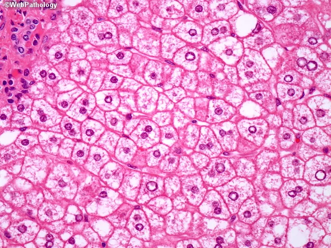
H&E section showing macrovesicular steatosis, ballooned hepatocytes, and Mallory-Denk (Mallory hyaline) inclusions - classic findings of alcoholic steatohepatitis (ASH).
3. Clinical Long Case Presentation
History
- Quantity, type, and duration of alcohol intake (>30 g/day sustained use is a rough threshold quoted in the Washington Manual of Medical Therapeutics); pattern of drinking (binge vs. daily); screen with CAGE questionnaire
- Symptoms depend heavily on stage:
- Fatty liver: often asymptomatic; occasionally mild right upper quadrant discomfort, fatigue
- Alcoholic hepatitis: anorexia, nausea/vomiting, malaise, fever, right upper quadrant pain, rapid onset jaundice; can present acutely after a binge, even in a previously undiagnosed cirrhotic
- Cirrhosis/decompensation: abdominal distension (ascites), confusion (encephalopathy), hematemesis/melena (variceal bleed), easy bruising, weight loss, muscle wasting
- Associated history: past withdrawal seizures/delirium tremens, pancreatitis, nutritional deficiency, psychiatric/social history, family history of liver disease
Examination
- General: malnutrition, temporal/thenar muscle wasting, fetor hepaticus
- Stigmata of chronic liver disease: spider angiomata (central arteriole with radiating vessels, blanch on pressure, mainly on trunk/face/upper limbs), palmar erythema, Dupuytren contracture, gynecomastia, testicular atrophy, parotid enlargement, caput medusae, leukonychia, clubbing
- Abdomen: tender hepatomegaly (alcoholic hepatitis) or shrunken nodular liver with splenomegaly (cirrhosis), ascites (shifting dullness/fluid thrill), umbilical hernia
- Decompensation signs: jaundice (scleral icterus), asterixis and altered mentation (hepatic encephalopathy), peripheral edema
- Vitals: fever and tachycardia may accompany acute alcoholic hepatitis or superimposed infection (e.g., spontaneous bacterial peritonitis)
4. Biochemical Tests
| Test | Typical finding in ALD |
|---|---|
| AST:ALT ratio (De Ritis ratio) | Characteristically >2 (often 2-4:1); AST rarely exceeds 300-400 IU/L, and ALT is often normal or only mildly raised - a key discriminator from viral/other hepatitis where ALT > AST |
| GGT (gamma-glutamyl transferase) | Raised; a sensitive but not very specific marker of heavy alcohol use (~65% sensitivity, ~80% specificity) |
| MCV | Macrocytosis, seen in most patients drinking 4+ drinks/day |
| Bilirubin | Elevated, predominantly conjugated; >3 mg/dL is part of the criteria for alcohol-associated hepatitis in some definitions |
| Albumin | Low, reflecting impaired synthetic function and/or malnutrition |
| PT/INR | Prolonged, reflects impaired hepatic synthetic function and correlates with severity |
| Platelet count | Reduced with portal hypertension/hypersplenism |
| Maddrey discriminant function (MDF) | = 4.6 x (patient PT - control PT) + serum bilirubin (mg/dL). MDF >= 32 defines severe alcoholic hepatitis and is used to decide on corticosteroid therapy (Harrison's Principles of Internal Medicine, 22nd ed). MELD score >20 is an alternative severity marker |
| Other | Neutrophilic leukocytosis common in alcoholic hepatitis; hypoglycemia and hypokalemia/hypomagnesemia may occur with malnutrition |
5. Urine Examination
Urine testing helps distinguish the pattern of jaundice, relevant because ALD causes a hepatocellular (mixed) picture:
| Jaundice type | Urine bilirubin | Urine urobilinogen |
|---|---|---|
| Hemolytic (pre-hepatic) | Absent | Markedly increased |
| Hepatocellular (e.g., ALD) | Present (conjugated bilirubin, dark/tea-colored urine) | Normal, reduced, or increased depending on severity |
| Obstructive (post-hepatic) | Present | Absent or markedly reduced |
Normal urine contains no bilirubin and only trace urobilinogen (normal excretion ~4 mg/day or less). In alcoholic hepatocellular disease, impaired hepatic bilirubin handling raises conjugated bilirubin in urine, and urobilinogen may be variably affected depending on how much bile flow/enterohepatic circulation is preserved (Goldman-Cecil Medicine; Harper's Illustrated Biochemistry). This test is a simple, low-cost bedside/laboratory tool useful in a long case to justify the type of jaundice.
6. Differential Diagnosis
For a patient presenting with hepatomegaly/cirrhosis/jaundice with a history of alcohol use, consider:
- Non-alcoholic fatty liver disease (NAFLD)/NASH: metabolic syndrome features (obesity, diabetes, dyslipidemia), typically ALT > AST (ratio <1), no significant alcohol history
- Chronic viral hepatitis B or C: risk factor history (transfusion, IV drug use), positive serology, ALT usually > AST
- Drug/toxin-induced liver injury: temporal relation to hepatotoxic drugs (e.g., paracetamol, isoniazid, methotrexate), improves on withdrawal
- Wilson disease: younger patient, low serum ceruloplasmin, Kayser-Fleischer rings, increased urinary/hepatic copper, may have neuropsychiatric features
- Hereditary hemochromatosis: raised serum ferritin and transferrin saturation, "bronze diabetes," family history, confirmed by genetic testing/liver iron studies
- Autoimmune hepatitis: positive ANA/anti-smooth muscle antibody, raised gamma-globulins, more common in younger women
- Primary biliary cholangitis/primary sclerosing cholangitis: predominantly cholestatic pattern (raised ALP disproportionate to transaminases), positive AMA (PBC)
- Budd-Chiari syndrome, right heart failure/cardiac cirrhosis: distinguished by history and imaging of hepatic venous outflow
- Alpha-1 antitrypsin deficiency: consider in early-onset liver disease, especially with coexisting lung disease
Recent Evidence Check
A quick search for recent systematic reviews/meta-analyses did not surface findings that contradict this core textbook framework; management of severe alcoholic hepatitis (corticosteroids guided by MDF/Lille score, MELD-based prognostication) remains consistent with current Harrison's 22nd edition and Goldman-Cecil guidance. If you want, I can run a focused PubMed search on a specific sub-area (e.g., corticosteroid vs. newer therapies for severe alcoholic hepatitis, or biomarkers for early alcohol-associated fibrosis).
If you'd like, I can also format this into a structured exam-style long case write-up (Chief Complaint -> History -> Examination -> Investigations -> Differential -> Diagnosis -> Management) as a downloadable document.
This is a shared conversation. Sign in to Orris to start your own chat.