
Ligaments of knee complex a,orgin insertion,blood supply,nerve supply
knee joint ligaments anatomy diagram ACL PCL MCL LCL
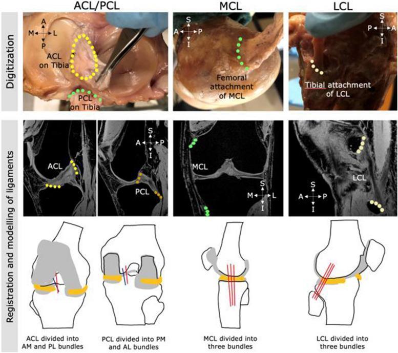
This composite educational image illustrates the multi-step process for digitizing and modeling human knee ligaments—ACL, PCL, MCL, and LCL—for orthopedic research. The top row features clinical photographs of cadaveric dissections where ligaments are digitized using anatomical landmarks: the ACL is highlighted as an oval area on the tibia, while the PCL, MCL, and LCL attachment sites are marked with colored point sequences. The middle row displays T1-weighted or Proton Density MRI scans in sagittal and coronal planes, showing the registration of these digitized points onto medical imaging to define ligamentous paths. The bottom row consists of schematic anatomical diagrams demonstrating the conceptual subdivision of ligaments into functional bundles: the ACL into anteromedial (AM) and posterolateral (PL) bundles; the PCL into posteromedial (PM) and anterolateral (AL) bundles; and the collateral ligaments (MCL and LCL) each into three distinct fiber bundles. This visualization supports biomechanical finite element modeling and the understanding of knee ligamentous stability and anatomy.
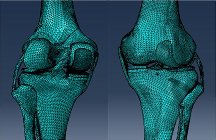
This image presents two views of a high-fidelity 3D computational model of a human knee joint featuring a Unicompartmental Knee Arthroplasty (UKA). The model is rendered using a complex triangular finite element mesh, providing a faceted visualization of the anatomical and prosthetic surfaces. The anatomy shown includes the distal femur, proximal tibia, and proximal fibula. Crucial soft tissue structures, including the anterior and posterior cruciate ligaments (ACL/PCL) and the medial and lateral collateral ligaments (MCL/LCL), are modeled as cable-like structures connecting the bony elements. The UKA is situated in the medial compartment, consisting of a metallic femoral condyle resurfacing component and a tibial baseplate platform. The model is designed for biomechanical stress analysis and surgical simulation, specifically investigating the impact of tibial component alignment and obliquity on load distribution. The teal-colored mesh highlights the geometry used for finite element analysis (FEA), demonstrating the interaction between the prosthetic implants and the native bone-cartilage complex.
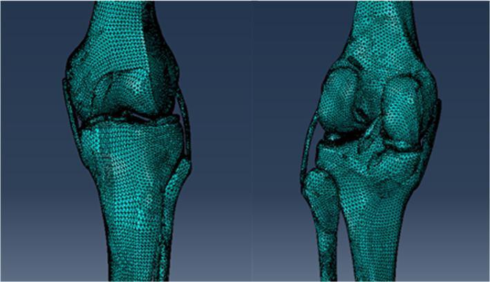
This diagnostic illustration presents two perspective views of a three-dimensional (3D) native human knee joint model. The visualization is a finite element or triangulated mesh representation derived from fused CT and MRI data. Anatomical structures shown include the distal femur, proximal tibia, and proximal fibula, with the mesh density clearly defining the cortical surfaces and contours. The model highlights soft tissue integration, specifically depicting the meniscus and the four major stabilizing ligaments: the anterior cruciate ligament (ACL), posterior cruciate ligament (PCL), medial collateral ligament (MCL), and lateral collateral ligament (LCL). The spatial relationships illustrate the tibiofemoral and proximal tibiofibular articulations. This digital model serves as a foundation for biomechanical simulations, such as Unicompartmental Knee Arthroplasty (UKA) planning and stress distribution analysis. The anatomical accuracy is tailored for orthopedic research and surgical education, emphasizing joint kinematics and structural integrity.
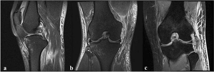
This set of three MRI images of the human knee joint illustrates combined ligamentous injuries. Image (a) is a sagittal view demonstrating an intraligamentous tear of the posterior cruciate ligament (PCL). The image shows discontinuity and high signal intensity within the PCL fibers, accompanied by peri-ligamentous edema. Image (b) is a coronal view displaying a medial collateral ligament (MCL) avulsion from its femoral attachment site, indicative of a posteromedial corner (PMC) injury. Image (c) is a coronal view highlighting a lateral collateral ligament (LCL) avulsion at the fibular attachment, representative of a posterolateral corner (PLC) injury. Both coronal images (b and c) reveal significant soft tissue edema and disruption of the normal dark, linear appearance of the collateral ligaments. These diagnostic images are essential for evaluating complex knee instability and planning surgical interventions such as PCL bracing or ligament augmentation.
Ligaments of the Knee Complex
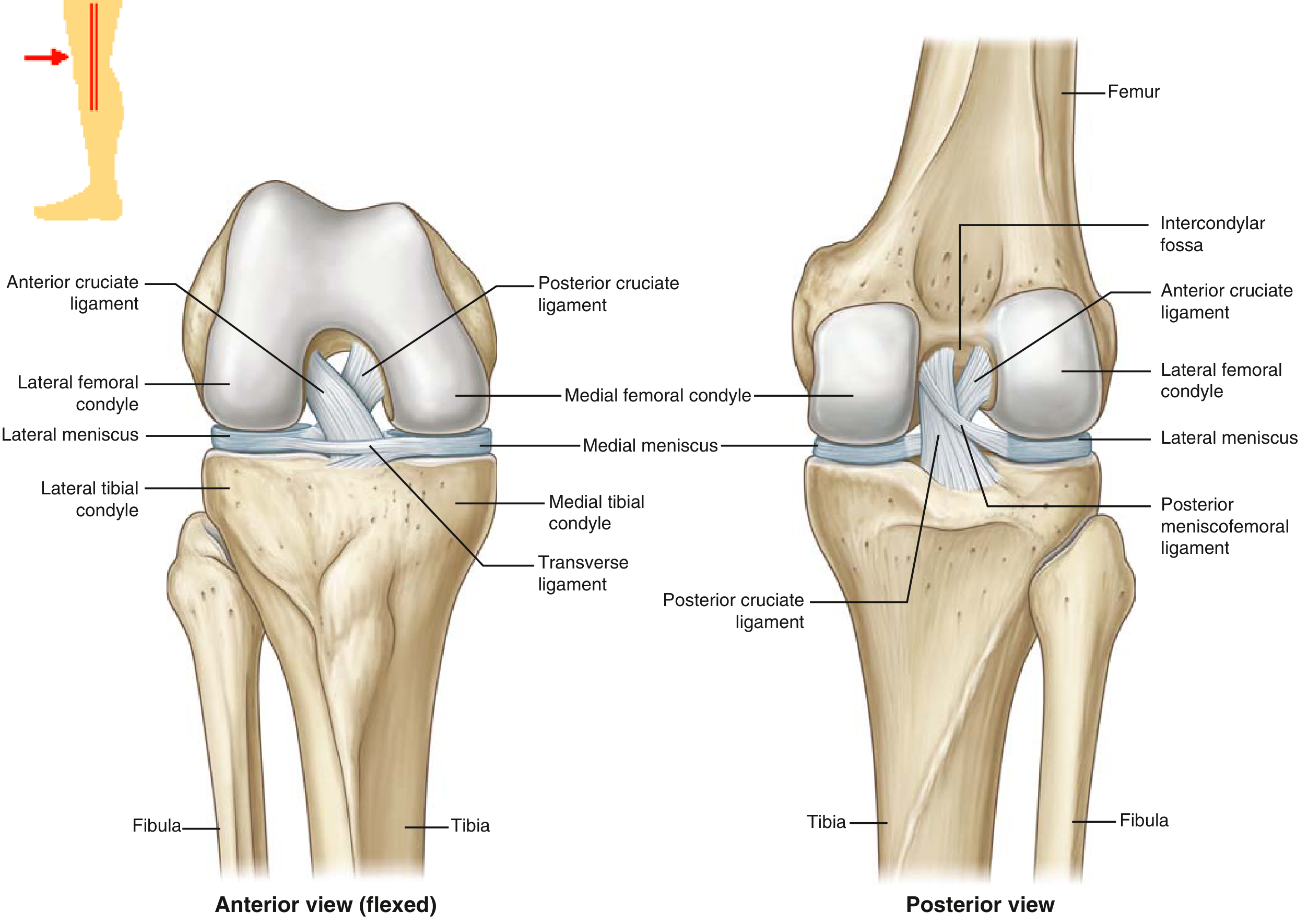
1. Anterior Cruciate Ligament (ACL)
- Anteromedial (AM) bundle - originates proximal to the bifurcate ridge; tight in flexion; primary restraint against anterior tibial translation (evaluated by Lachman and anterior drawer tests)
- Posterolateral (PL) bundle - originates distal to the bifurcate ridge; tight in extension; primarily a rotatory restraint (evaluated by pivot shift test)
2. Posterior Cruciate Ligament (PCL)
- Anterolateral (AL) bundle - originates anterior to the bifurcate prominence; tight in flexion (mnemonic: PCL is your "PAL" - important AL bundle is tight in flexion)
- Posteromedial (PM) bundle - originates posterior to the bifurcate prominence; tight in extension
3. Medial Collateral Ligament (MCL)
A. Superficial MCL (sMCL)
- Proximal: onto soft tissue overlying the anterior arm of semimembranosus
- Distal: directly to bone, just anterior to the posterior cortex of the tibia, 6-7 cm distal to the joint line
- Primary stabilizer against valgus stress throughout the flexion arc
- Secondary stabilizer to rotatory stress
- Anterior fibers tighten during the first 90 degrees of flexion; posterior fibers tighten in extension
B. Deep MCL (dMCL) / Mid-third Medial Capsular Ligament
- Thickening of the medial joint capsule
- Has distinct meniscofemoral and meniscotibial components
- Intimately associated with the medial meniscus
- Tensile strength: ~100 N
4. Lateral Collateral Ligament (LCL / Fibular Collateral Ligament)
- Primary restraint to varus stress at all degrees of knee flexion
- Tight in extension, lax in flexion (due to its location posterior to the axis of knee rotation)
5. Posterolateral Corner (PLC) Structures
- LCL
- Popliteus tendon - muscle originates on posterior tibia above the soleal line; femoral insertion is distal, anterior, and deep to the LCL; internally rotates the tibia
- Popliteofibular ligament - originates from the musculotendinous junction of popliteus; inserts on the medial aspect of the fibular head and styloid
6. Posteromedial Corner (PMC) Structures
- Capsular thickenings from semimembranosus insertions
- Posterior horn of medial meniscus
- Posterior Oblique Ligament (POL)
- Oblique Popliteal Ligament (OPL)
Posterior Oblique Ligament (POL)
- Origin: Proximal and posterior to the sMCL origin; distal and posterior to the adductor tubercle
- Insertion (central arm): Posteromedial tibia
- Three components: superficial, central, and capsular arms
- Primary stabilizer against internal rotation between 0-30 degrees of knee flexion
- Secondary stabilizer to valgus stress, particularly in extension
7. Anterolateral Ligament (ALL)
8. Medial Patellofemoral Ligament (MPFL)
9. Meniscofemoral Ligaments (Variably Present)
- Humphrey ligament (anterior meniscofemoral ligament): Passes anterior to the PCL
- Wrisberg ligament (posterior meniscofemoral ligament): Passes posterior to the PCL
- Both insert on the posterior horn of the lateral meniscus and originate on either side of the posteromedial bundle of the PCL on the medial femoral condyle
Summary Table: Blood Supply and Nerve Supply
| Ligament | Blood Supply | Nerve Supply |
|---|---|---|
| ACL | Middle genicular artery + fat pad | Posterior articular nerve (tibial n.); proprioceptive mechanoreceptors |
| PCL | Middle genicular artery + fat pad | Posterior articular nerve (tibial n.) |
| sMCL / dMCL | Medial superior & inferior genicular arteries | Saphenous nerve (femoral n.) + posterior articular nerve |
| LCL | Lateral superior & inferior genicular arteries | Common peroneal nerve territory; posterior articular nerve |
| POL / PMC | Medial genicular branches | Posterior articular nerve (tibial n.) |
| MPFL | Medial superior genicular artery | Saphenous nerve branches |
- Both cruciate ligaments are intra-articular but extrasynovial - covered by a synovial fold; this limits their healing capacity after rupture
- The saphenous nerve (medial) and common peroneal nerve (lateral) are at risk during medial and lateral knee repairs respectively
- The middle genicular artery is the dominant vessel to both cruciates; it arises directly from the popliteal artery and pierces the posterior capsule
- Mechanoreceptors (Ruffini endings, Pacinian corpuscles, Golgi tendon organ-like endings) in the ACL and PCL provide proprioceptive feedback