
Hisyopathegenesis of nodular scleritis and diff between scleritis and episcleritis
nodular scleritis episcleritis eye clinical
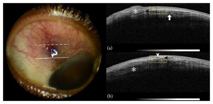
This composite educational image features a color photograph and corresponding Anterior Segment Optical Coherence Tomography (AS-OCT) scans of a right eye, illustrating clinical signs of nodular episcleritis and scleritis. The color photograph reveals marked violaceous ocular injection, superficial blood vessel congestion, and a localized nodular thickening approximately 4mm from the limbus. AS-OCT cross-sections (a) and (b) provide detailed visualization of the underlying tissue architecture. Key findings include nodular episcleral thickening (asterisk), a subepiscleral fluid level presenting as a hyporeflective space (black arrow), and intlamellar scleral edema (white arrow/asterisk). These OCT features correlate with the clinical appearance of vessel dilation and deep tissue inflammation. The image serves as a diagnostic reference for identifying inflammatory eye conditions using both external photography and non-invasive cross-sectional imaging, highlighting the relationship between visible clinical signs and internal structural alterations like scleral lamellae bisection by hyporeflective fluid.
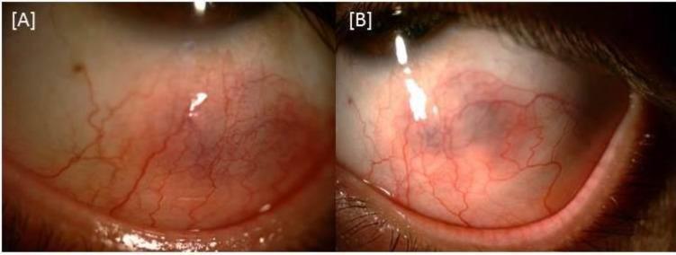
This clinical comparison chart consists of two side-by-side slit-lamp photographs (labeled A and B) documenting the progression and resolution of nodular anterior scleritis in a patient's left eye. Image A shows the eye one week after starting systemic treatment, demonstrating a distinct, elevated scleral nodule characterized by a deep purplish/violaceous hue. There is significant overlying episcleral and conjunctival vascular injection, with engorged, tortuous vessels radiating from the inflammatory site. Image B depicts the same eye three weeks after treatment initiation, illustrating a notable clinical improvement. The purplish hue has significantly faded, the nodular elevation has flattened, and there is a visible reduction in vascular engorgement and perilimbal hyperemia. This comparison serves as a clinical timeline for the response of scleritis associated with Behçet’s disease to immunosuppressive and corticosteroid therapy. The visual features highlighted are critical for differentiating nodular scleritis from simpler episcleritis, specifically the characteristic 'bluish' tint visible in natural light indicating deep scleral involvement.
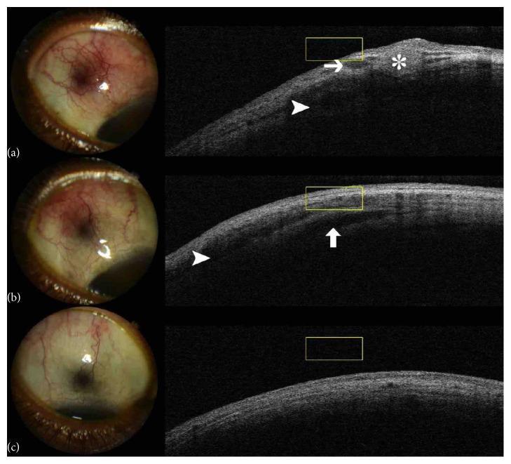
A longitudinal comparison series showing the clinical progression of tuberculous nodular episcleritis and anterior scleritis in the right eye. The image is structured into three temporal stages (a, b, c), each paired with a slit-lamp photograph and a corresponding anterior segment optical coherence tomography (AS-OCT) scan. Stage (a) displays intense superotemporal sector hyperaemia, a subconjunctival nodule, and violaceous injection; AS-OCT reveals a hyperreflective episcleral nodule (asterisk) and hyporeflective spaces indicating subepiscleral (arrow) and scleral (arrowhead) oedema. Stage (b), following initiation of antituberculous chemotherapy, demonstrates reduced hyperaemia and clinical resolution of the nodule, though AS-OCT shows persistent hyporeflective intrascleral spaces (arrow) and subepiscleral oedema (arrowhead). Stage (c), upon completion of therapy, shows total clinical remission with no hyperaemia and a restored, healthy anatomical structure on AS-OCT with complete elimination of prior lesions. This comparison illustrates the utility of AS-OCT in monitoring deep tissue resolution of inflammatory eye disease that may persist after superficial clinical signs have improved.
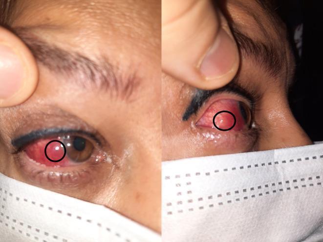
Content Type: Clinical Photograph. This composite image shows two views of a patient's right eye, highlighting inflammatory ocular signs. The left panel shows a frontal view of the eye with significant violaceous (purplish) scleral hyperemia and a distinct, circumscribed nodule (circled) on the temporal sclera. The right panel provides a lateral view, further illustrating the elevation and localized nature of the nodular lesion against the hyperemic scleral background. The purplish hue is characteristic of deep episcleral or scleral vessel involvement, often seen in scleritis. The iris appears brown and the cornea remains clear in these views. The images demonstrate the clinical presentation of nodular scleritis, an inflammatory condition of the eye's outer protective layer. These findings are pedagogically useful for distinguishing between superficial episcleritis (which typically presents with a brighter red hue) and deeper scleral inflammation (which exhibits a darker, violaceous color).
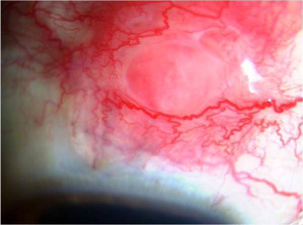
This clinical photograph, captured via slit-lamp biomicroscopy, illustrates a focal scleral nodule in a patient with systemic sarcoidosis. The image focuses on the anterior segment of the eye, specifically the bulbar conjunctiva and underlying sclera. The primary feature is a well-circumscribed, elevated, pinkish-red nodular mass. Surrounding the lesion is intense episcleral and conjunctival injection, characterized by dilated, tortuous blood vessels radiating towards the nodule, indicating significant localized inflammation and vascular engorgement. The central portion of the nodule appears slightly paler than its hyperemic periphery. This finding represents a rare ocular manifestation of sarcoidosis, showcasing granulomatous infiltration of the sclera. Key educational concepts include the identification of inflammatory scleral lesions, differentiating episcleritis from nodular scleritis, and recognizing ocular markers of systemic granulomatous diseases. The image is a valuable diagnostic reference for ophthalmologists and rheumatologists managing multi-system inflammatory conditions.
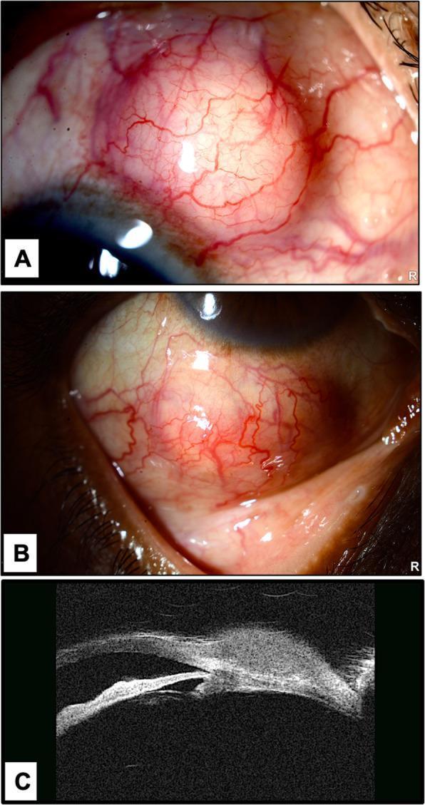
This composite medical image provides a multi-modal clinical and diagnostic evaluation of ocular scleral nodules. Panels A and B are clinical photographs of a right eye showing two distinct, raised, round scleral nodules located in the superior and inferior bulbar regions. The nodules are well-circumscribed and characterized by prominent, dilated episcleral 'feeder' vessels crossing their surfaces, while the surrounding conjunctiva remains relatively white and non-inflamed. Panel C presents an Ultrasound Biomicroscopy (UBM) cross-sectional image of a nodule. The UBM reveals a homogenous, hypo-reflective subconjunctival mass. Notably, the imaging demonstrates localized scleral thinning (scleral melting) beneath the lesion, without evidence of full-thickness perforation. These findings are clinically relevant for the differential diagnosis of infectious scleritis, particularly rare manifestations like Tropheryma whipplei, or inflammatory conditions such as nodular episcleritis. The images serve as an educational resource for identifying ocular mass lesions and the utility of UBM in assessing scleral integrity.
Histopathogenesis of Nodular Scleritis & Comparison with Episcleritis
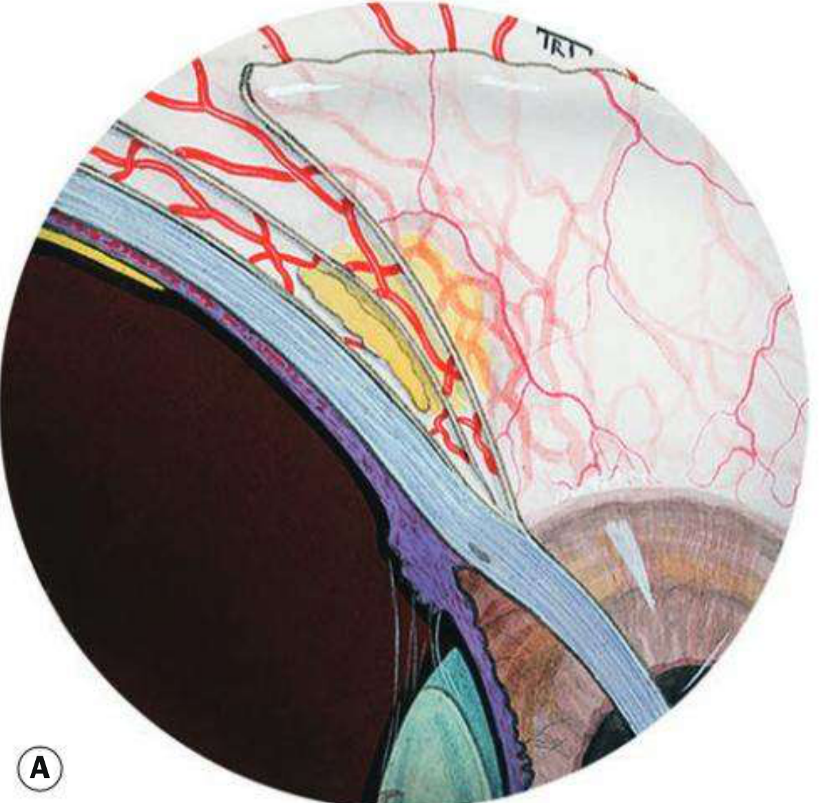
Anatomy Primer (foundation for understanding pathogenesis)
- Conjunctival vessels - most superficial, tortuous arteries/straight veins
- Superficial episcleral plexus - straight, radially configured; congested in episcleritis
- Deep vascular plexus - within superficial scleral stroma; congested in scleritis (gives the characteristic violaceous/purplish hue)
Histopathogenesis of Nodular Scleritis
Classification Context
| Type | Notes |
|---|---|
| Anterior Non-necrotizing | Diffuse or Nodular |
| Anterior Necrotizing with inflammation | Most destructive |
| Anterior Necrotizing without inflammation (Scleromalacia perforans) | Silent, RA-related |
| Posterior scleritis | Posterior to rectus insertions |
Histopathogenesis - Step by Step
- Central area of collagen necrosis (fibrinoid necrosis of scleral collagen bundles)
- Surrounded by palisading epithelioid histiocytes and giant cells (granulomatous reaction)
- Peripheral zone of lymphocytes and plasma cells
- Vascular occlusion and ischaemia contribute to collagen destruction This zonal pattern resembles the rheumatoid nodule, and in RA-associated scleritis the pathogenesis mirrors the rheumatoid granuloma - immune complex deposition -> complement activation -> vasculitis -> tissue necrosis. - Kanski's; Rheumatology 2-Volume Set (Elsevier 2022)
- Oedema and inflammatory cell infiltration involve the full thickness of the sclera but are localized, producing a discrete, elevated, tender nodule
- The nodule has a deeper blue-red colour than episcleral nodules (because the deep plexus is involved)
- On slit-lamp examination, the anterior scleral surface is elevated (vs. episcleritis where the scleral surface is flat)
- Nodules are immobile - they cannot be displaced over the underlying sclera (unlike episcleral nodules)
- Most frequently develop in the interpalpebral region close to the limbus
- Instillation of 10% phenylephrine blanches conjunctival and superficial episcleral vessels but NOT the deep plexus overlying the nodule
- Multiple nodules may expand and coalesce if treatment is delayed
- As inflammation subsides, increased scleral translucency (grey/bluish appearance) develops - due to rearrangement of collagen fibers
- More than 10% of nodular scleritis cases progress to necrotizing disease; if treated early, superficial necrosis does not occur and the nodule heals centrally leaving a small atrophic scar
Clinical Images of Nodular Scleritis
Comparison: Scleritis vs. Episcleritis
| Feature | Episcleritis | Scleritis |
|---|---|---|
| Layer involved | Episclera (superficial connective tissue layer between scleral stroma and Tenon capsule) | Full thickness of sclera + deep vascular plexus |
| Vascular plexus | Superficial episcleral plexus (congested) | Deep vascular plexus (congested) |
| Color | Bright/salmon pink, peri-limbal injection | Deeper violaceous/purple hue - best seen in daylight |
| Pain | Mild discomfort or absent (up to 50% painless); grittiness | Severe boring/gnawing pain, often wakes patient at night; radiates to temple/face; responds poorly to common analgesics |
| Phenylephrine test | 10% phenylephrine blanches vessels (positive blanch) | 10% phenylephrine does NOT blanch deep scleral vessels (negative blanch) |
| Slit-lamp surface | Flat anterior scleral surface | Elevated anterior scleral surface (scleral oedema) |
| Nodule mobility | Episcleral nodule is movable over sclera | Scleral nodule is immovable |
| Vision threat | NOT vision-threatening | Can threaten vision (glaucoma, peripheral keratitis, uveitis, scleral perforation) |
| Systemic association | Rare (majority idiopathic) | ~50% associated with systemic disease (RA, ANCA vasculitis, SLE, gout, IBD) |
| Cotton swab test | Vessels move with conjunctiva | Vessels do NOT move with conjunctiva |
| Bilaterality | Often bilateral (>50%) | Bilateral in 60% for necrotizing; variable for others |
| Course | Self-limiting (days to 3 weeks), recurrent but benign | Requires systemic therapy; mean duration ~6 years; can progress |
| Histopathology | Non-specific vascular dilation + mild perivascular lymphocytic infiltrate | Zonal granulomatous necrosis, fibrinoid collagen necrosis, epithelioid histiocytes, palisading giant cells, vasculitis |
| Scleral thinning | Does NOT occur | Can occur - choroidal show/bulge in severe cases |
| IOP | Rarely elevated | Glaucoma is the most common cause of visual loss |
| Treatment | Cool compresses; topical NSAIDs or mild topical steroids; oral NSAIDs rarely needed | Systemic NSAIDs (flurbiprofen, indomethacin) first-line; systemic corticosteroids; immunosuppressives (methotrexate, azathioprine); biologics for refractory cases |
Key Memory Tricks
- "Epi" = superficial - episclera only, salmon pink, blanches, self-limits
- "Sclera" = deep - violaceous, doesn't blanch, painful at night, systemic disease, vision threat
- Nodular scleritis vs. nodular episcleritis: immovable + elevated scleral surface + blue-red colour = scleritis; movable + flat scleral surface + pink = episcleritis
- The phenylephrine test is the single most useful bedside test to distinguish the two
?
- Did something in my previous answer need more explanation?
- Do you have a new question?
- Was there a typo or incomplete message?
Got no answer for previous question
Histopathogenesis of Nodular Scleritis
Anatomy Foundation
- Conjunctival vessels - most superficial
- Superficial episcleral plexus - straight, radially configured; congested in episcleritis
- Deep vascular plexus - within superficial scleral stroma; congested in scleritis (gives the violaceous/purple hue)
Pathogenesis - Step by Step
- Central zone of fibrinoid necrosis of scleral collagen
- Surrounded by palisading epithelioid histiocytes and giant cells
- Peripheral zone of lymphocytes and plasma cells
- Vasculitis and vascular occlusion cause ischaemia, amplifying collagen destruction
- This mirrors the rheumatoid nodule histologically - immune complex deposition → complement activation → vasculitis → tissue necrosis
- Inflammation is localized but involves the full thickness of the sclera
- Produces a discrete, elevated, immovable tender nodule
- Characteristically deep blue-red in colour (because the deep plexus is involved)
- Slit-lamp shows an elevated anterior scleral surface (distinguishes it from episcleritis nodule where the surface is flat)
- Most commonly in the interpalpebral region, close to the limbus
- 10% phenylephrine blanches superficial/episcleral vessels but does NOT blanch the deep vessels overlying the nodule
- Nodules expand and coalesce if untreated
- As inflammation resolves, scleral translucency increases (grey-blue sclera) due to collagen fiber rearrangement
- >10% progress to necrotizing scleritis if untreated
- With early treatment, the nodule heals from the centre leaving a small atrophic scar
Clinical Images
Difference: Scleritis vs. Episcleritis
| Feature | Episcleritis | Scleritis |
|---|---|---|
| Layer involved | Episclera only (superficial) | Full thickness sclera + deep vascular plexus |
| Colour | Salmon pink, bright red | Deep violaceous/purple (best seen in daylight) |
| Pain | Mild or absent (50% painless); grittiness | Severe boring pain, wakes at night, radiates to face/temple, poorly controlled by analgesics |
| Phenylephrine 10% test | Vessels blanch (positive) | Vessels do NOT blanch (negative) |
| Slit-lamp surface | Flat scleral surface | Elevated scleral surface (scleral oedema) |
| Nodule mobility | Episcleral nodule - movable over sclera | Scleral nodule - immovable |
| Cotton swab test | Vessels move with conjunctiva | Vessels do NOT move with conjunctiva |
| Vision threat | NOT vision-threatening | YES - glaucoma, keratitis, uveitis, scleral thinning |
| Systemic association | Majority idiopathic | ~50% with systemic disease (RA, vasculitis, SLE, gout) |
| Histology | Mild perivascular lymphocytic infiltrate, vascular dilation | Zonal granulomatous necrosis, fibrinoid collagen necrosis, palisading histiocytes/giant cells, vasculitis |
| Scleral thinning | Does NOT occur | Occurs in severe/necrotizing cases (choroidal show) |
| IOP | Rarely elevated | Glaucoma = most common cause of visual loss |
| Course | Self-limiting (days-3 weeks), recurrent but benign | Requires systemic therapy; mean duration ~6 years |
| Treatment | Topical NSAIDs/mild steroids; oral NSAIDs occasionally | Systemic NSAIDs (flurbiprofen, indomethacin); oral corticosteroids; immunosuppressives; biologics for refractory cases |
Key Memory Points
- Phenylephrine test is the single most useful bedside distinguishing test
- Colour: pink = episcleritis (superficial); purple/violet = scleritis (deep)
- Nodule: movable + flat scleral surface = episcleral; immovable + elevated surface = scleral
- Pain at night strongly suggests scleritis
- Episcleritis = benign, self-limiting, rarely systemic; Scleritis = serious, vision-threatening, systemic association in 50%