


How to treat this
truncal acne bacne folliculitis treatment topical oral 2024 2025
folliculitis back papules pustules acne truncal
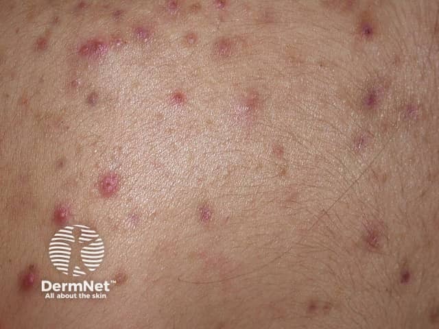
High‑resolution clinical photograph of the dorsal trunk demonstrating truncal acne lesions. This close‑up dermatologic image records the back skin in posterior view, with a focus on follicular papules and pustules at varying stages of inflammation. The observable features include small erythematous papules, superficial pustules with central necrosis, and subtle perifollicular redness against normal skin texture. No large nodules or cystic lesions are evident in the field of view. The distribution is scattered across the upper and mid back with a mixed inflammatory pattern, consistent with acne vulgaris affecting the trunk. The skin shows mild oiliness and tiny follicular openings; scale is minimal. The image emphasizes inflammatory sequelae rather than postinflammatory scarring. Clinically, these findings correlate with active sebaceous gland activity and Propionibacterium acnes–driven folliculitis. Differentials include folliculitis, keratosis pilaris, and less likely bacterial infection; clinical correlation and distribution help distinguish among etiologies. Diagnostically, this image supports a diagnosis of mild to moderate truncal acne and aids in patient education, assessment of treatment response, and dermatologic research on lesion morphology. Potential uses include educational demonstrations, photography‑based scoring, and outcome tracking in clinical trials of topical retinoids, benzoyl peroxide, and combination therapies.
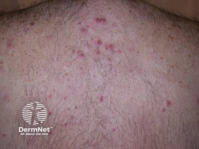
Clinical photography of the patient's upper back illustrating an inflammatory acneiform eruption consistent with folliculitis or acne related lesions. Modality: digital color photograph, posterior dorsal view of the thoracic back. The image shows multiple erythematous papules and a few pustules concentrated on the upper back and near the shoulders, with mild superficial crusting in a few lesions. Lesions are discrete, roughly 2-4 mm in diameter, with perifollicular erythema and a central palpably raised area. No large nodules or abscesses are seen on this field; the skin texture shows mild post-inflammatory hyperpigmentation in places. Hair follicles appear conspicuously involved, suggesting folliculitis or an acneiform eruption rather than a vesicular or scaly dermatosis. The distribution is truncal, predominantly in the upper back, with bilateral symmetry but no midline predilection. Clinically, this pattern favors bacterial folliculitis or acneiform dermatitis; fungal folliculitis (Pityrosporum) or pityrosporum-centric folliculitis could be considered in persistent cases, particularly with pruritus. Management implications include topical antiseptics, benzoyl peroxide, or topical/oral antibiotics depending on severity. Correlate with patient history: recent sweating, occlusive clothing, shaving, or trauma. This image is useful for education on trunk acneiform eruptions, differential diagnosis of folliculitis, and treatment planning.
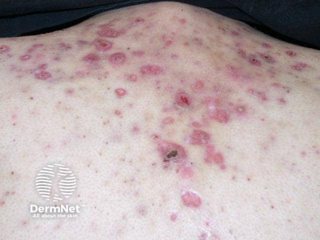
This is a clinical photograph of the back/trunk showing an extensive inflammatory acneiform eruption with scattered erythematous papules and pustules, some coalescing into superficial plaques. Lesions are follicular in distribution, centered around hair follicles, with perifollicular erythema and crusting. The skin appears slightly edematous with mild scaling. The pattern is diffuse across the upper and mid-back, with higher density in the central to lateral paraspinal regions. Several lesions show a central pustule or crust, suggesting secondary infection or rupture. No visible comedones are clearly evident in this view, but follicular involvement is evident. The appearance is compatible with folliculitis or acne vulgaris involving the trunk; differential includes pityrosporum folliculitis or a contact dermatitis-driven papulopustular eruption. There is no edema of deeper tissues; no ulceration or necrosis except one crusted lesion with small crater. The image is taken under standard clinical lighting with a neutral background; no contrast or magnification. Clinically relevant significance: trunk acneiform eruptions can be painful, pruritic, and cosmetically distressing; management typically includes topical retinoids, benzoyl peroxide, antibiotics (topical or oral for moderate to severe disease), and addressing inflammatory components. This image is useful for educational discussion of differential diagnosis, truncal skin infections, and dermatologic therapy planning and education.
What This Looks Like
- Multiple small (1-3 mm), discrete, erythematous follicular papules (bumps centered on hair follicles)
- Some with pinpoint pustule heads
- Scattered post-inflammatory hyperpigmented macules (brownish marks from older healed lesions)
- Lesions are widespread across the upper and mid back, fairly uniform in size
- If itchy - strongly favors Malassezia (Pityrosporum) folliculitis (fungal)
- If tender/painful but not itchy - favors acne vulgaris or bacterial folliculitis
Treatment Options
1. If Malassezia (Pityrosporum/Fungal) Folliculitis
- Ketoconazole 2% shampoo or selenium sulfide 2.5% lotion - apply to the back, leave on for 5-10 minutes, then rinse. Use daily for 2-4 weeks
- Topical imidazole creams (clotrimazole, miconazole, econazole) applied once or twice daily
- Oral fluconazole - 150-300 mg once weekly for 1-2 months, OR a single dose of 400 mg
- Oral itraconazole - 200 mg/day for 5-7 days
-
Monthly applications of selenium sulfide lotion or topical econazole, since relapses are very common
-
Andrews' Diseases of the Skin, p. 2493-2495
-
Dermatology 2-Volume Set 5e, p. 4058
2. If Bacterial Folliculitis (Staphylococcus aureus)
- Mupirocin 2% ointment applied to affected areas
- Fusidic acid cream
- Benzoyl peroxide wash (5-10%) - antibacterial and helps prevent resistance
- Clindamycin 1% lotion/gel
- Cephalexin 500 mg twice daily for 7-10 days
- Doxycycline 100 mg twice daily (also covers gram-negative folliculitis)
- For MRSA: trimethoprim-sulfamethoxazole or doxycycline
3. If Truncal Acne Vulgaris
- Benzoyl peroxide body wash (5-10%) - used as a wash in the shower daily; very effective for back acne
- Topical retinoid (adapalene 0.1% gel) applied at night - trifarotene 0.005% cream is FDA-approved specifically for truncal acne
- Topical clindamycin or dapsone 7.5% gel applied once daily
- Doxycycline 100 mg/day - most commonly used oral antibiotic for truncal acne
- Sarecycline - a newer narrow-spectrum tetracycline with evidence for truncal acne efficacy within 3 weeks
- Isotretinoin - reserved for severe, nodular, or scarring acne; requires a dermatologist prescription
General Skin Care Tips (Apply to All Cases)
| What to do | Why |
|---|---|
| Shower promptly after sweating | Prevents yeast/bacteria overgrowth |
| Wear loose, breathable clothing | Reduces friction and occlusion |
| Use a gentle, non-comedogenic body wash | Avoids pore clogging |
| Do NOT pick or squeeze lesions | Prevents scarring and spread |
| Change pillowcases/bed sheets frequently | Reduces bacterial recolonization |
| Avoid heavy oils or occlusive body lotions | Worsens follicular plugging |
Bottom Line
⚠️ This is educational information only and not a substitute for a clinical evaluation. A dermatologist can diagnose this definitively and prescribe the most effective treatment.
Non itchy
Treatment Plan for Non-Itchy Truncal Acne / Bacterial Folliculitis
Step 1 - Daily Wash (Start Immediately)
- Benzoyl peroxide 5-10% body wash - use in the shower daily, lather on the back/shoulders, leave on for 1-2 minutes before rinsing. This is the single most effective OTC step for back acne. Brands: PanOxyl, Neutrogena Body Clear.
- Alternatively, a salicylic acid 2% body wash if your skin is sensitive to benzoyl peroxide.
Step 2 - Topical Treatment (Apply After Shower)
| Option | What to apply | Frequency |
|---|---|---|
| Adapalene 0.1% gel (Differin) | Thin layer on back/shoulders | Every night |
| Clindamycin 1% gel/lotion | Thin layer on affected areas | Twice daily |
| Benzoyl peroxide 2.5-5% gel | Thin layer | Once daily (morning) |
Step 3 - Oral Antibiotics (If Widespread / Not Improving in 6-8 Weeks)
- Doxycycline 100 mg once or twice daily for 3 months - most commonly used for truncal acne
- Sarecycline - newer narrow-spectrum tetracycline, shown to reduce truncal lesions within 3 weeks in phase 3 trials
- Always combine oral antibiotics with benzoyl peroxide to prevent resistance
Step 4 - For Stubborn / Scarring Cases
- Isotretinoin (Accutane) - dermatologist-prescribed oral retinoid; highly effective for severe or recurrent truncal acne; requires blood monitoring
Lifestyle Adjustments That Actually Matter
- Shower within 30 minutes of sweating - sweat sitting on the back is a major trigger
- Wear cotton/loose shirts - synthetic fabrics trap heat and increase follicular plugging
- Change bed sheets weekly - the back rests on sheets for hours each night
- Avoid heavy body lotions or oils on the back
- Do not squeeze or pop - this causes the dark spots (PIH) you already have, and can scar
For the Post-Inflammatory Dark Spots (Brown Marks)
- Niacinamide 10% serum on the back
- Azelaic acid 10-20% - also treats active acne simultaneously
- Avoid sun exposure on the back (UV darkens PIH)
Summary Priority Order
- Start benzoyl peroxide body wash today (available OTC)
- Add adapalene 0.1% gel at night (OTC in many countries)
- If no improvement in 6-8 weeks, see a dermatologist for oral doxycycline
- For severe/widespread disease, ask about isotretinoin
⚠️ A dermatologist visit is still recommended for a firm diagnosis and prescription-strength treatment if OTC options don't work within 2 months.
How long will treatment take to show results?
Realistic Treatment Timeline
Weeks 1-2: Possible Initial Worsening ("Purging")
- Benzoyl peroxide and retinoids (adapalene) can cause an initial flare — more breakouts, dryness, or irritation
- This is normal and expected — the skin is turning over faster and pushing out clogged follicles
- Do not stop treatment during this phase
Weeks 4-6: First Signs of Improvement
- New active lesions start appearing less frequently
- Existing pustules begin to flatten and dry out
- The benzoyl peroxide wash usually shows the earliest results here
Weeks 8-12: Noticeable Improvement
- This is the minimum time you need to fairly judge whether a treatment is working
- Roughly 50-60% reduction in active lesions is a realistic expectation by week 12 with consistent topical treatment
- Oral doxycycline can show meaningful results slightly faster — often by week 6-8
Months 4-6: Near-Clear Skin
- With consistent treatment (topical + oral if prescribed), the back can be largely clear
- The dark spots (PIH) from healed lesions take the longest — typically 3-6 months to fade significantly, even after active acne is gone
Month 6+: Maintenance Phase
- Acne often returns if you stop all treatment
- Continue a maintenance routine (benzoyl peroxide wash 2-3x/week + adapalene 2-3x/week) long-term to prevent relapse
Summary Table
| Timeframe | What to Expect |
|---|---|
| Week 1-2 | Possible purging/dryness — normal |
| Week 4-6 | Fewer new spots forming |
| Week 8-12 | Visible reduction in active lesions |
| Month 4-6 | Mostly clear skin |
| Month 3-6 | Dark spots (PIH) gradually fading |
| Ongoing | Maintenance needed to stay clear |
Key Things That Slow Down Results
- Inconsistency — skipping days is the #1 reason treatment fails
- Sweating without showering shortly after
- Squeezing lesions — adds weeks to healing and causes more PIH
- Using comedogenic body products (oily lotions, hair products that run down the back)