
Collagen for cartilage
I'll look up collagen in cartilage from the medical library while searching for recent evidence simultaneously.
Searching sources...
Reading File
Searching PubMed
collagen cartilage structure function
Now let me search the medical library for collagen in cartilage content:
Finding Sources
Finding Sources
Excellent results. Let me now read the key sections from Harper's Biochemistry and the Histology text in parallel:
Reading File
Reading File
Reading File
Let me also fetch the cartilage matrix diagram and read the Rheumatology content on collagen types:
Loading Image
Reading File
I now have comprehensive information from multiple authoritative textbooks. Here is the full answer:
Collagen in Cartilage
Cartilage is a specialized connective tissue whose mechanical properties depend directly on the type and arrangement of its collagen molecules. The overall composition is roughly: water (75%) > collagen (25% wet weight) > proteoglycans (10% wet weight) > noncollagenous proteins > chondrocytes (the only cell type present). - Miller's Review of Orthopaedics, 9th Ed
Types of Cartilage and Their Collagen
| Cartilage Type | Dominant Collagen | Additional Notes |
|---|---|---|
| Hyaline (articular) | Type II (90-98% of total collagen) | Also contains types V, VI, IX, X, XI |
| Elastic cartilage | Type II + elastin | Elastin gives flexibility |
| Fibrocartilage | Type I | Dense fibrous structure |
Collagen Types in Hyaline Cartilage - Roles
The matrix of hyaline cartilage contains five "cartilage-specific" collagen types (II, VI, IX, X, XI) found in significant amounts only in cartilage:
Type II Collagen - the dominant structural protein
- Composed of three identical α1(II) chains (homotrimer)
- Forms the 3D fibril meshwork (~20 nm diameter fibrils)
- Provides tensile strength; restrains the "swelling" of aggrecan (contributing to viscoelastic behavior)
- Highest concentration in the superficial zone (Zone 1)
Type IX Collagen (FACIT collagen - fibril-associated collagen with interrupted triple helices)
- Bound at the surface of type II collagen fibrils
- Has three alpha chains forming three triple-helical domains (col1, col2, col3)
- Its NC4 domain protrudes outward and interacts with COMP and fibromodulin
- Covalently cross-linked to type II collagen in adults
- Facilitates fibril interaction with proteoglycans (often carries a chondroitin sulfate side chain)
- Mutations → pseudoachondroplasia, multiple epiphyseal dysplasia
Type XI Collagen
- Co-assembles with type II to form the initial microfibrils that regulate further fiber assembly
- Its N-terminal propeptides are retained and exposed at fiber surface
- Regulates fibril diameter and controls fiber growth
- Cross-links primarily to other type XI molecules
- Mutations → major growth disturbances (Stickler syndrome, Marshall syndrome)
Type X Collagen
- Found specifically in the calcified zone (Zone 4) of articular cartilage
- Organizes collagen fibrils into a hexagonal lattice - critical for mechanical function
- Marker of chondrocyte hypertrophy
Type VI Collagen
-
Concentrated at the periphery of chondrocytes (pericellular matrix)
-
Anchors chondrocytes to the matrix framework
-
Histology: A Text and Atlas with Correlated Cell and Molecular Biology / Rheumatology, 2-Volume Set (2022, Elsevier)
Molecular Architecture - The Matrix
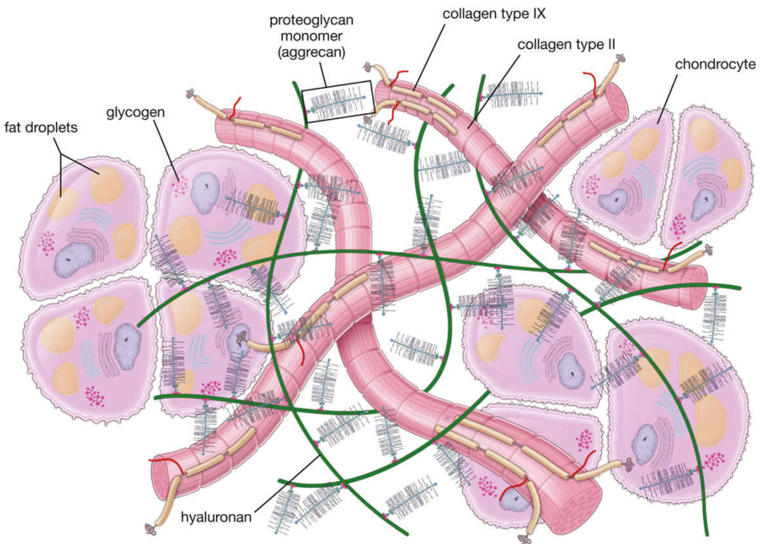
The collagen fibril network interlocks with aggrecan (the major proteoglycan, ~2000 kDa), which carries:
- Chondroitin sulfate chains (domain C of core protein)
- Keratan sulfate chains (domain B)
- Binds hyaluronan non-covalently via link protein
The sulfate groups of aggrecan give the matrix a high negative charge, attracting water molecules (responsible for compressibility). Collagen fibrils physically restrain this swelling tendency. The interplay between collagen tension and proteoglycan osmotic pressure gives cartilage its unique viscoelastic properties.
An additional non-collagen protein, chondronectin, promotes chondrocyte attachment specifically to type II collagen.
- Harper's Illustrated Biochemistry, 32nd Ed
Zonal Distribution in Articular Cartilage
| Zone | Collagen Characteristics |
|---|---|
| Zone 1 - Superficial | Highest collagen concentration, lowest proteoglycan; collagen fibers run parallel to surface |
| Zone 2 - Middle/Transitional | High proteoglycan and water content |
| Zone 3 - Deep | Highest proteoglycan concentration; collagen fibers perpendicular to surface |
| Zone 4 - Calcified | Type X collagen present; anchors to subchondral bone |
Pathological Relevance
-
Osteoarthritis: Chondrocytes switch from synthesizing type II to type I collagen - this marks dedifferentiation and cartilage degradation
-
IL-1 stimulates MMPs and COX-2, degrading the collagen matrix
-
TGF-β promotes ECM synthesis and counteracts IL-1/MMP activity
-
Aging: Smaller proteoglycan molecules lose water-binding capacity, increasing stiffness; collagen cross-linking increases via advanced glycation end-products
-
Excess mechanical stress or underloading both lead to cartilage thinning and proteoglycan loss
-
Miller's Review of Orthopaedics, 9th Ed
Summary: Type II collagen is the backbone of cartilage (90-98% of collagen content in hyaline cartilage), with types IX, XI, X, and VI serving as structural organizers, fibril regulators, zone-specific scaffolds, and cell-anchoring proteins respectively. Fibrocartilage replaces type II with type I collagen, giving it a far denser, less compressible character.
This is a shared conversation. Sign in to Orris to start your own chat.