
Rosai-Dorfman disease hallmark microscopic features
Reading File
The hallmark of Rosai-Dorfman disease (sinus histiocytosis with massive lymphadenopathy) is emperipolesis - the presence of intact lymphocytes, plasma cells, and other hematopoietic cells within the cytoplasm of large histiocytes. Let me pull the full textbook coverage now.
Searching sources...
Finding Sources
Finding Sources
Good results. Let me read the most detailed pathology sections from multiple books simultaneously.
Reading File
Reading File
Reading File
Excellent detail. Now let me fetch the histology images and also read the Dermatology 5e section for more microscopic detail.
Reading File
Loading Image
Loading Image
I now have comprehensive, multi-source information. Here is the full answer:
Rosai-Dorfman Disease (RDD) - Hallmark Microscopic Features
1. The Defining Hallmark: Emperipolesis
The absolute hallmark of RDD is emperipolesis - the presence of intact, viable hematopoietic cells within the cytoplasm of large histiocytes. The engulfed cells include:
- Lymphocytes (most common)
- Plasma cells
- Erythrocytes
- Neutrophils
This is distinct from phagocytosis - in emperipolesis the engulfed cells remain intact and viable (not degraded), often appearing to "swim" within the large pale cytoplasm of the host histiocyte.
Fitzpatrick's Dermatology describes it precisely: "different types of bone marrow cells, such as lymphocytes or neutrophils, are found in the cytoplasm of histiocytes."
2. The RDD Histiocytes Themselves
The large histiocytes that characterize RDD have:
- Abundant pale/clear cytoplasm (sometimes described as foamy or vacuolated)
- Large vesicular nuclei with prominent nucleoli
- Hypochromatic nuclei (Goldman-Cecil)
- They proliferate within lymph node sinuses (hence "sinus histiocytosis")
3. Lymph Node Architecture (Nodal Disease)
- Dilated sinuses packed with the large histiocytes showing emperipolesis
- Cortical regions contain abundant plasma cells (often polyclonal) and activated B lymphocytes
- Background inflammatory infiltrate of mature lymphocytes and plasma cells
4. Immunohistochemistry Profile
| Marker | Result | Significance |
|---|---|---|
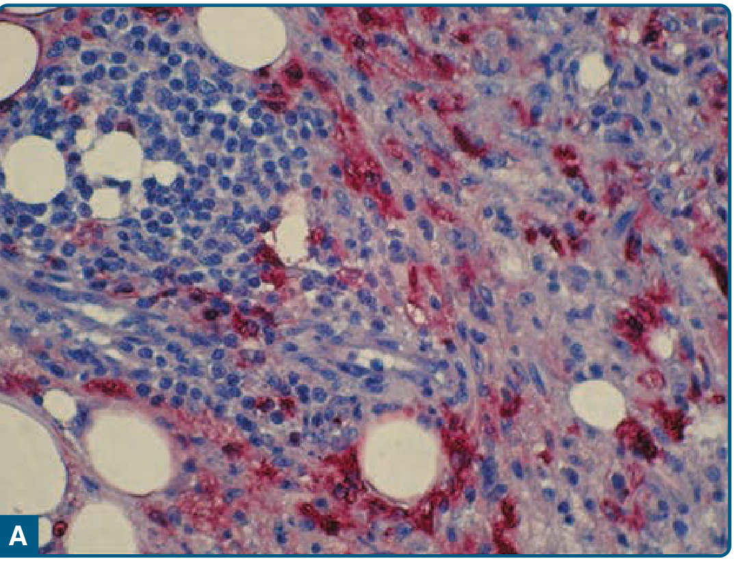
| S100 | Positive | Key diagnostic marker; also used as serum marker |
| CD68 | Positive | Macrophage lineage |
| CD163 | Positive | Macrophage/monocyte marker |
| CD14 | Positive | Macrophage marker |
| HLA-DR | Positive | |
| α₁-antitrypsin | Positive | |
| α₁-antichymotrypsin | Positive | |
| Fascin | Positive | |
| HAM-56 | Positive | Human alveolar macrophage 56 |
| Stabilin-1 | Positive | |
| Cyclin D1 + pERK | Positive | MAPK pathway activation |
| OCT2 (nuclear) | Positive | Recent finding (51/51 cases) |
| CD1a | Negative | Key distinction from LCH |
| CD207 (Langerin) | Negative | Key distinction from LCH |
The S100+/CD1a- combination is the cardinal IHC signature.
5. Photomicrographs from Fitzpatrick's Dermatology
Panel A - S100 immunostain showing histiocyte positivity:
Panel B - Stabilin-1 staining:
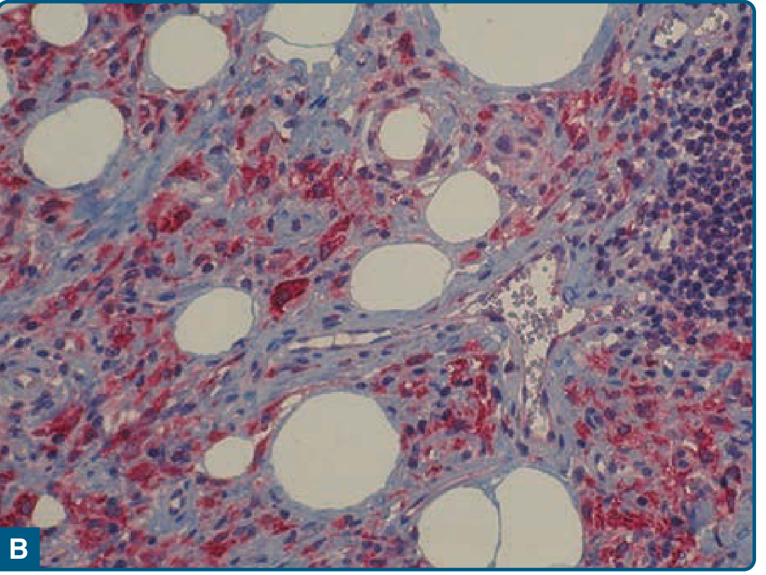
Figures 117-13A and 117-13B from Fitzpatrick's Dermatology - histiocytes are positive for both S100B and stabilin-1
6. Cutaneous RDD (Extranodal)
In skin biopsies, the same features apply but the infiltrate is typically in the dermis and subcutis:
- Dense infiltrate of large pale histiocytes with emperipolesis
- Abundant polyclonal plasma cells
- IgG4-positive plasma cells may be present within the infiltrate (though RDD ≠ IgG4-related disease)
7. Key Differential Diagnosis by Microscopy
| Feature | RDD | Langerhans Cell Histiocytosis | Juvenile Xanthogranuloma |
|---|---|---|---|
| Emperipolesis | Yes (hallmark) | Rare | Rare (occasionally) |
| S100 | + | + | Can be + (large macrophages) |
| CD1a | Negative | Positive | Negative |
| Langerin (CD207) | Negative | Positive | Negative |
| Birbeck granules (EM) | Absent | Present | Absent |
| Touton giant cells | Absent | Absent | Present |
RDD is distinguished from LCH by the absence of CD1a/CD207 staining.
Summary: The microscopic signature of Rosai-Dorfman disease is large, pale S100+/CD1a- histiocytes showing emperipolesis (intact lymphocytes, plasma cells, and erythrocytes within their cytoplasm), set against a background rich in plasma cells, filling lymph node sinuses or dermal/subcutaneous tissue.
Sources:
- Fitzpatrick's Dermatology, Vol. 1-2, p. 2060 (Histopathologic Findings)
- Dermatology 2-Volume Set 5e (Dermatology 5e), "R" Group: Rosai-Dorfman Disease, Pathology section
- Goldman-Cecil Medicine, Pathobiology of Rosai-Dorfman-Destombes
This is a shared conversation. Sign in to Orris to start your own chat.