
Leptospirosis
I'll start by reading the medical library skill, then search for comprehensive leptospirosis content in parallel.
Reading File
Now I'll search the library and PubMed in parallel for comprehensive leptospirosis coverage.
Finding Sources
Searching PubMed
leptospirosis
Searching Images
leptospirosis clinical features spirochete Leptospira
Good results. Now I'll read the main textbook sections in parallel - Goldman-Cecil Medicine (most comprehensive clinical coverage) and Microbiology (pathogen details), plus Andrews' Dermatology.
Reading File
Reading File
Reading File
Excellent. Now let me read more from Goldman-Cecil (the main clinical reference) to get pathogenesis, diagnosis, and treatment details.
Reading File
Reading File
Good. I have a Leptospira SEM image URL in the Goldman-Cecil text. Let me read more clinical manifestations, diagnosis and treatment from Goldman-Cecil, and fetch the image.
Reading File
Loading Image
I have the SEM image of Leptospira. Now let me get the rest of the clinical manifestations, diagnosis, and treatment sections from Goldman-Cecil.
Reading File
Reading File
I now have comprehensive information. Let me compile the full, well-structured response.
Leptospirosis
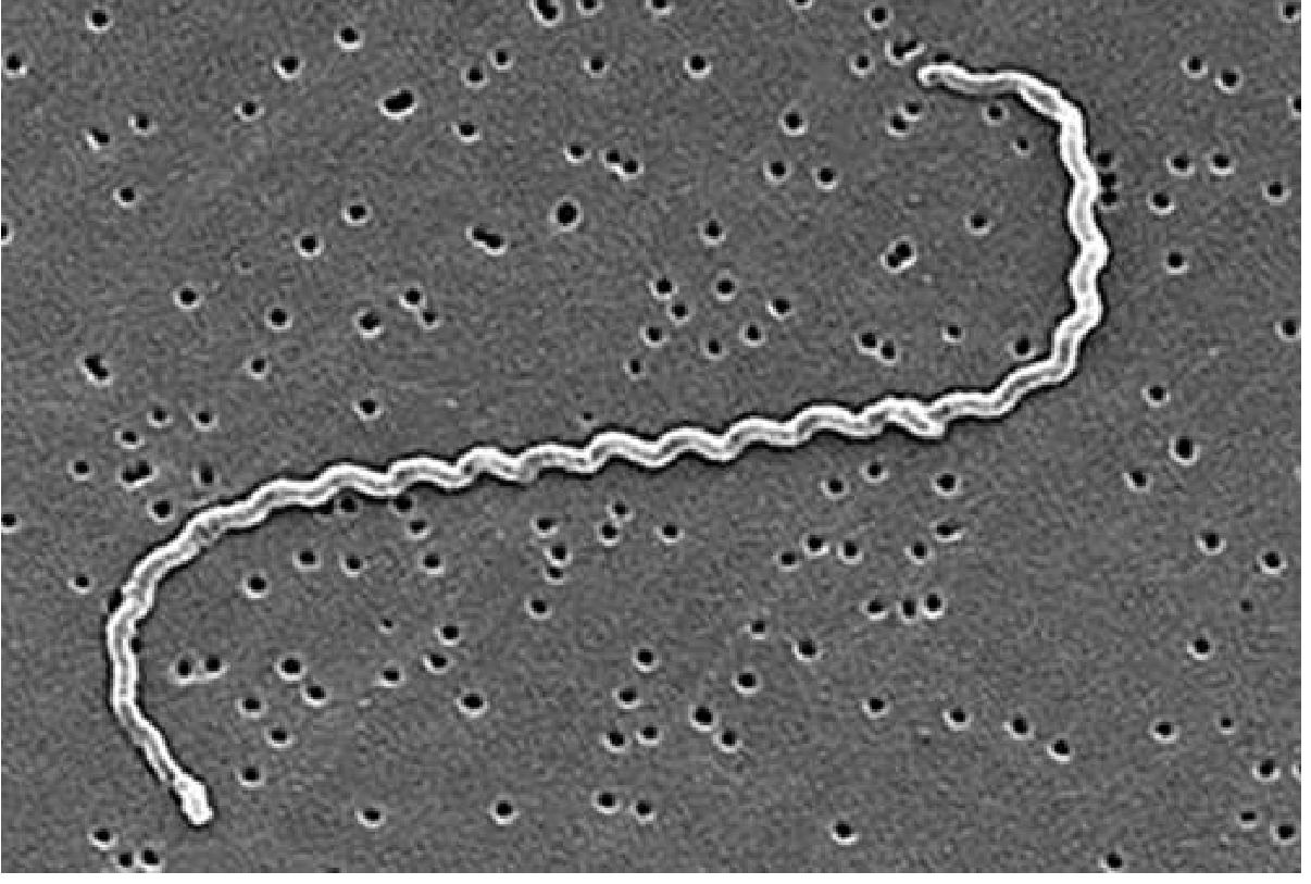
Definition
Leptospirosis is a zoonotic infection caused by pathogenic spirochetes of the genus Leptospira. It ranges from a mild self-limited febrile illness to a life-threatening sepsis-like syndrome characterized by the classic triad of jaundice, acute renal insufficiency, and bleeding - known as Weil disease. It is the most widespread zoonosis globally and a leading cause of morbidity and mortality in tropical regions.
- Goldman-Cecil Medicine, Ch. 298
The Pathogen
- Leptospira comes from the Greek "leptos" (thin) + "speira" (coil)
- Tightly coiled, highly motile spirochetes: 0.1 µm diameter, 6-20 µm length
- The trailing end is hooked when moving through liquid, giving it a question-mark shape - hence Leptospira interrogans
- Over 64 species described; pathogenic species include >25 serogroups and 250 serovars
- L. interrogans serovar Icterohaemorrhagiae (via rats) is the most common cause of Weil disease
- L. interrogans serovar autumnalis is associated with pretibial (Fort Bragg) fever
- Survives days to weeks in water at pH >7.0; killed rapidly by acidic conditions, drying, and common disinfectants
- Periplasmic flagella drive motility and enable tissue burrowing
- Sherris & Ryan's Medical Microbiology, 8th Ed.
Epidemiology
| Feature | Detail |
|---|---|
| Global burden | ~1 million clinical cases and ~60,000 deaths/year |
| Distribution | Worldwide; highest in tropical areas with poor infrastructure and heavy rainfall |
| Endemic regions | India, Malaysia, Brazil, other developing nations |
| Main reservoir | Urban brown rat (Rattus norvegicus) - sheds leptospires persistently in urine |
| Other reservoirs | Dogs, cattle, pigs, wild animals |
| Transmission | Contact with urine-contaminated water or soil; rarely human-to-human |
| Occupational risk | Farmers, veterinarians, slaughterhouse workers, sewer workers |
| Recreational risk | Swimmers, kayakers, triathletes in contaminated water |
| Risk factors | Skin abrasions, prolonged water exposure, HLA-DQ6 haplotype |
Leptospirosis is frequently misdiagnosed as dengue, malaria, or influenza due to its nonspecific early presentation. Cases spike after heavy rainfall and flooding. Climate change is expected to increase incidence.
- Goldman-Cecil Medicine, Ch. 298
Pathogenesis
Entry routes: Abraded or diseased skin, conjunctiva, upper alimentary/respiratory tract mucosa.
Dissemination: Active motility allows burrowing into tissues. Leptospires spread hematogenously to all organs including kidneys, liver, lungs, brain, and CSF.
Immune evasion mechanisms:
- Leptospiral proteins bind LPS and peptidoglycan, shielding them from innate immune detection
- Surface proteins bind complement regulators, resisting complement-mediated killing
- Disruption of endothelial cell junctions facilitates multi-organ invasion
Organ-specific effects:
- Kidney: Renal tubular colonization → interstitial nephritis → impaired Na/water reabsorption → potassium wasting and fluid loss (proximal tubular dysfunction)
- Liver: Hepatic sinusoidal invasion → hepatocellular disruption → bile leakage → jaundice
- Lungs: Impaired hemostasis + thrombocytopenia → pulmonary hemorrhage
- Hemostasis: Thrombocytopenia and coagulopathy → bleeding at multiple sites
Biphasic illness:
- Leptospiremic phase - bacteremia, organism in blood, CSF, urine
- Immune phase - agglutinating antibodies appear, bacteremia clears, but immune activation can produce a sepsis-like syndrome. Antibiotics are less effective in this phase.
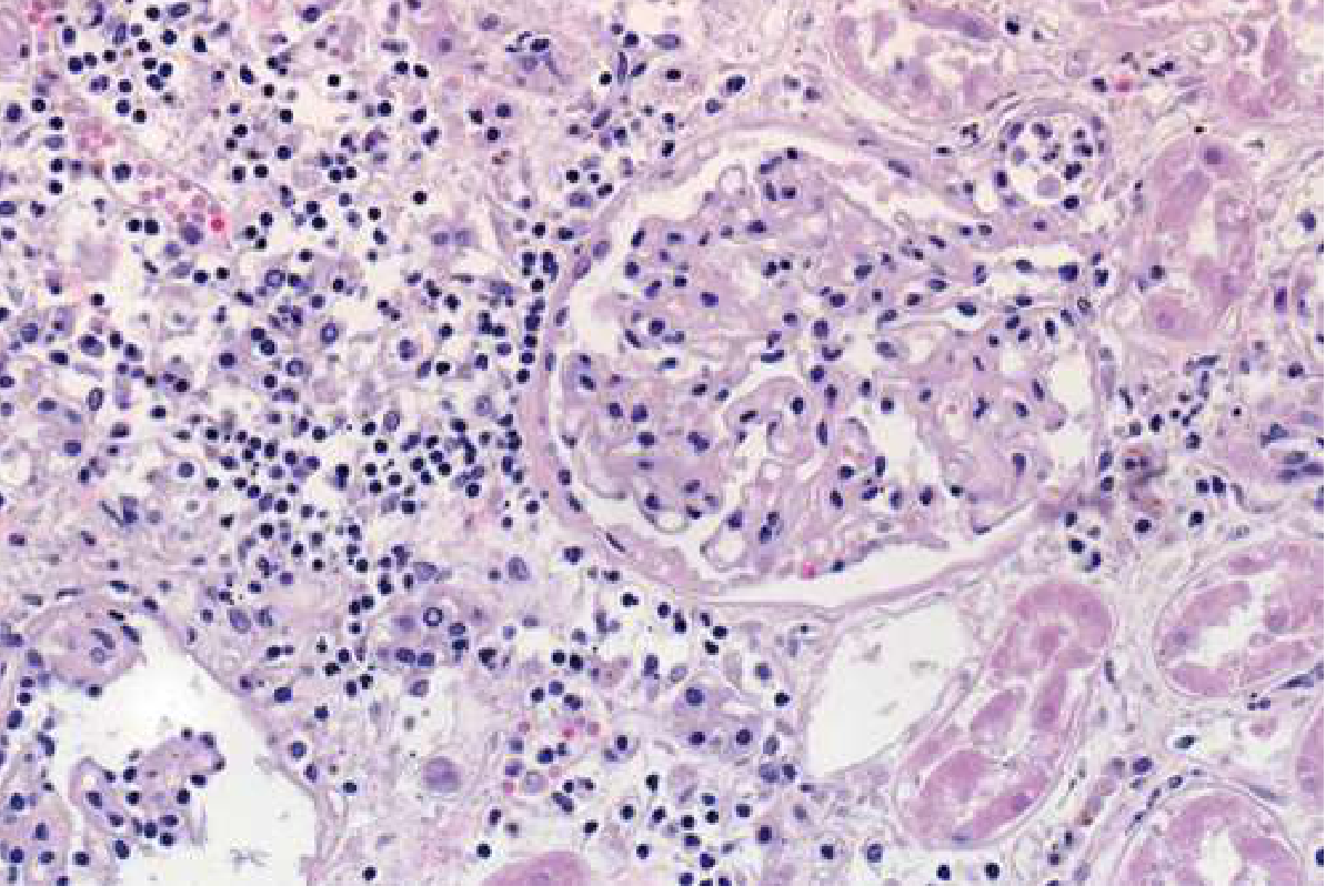
Clinical Manifestations
Incubation period: 6-29 days (median ~15 days)
Phase 1 - Leptospiremic (Septicemic) Phase (days 1-7)
Abrupt onset with:
- High fever and chills
- Severe bitemporal/frontal throbbing headache with retro-orbital pain and photophobia
- Myalgia - characteristically involving calves and lower back (calf tenderness is a key clinical clue)
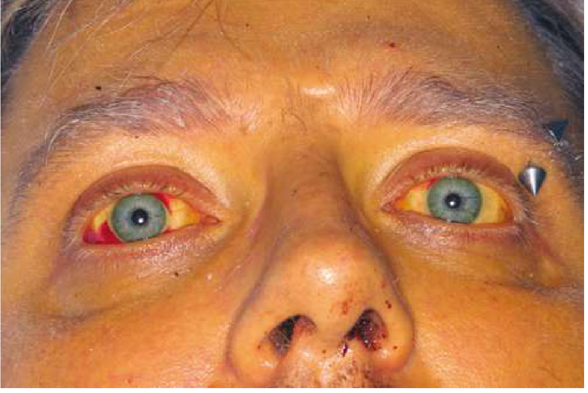
- Conjunctival suffusion - pathognomonic injection without exudate
- Subconjunctival hemorrhages
- Nonproductive cough (~50% of cases)
- Nausea, vomiting, diarrhea, abdominal pain
- Maculopapular erythematous skin rash (especially pretibial in Fort Bragg fever)
Phase 2 - Immune Phase (days 7-14+)
- Fever may transiently resolve then return
- Aseptic meningitis - headache, neck stiffness, CSF lymphocytosis (Leptospira rarely recovered from CSF at this stage)
- Uveitis, iritis (can be a late complication, weeks to months after acute illness)
Weil Disease (Severe Leptospirosis, ~10% of cases)
Classic triad:
- Jaundice (direct hyperbilirubinemia from hepatocellular + cholestatic injury)
- Acute renal failure (non-oliguric early → oliguric late; hypokalemia)
- Hemorrhage - petechiae, ecchymoses, GI bleed, pulmonary hemorrhage (most feared complication - diffuse alveolar hemorrhage with hemoptysis; high mortality)
Other severe features:
- Acute hemolytic anemia (especially in G6PD deficiency)
- Multiorgan failure (liver, kidney, lung, brain)
- Cardiac arrhythmias, myocarditis
- Pancreatitis
Weil disease case fatality rate: 5-10% (from renal failure, vascular collapse, hemorrhage)
Case Definitions (WHO/PAHO)
| Category | Criteria |
|---|---|
| Suspect | Fever ≥38.5°C and/or severe headache + myalgia/prostration/conjunctival suffusion + exposure history |
| Probable (clinical) | Suspect + any 2 of: calf tenderness, cough ± hemoptysis, dyspnea, rash, jaundice, hemorrhage, meningism, oliguria/proteinuria, arrhythmia |
| Probable (lab) | Suspect + positive rapid IgM test OR any 3 of: abnormal urinalysis, neutrophilia >80% + lymphopenia, platelets <100,000/µL, elevated bilirubin/liver enzymes |
| Confirmed | Suspect/probable + isolation, positive PCR, seroconversion, or MAT titer ≥1:400 |
Diagnosis
Clinical approach: A high index of suspicion is essential. Exposure history + acute febrile illness + calf tenderness + conjunctival suffusion in a tropical/flood setting should trigger workup.
Laboratory Tests
| Test | Notes |
|---|---|
| PCR (serum/urine/CSF) | Highest sensitivity for early infection; increasingly available in resource-limited settings |
| MAT (Microscopic Agglutination Test) | Gold standard serology; reference lab only; titer ≥1:400 or 4-fold rise is diagnostic |
| IgM ELISA / Rapid IgM test | Detectable from ~day 5-7; useful for field diagnosis |
| Blood culture | Specialized media (EMJH); takes weeks; useful early before antibiotics |
| Urine culture | Useful after first week |
| Darkfield microscopy | Not recommended - low yield, high false-positive rate with fibrin/debris |
Non-specific labs:
- Leukocytosis (>80% neutrophils) with thrombocytopenia
- Elevated INR, aPTT, D-dimer
- Elevated creatinine, BUN, direct bilirubin
- Moderate transaminase elevation (AST/ALT usually <200 IU/L - lower than viral hepatitis)
- Pyuria, hematuria, proteinuria
- Elevated procalcitonin and CRP (helps distinguish from viral illness like dengue)
- CSF: aseptic meningitis pattern (lymphocytes, raised protein, normal glucose)
- CT chest: ground-glass opacities/airspace nodules in pulmonary hemorrhage
Differential Diagnosis
- Dengue fever (most common mimic in tropics)
- Malaria
- Typhoid fever
- Viral hepatitis
- Hantavirus
- Melioidosis
- Rickettsial disease
- Influenza
Treatment
Antibiotic Therapy
| Severity | Regimen |
|---|---|
| Mild disease | Doxycycline 100 mg PO twice daily for 7 days or Azithromycin |
| Severe disease | Penicillin G 1.5 million units IV q6h or Ampicillin 1 g IV q6h or Ceftriaxone 1 g IV daily |
- Antibiotics are most effective when started early (leptospiremic phase)
- IV penicillin remains first-line for severe/Weil disease; ceftriaxone is equally effective and more convenient
Supportive Care
- Hypokalemia + non-oliguric renal failure (early): Aggressive IV fluids and potassium supplementation
- Oliguric renal failure: Early initiation of hemodialysis or peritoneal dialysis
- Pulmonary hemorrhage: Mechanical ventilation, consider corticosteroids (limited evidence)
- Monitor for arrhythmias, bleeding, and multiorgan failure
2024 Cochrane Evidence
Two 2024 Cochrane systematic reviews assessed antibiotics for treatment (PMID 38483092) and prophylaxis (PMID 38483067) of leptospirosis. The evidence base remains limited by small RCTs with high bias risk, but current practice supports doxycycline, penicillin, and ceftriaxone as effective agents.
Chemoprophylaxis
| Indication | Regimen |
|---|---|
| High-risk short-term exposure (adventure travel, flood workers) | Doxycycline 200 mg PO once weekly during and 1 week after exposure |
Prevention
- Rodent control - particularly in urban/peri-urban settings
- Avoid swimming/wading in potentially contaminated floodwaters
- Use protective clothing and boots in occupational settings
- Drain contaminated water sources
- Vaccination of livestock and pets (reduces human exposure)
- Human vaccines available in some countries (Cuba, China, France) - not widely used elsewhere
- Avoid ingestion or skin contact with animal urine
Key Points Summary
| Feature | Detail |
|---|---|
| Causative organism | Leptospira interrogans (most common) - spirochete |
| Transmission | Rodent urine-contaminated water/soil through skin breaks |
| Pathognomonic sign | Conjunctival suffusion (injection without exudate) |
| Classic severe triad (Weil disease) | Jaundice + renal failure + hemorrhage |
| Most feared complication | Pulmonary hemorrhage (high mortality) |
| Best early diagnosis | PCR (serum/urine) |
| Gold standard serology | MAT (titer ≥1:400) |
| Treatment - mild | Doxycycline 100 mg PO BD x 7 days |
| Treatment - severe | IV Penicillin G or Ceftriaxone |
| Prophylaxis | Doxycycline 200 mg once weekly |
Sources: Goldman-Cecil Medicine International Edition (Ch. 298) | Sherris & Ryan's Medical Microbiology 8th Ed. | Andrews' Diseases of the Skin | Cochrane Reviews 2024 (PMIDs 38483092, 38483067)
This is a shared conversation. Sign in to Orris to start your own chat.