
I am 2nd yr mbbs Student from knruhs University i wanna learn pathology from scratch i have my final exam in sept firsfirst week but I also have my pre final exam in July 1st week for that also I have to complete my whole syllabus and I am starting patho now it self can u help me to study patho and give me the correct practical time table to study patho with topics per day to complete syllabus easily n I also have my clg 9-5 and also have to study 2 more subjects like Pharmacology and microbiology... Guide me please to study for my university and also build my concept
Which pathology syllabus are you following? This helps me build the exact topic list. · How many hours per day can you realistically study OUTSIDE college (9-5)? · What is your current level in Pathology?
KNRUHS 2nd year MBBS pathology syllabus topics list
https://www.scribd.com/document/529010822/Second-MBBS-Pathol…
🩺 Complete Pathology Study Plan - KNRUHS 2nd MBBS
- Today: May 29, 2026 (Friday)
- Pre-final exam: July 1st week (~33 days away)
- Final exam: September 1st week (~95 days away)
- College hours: 9 AM - 5 PM (not available for study)
- Study time per day: 3-4 hours (evenings/night + early morning)
- Subjects: Pathology + Pharmacology + Microbiology
- Pathology level: Starting from zero
PHASE 1 - YOUR DAILY TIME BUDGET
| Slot | Time | Subject |
|---|---|---|
| Evening (5:30-6:30 PM) | 1 hour | Pharmacology |
| Evening (6:30-8:30 PM) | 2 hours | Pathology (main focus) |
| Night (9:00-9:45 PM) | 45 min | Microbiology |
| Morning (6:00-6:30 AM) | 30 min | Quick revision of previous day's patho |
PHASE 2 - THE FULL PATHOLOGY SYLLABUS
GENERAL PATHOLOGY (GP) - ~16 topics
| # | Topic | Approximate Weight |
|---|---|---|
| 1 | Introduction to Pathology + Cell Injury | High |
| 2 | Cell Death: Necrosis, Apoptosis, Gangrene | High |
| 3 | Cellular Adaptations (Atrophy, Hypertrophy, Hyperplasia, Metaplasia, Dysplasia) | High |
| 4 | Intracellular accumulations + Calcification | Medium |
| 5 | Acute Inflammation | Very High |
| 6 | Chronic Inflammation + Granulomatous Inflammation | Very High |
| 7 | Healing and Repair (Wound healing) | High |
| 8 | Hemodynamic disorders (Edema, Congestion, Hyperemia, Thrombosis, Embolism, Infarction) | Very High |
| 9 | Shock | High |
| 10 | Neoplasia - Part 1 (Nomenclature, Carcinogenesis, Tumor markers) | Very High |
| 11 | Neoplasia - Part 2 (Spread, Grading, Staging, Paraneoplastic syndromes) | Very High |
| 12 | Immunity and Immunopathology (Hypersensitivity types I-IV) | High |
| 13 | Autoimmune diseases (SLE, RA basics) | Medium |
| 14 | Amyloidosis | Medium |
| 15 | Genetic and pediatric disorders (Down syndrome, Turner, Klinefelter, etc.) | Medium |
| 16 | Environmental and Nutritional pathology | Low-Medium |
SYSTEMIC PATHOLOGY (SP) - ~12 topics
| # | Topic | Approximate Weight |
|---|---|---|
| 17 | Hematology - Anemias (IDA, megaloblastic, hemolytic, aplastic) | Very High |
| 18 | Hematology - Leukemias + Lymphomas | Very High |
| 19 | Cardiovascular Pathology (Atherosclerosis, MI, Hypertension, Valvular diseases) | Very High |
| 20 | Respiratory Pathology (Pneumonia, TB, COPD, Lung carcinoma) | Very High |
| 21 | Gastrointestinal Pathology (Peptic ulcer, Carcinoma stomach, IBD, Colorectal Ca) | High |
| 22 | Hepatobiliary Pathology (Hepatitis, Cirrhosis, Jaundice, Carcinoma) | Very High |
| 23 | Renal Pathology (Glomerulonephritis, Nephrotic/Nephritic, RCC, Wilms) | High |
| 24 | Endocrine Pathology (Diabetes, Thyroid, Adrenal) | High |
| 25 | Female Reproductive Pathology (Cervix, Endometrium, Ovary, Breast) | High |
| 26 | CNS Pathology (Meningitis, Gliomas, Metastasis) | Medium |
| 27 | Bone and Soft tissue tumors | Medium |
| 28 | Practical Pathology (Histology slides, Blood films, Urine analysis, LFT, RFT, TFT) | High (practical exam) |
PHASE 3 - DAY-BY-DAY TIMETABLE
PRE-FINAL PREPARATION: May 29 - June 30 (33 days)
Book to use: Start with Harsh Mohan's Textbook of Pathology (concise, India-exam-oriented). Supplement with Robbins Basic Pathology for concepts. For quick reads: Pathoma by Dr. Husain Sattar (free videos on YouTube - HIGHLY recommended for concept building).
WEEK 1 (May 29 - June 4): Cell Injury, Death, Adaptations
| Day | Date | Topic | What to Do |
|---|---|---|---|
| 1 | May 29 (Fri) | Introduction to Pathology + Overview | Read what is pathology, types of pathology, how disease occurs. Make a 1-page overview. |
| 2 | May 30 (Sat) | Cell Injury - Causes & Mechanisms | Study reversible vs irreversible injury, free radical injury, ischemic injury. Draw the injury cascade. |
| 3 | May 31 (Sun) | FULL DAY - Cell Injury Deep Dive | Sunday = no college. Give 4-5 hours. Revise Day 1+2, then study apoptosis mechanisms in detail. |
| 4 | June 1 (Mon) | Necrosis - All types | Coagulative, liquefactive, caseous, fat necrosis, fibrinoid, gangrenous. Draw a comparison table. |
| 5 | June 2 (Tue) | Apoptosis vs Necrosis + Gangrene | Compare in a table. Wet vs dry vs gas gangrene. |
| 6 | June 3 (Wed) | Cellular Adaptations | Atrophy, hypertrophy, hyperplasia, metaplasia, dysplasia - definitions, examples, clinical significance. |
| 7 | June 4 (Thu) | Intracellular accumulations + Calcification | Fatty change (steatosis), glycogen, proteins, pigments (melanin, hemosiderin, bilirubin). Dystrophic vs metastatic calcification. |
WEEK 2 (June 5 - June 11): Inflammation
| Day | Date | Topic | What to Do |
|---|---|---|---|
| 8 | June 5 (Fri) | Acute Inflammation - Vascular events | Vasodilation, increased permeability, exudate vs transudate. |
| 9 | June 6 (Sat) | Acute Inflammation - Cellular events | Margination, rolling, adhesion, diapedesis, chemotaxis. Know the molecules (selectins, integrins, ICAM). |
| 10 | June 7 (Sun) | FULL DAY - Acute Inflammation complete | Mediators of inflammation (histamine, prostaglandins, leukotrienes, complement, cytokines). Write a flowchart. |
| 11 | June 8 (Mon) | Outcomes of acute inflammation | Resolution, suppuration, organization, chronic inflammation. Exudate types. |
| 12 | June 9 (Tue) | Chronic Inflammation | Features, cells involved (macrophages, lymphocytes, plasma cells, fibroblasts), causes. |
| 13 | June 10 (Wed) | Granulomatous Inflammation | Definition, types, TB granuloma vs sarcoid vs foreign body. Draw and label a granuloma. |
| 14 | June 11 (Thu) | Wound Healing + Repair | Primary vs secondary intention, phases of healing, factors affecting, complications. |
WEEK 3 (June 12 - June 18): Hemodynamics + Shock + Neoplasia Part 1
| Day | Date | Topic | What to Do |
|---|---|---|---|
| 15 | June 12 (Fri) | Edema + Hyperemia + Congestion | Mechanisms of edema (Starling forces), types, cardiac vs renal vs hepatic edema, nutmeg liver. |
| 16 | June 13 (Sat) | Thrombosis + Embolism | Virchow's triad, thrombus vs embolus, types of emboli - pulmonary, fat, air, amniotic. |
| 17 | June 14 (Sun) | FULL DAY - Infarction + Shock | Red vs white infarcts, zones, shock types, stages of shock, organ changes in shock. |
| 18 | June 15 (Mon) | Neoplasia - Definitions + Nomenclature | Benign vs malignant, naming conventions, differences in a comparison table. |
| 19 | June 16 (Tue) | Carcinogenesis | Chemical, radiation, viral carcinogenesis. Oncogenes, tumor suppressor genes (TP53, Rb). |
| 20 | June 17 (Wed) | Tumor spread + Grading + Staging | Local invasion, lymphatic, hematogenous. TNM staging, grading systems. |
| 21 | June 18 (Thu) | Tumor markers + Paraneoplastic syndromes | AFP, CEA, PSA, CA-125, CA 19-9, BHCG. Paraneoplastic: PTHrP, SIADH, ACTH. |
WEEK 4 (June 19 - June 25): Immunopathology + Genetic Disorders + Hematology
| Day | Date | Topic | What to Do |
|---|---|---|---|
| 22 | June 19 (Fri) | Hypersensitivity Type I & II | Anaphylaxis, atopy, cytotoxic reactions, examples for each. |
| 23 | June 20 (Sat) | Hypersensitivity Type III & IV + Autoimmunity | Immune complex (SLE), delayed type (TB test), autoimmune diseases, SLE criteria. |
| 24 | June 21 (Sun) | FULL DAY - Amyloidosis + Genetic disorders | Amyloid types, Congo red stain, apple-green birefringence. Down, Turner, Klinefelter, Marfan, NF. |
| 25 | June 22 (Mon) | Anemias - Classification + IDA | Classify by MCV, by mechanism. Iron deficiency anemia - causes, blood picture, treatment. |
| 26 | June 23 (Tue) | Megaloblastic anemia + Hemolytic anemias | B12/Folate deficiency, hypersegmented neutrophils. Sickle cell, thalassemia, G6PD, hereditary spherocytosis. |
| 27 | June 24 (Wed) | Aplastic anemia + Leukemias overview | Aplastic anemia pathogenesis. ALL vs AML vs CLL vs CML - key differences, auer rods, Philadelphia chromosome. |
| 28 | June 25 (Thu) | Lymphomas | Hodgkin (RS cell, subtypes) vs Non-Hodgkin. |
WEEK 5 (June 26 - June 30): High-yield Systemic for Pre-final
| Day | Date | Topic | What to Do |
|---|---|---|---|
| 29 | June 26 (Fri) | Cardiovascular - Atherosclerosis + MI | Pathogenesis of atherosclerosis, MI zones, complications. |
| 30 | June 27 (Sat) | Respiratory - Pneumonia + TB | Lobar vs bronchopneumonia, TB pathology, ghon complex, military TB. |
| 31 | June 28 (Sun) | FULL DAY - Liver Pathology + Hepatitis | Hepatitis types, chronicity, cirrhosis, portal hypertension, jaundice types. |
| 32 | June 29 (Mon) | GIT Pathology | Peptic ulcer, gastric carcinoma, IBD (Crohn vs UC table), colorectal carcinoma. |
| 33 | June 30 (Tue) | PRE-FINAL RAPID REVISION | Only flowcharts, comparison tables, diagrams. No new reading. |
Pre-final is roughly July 1st week. After June 30, the first 2-3 days of July are pure revision and past paper practice.
JULY - AFTER PRE-FINAL: Complete Systemic Pathology (July 10 - August 15)
| Week | Dates | Topics |
|---|---|---|
| Week 1-post | July 10-16 | Renal pathology (GN, nephrotic/nephritic, RCC, Wilms) |
| Week 2-post | July 17-23 | Endocrine (DM types, thyroid disorders, adrenal) |
| Week 3-post | July 24-30 | Female reproductive + Breast pathology |
| Week 4-post | July 31-Aug 6 | CNS pathology + Bone tumors |
| Week 5-post | Aug 7-15 | CVRS/Respiratory full revision + Practical pathology (slides, blood films) |
AUGUST 16 - SEPTEMBER 1: Final Exam Revision Sprint
- Week 1: Revise ALL General Pathology (notes only, no rereading)
- Week 2: Revise ALL Systemic Pathology with past papers
- Week 3: Mock self-tests, writing practice, important diagrams
- Final 3-4 days: Only flashcard-style revision of key facts
PHASE 4 - HOW TO ACTUALLY STUDY EACH TOPIC
The 4-Step System for Every Topic
PHASE 5 - CONCEPT-BUILDING SHORTCUTS
Most Important Concepts for KNRUHS Exams (these come every year):
- Cell injury cascade - draw it until you can do it in 2 minutes
- Virchow's triad - always comes with clinical scenarios
- Types of necrosis - with examples, very high yield
- Granuloma - definition + draw a labeled granuloma (TB vs sarcoid)
- Apoptosis pathway - intrinsic vs extrinsic with caspases
- Hypersensitivity types - table with examples (comes EVERY year)
- TP53 and cell cycle - for neoplasia understanding
- Blood film descriptions - for practical exams
- Comparison tables - always: Crohn vs UC, Hodgkin vs NHL, Nephrotic vs Nephritic, Type 1 vs Type 2 DM
Diagrams you MUST be able to draw:
- Granuloma (labeled)
- Wound healing (primary + secondary intention)
- Coagulation cascade (connects with hemodynamics)
- Leukemia classification tree
- Portal hypertension consequences
PHASE 6 - PHARMACOLOGY + MICROBIOLOGY PARALLEL PLAN
Pharmacology (1 hour/day, 5 PM - 6 PM):
- When studying cardiovascular patho → study anti-hypertensives, anti-anginals
- When studying respiratory patho → study bronchodilators, steroids
- When studying hematology → study iron, B12, folate, anticoagulants This creates cross-linking between subjects, which is the fastest way to learn.
Microbiology (45 min/day, 9 PM):
- Respiratory week in patho → study TB, pneumococcal pneumonia, influenza in micro
- Liver week in patho → study Hepatitis viruses (A, B, C, E) in micro
- GIT week in patho → study H. pylori, Salmonella, Shigella in micro
PHASE 7 - PRACTICAL PATHOLOGY
- Histology slides - know the basic 10 slides (TB, carcinoma, hepatitis, fibroadenoma, etc.)
- Blood films - IDA (hypochromic microcytic), megaloblastic (macro-ovalocytes, hypersegmented neutrophils), sickle cell, malaria
- Urine analysis - RBC casts (GN), granular casts, bilirubin
- Spot specimens - Kidney (hydronephrosis, stones), liver (cirrhosis, nutmeg), spleen, lung
QUICK REFERENCE - TIMELINE SUMMARY
May 29 → June 7      : General Pathology Block 1 (Cell injury, Death, Adaptations)
June 8 → June 14     : General Pathology Block 2 (Inflammation, Healing)
June 15 → June 21    : General Pathology Block 3 (Hemodynamics, Neoplasia)
June 22 → June 30    : Immunopathology + Hematology + High-yield Systemic
July 1st week        : PRE-FINAL EXAM
July 10 → Aug 15     : Complete Systemic Pathology
Aug 16 → Sept 1      : Final Exam Revision Sprint
Sept 1st week        : FINAL EXAM
FINAL TIPS SPECIFICALLY FOR YOU
-
Do NOT try to read Robbins cover to cover. Use Harsh Mohan as your primary text. Use Robbins only when you need to understand a mechanism better.
-
Sunday is your power day. No college = use it for 4-5 hours. This one day every week can cover 2 normal days of content.
-
Write, do not just read. Every important mechanism should be written out as a flowchart at least once. Writing creates stronger memory than reading.
-
Past papers are gold. After July, solve the last 5 years of KNRUHS Pathology papers. Most questions repeat. The pattern will become obvious.
-
The morning 30-min slot is sacred. Every morning, just rewrite yesterday's 5-point notes without looking at them. This is spaced repetition and it dramatically reduces forgetting.
-
Do not skip practical classes. In Indian universities, the practical viva and internal marks make up a significant portion. Every slide you observe properly in college saves you 30 minutes of studying at home.
-
For concept clarity: Pathoma videos by Husain Sattar (available on YouTube) are the single best free resource for building pathology concepts. They are specifically designed to explain mechanisms, not just list facts. Watch the video before you read the chapter.
Let's start pathology now
📚 DAY 1 - PATHOLOGY LESSON
Introduction to Pathology + Cell Injury
Tonight's goal: Read this fully, draw the flowchart at the end, and write your 5 key points in your notebook.
PART 1 - WHAT IS PATHOLOGY?
| Term | Definition | Memory trick |
|---|---|---|
| Etiology | The cause (origin) of disease - what triggers it | Etiology = "E" for "Elicit/cause" |
| Pathogenesis | The mechanism - how disease develops step by step after the trigger | Patho = disease, genesis = origin/development |
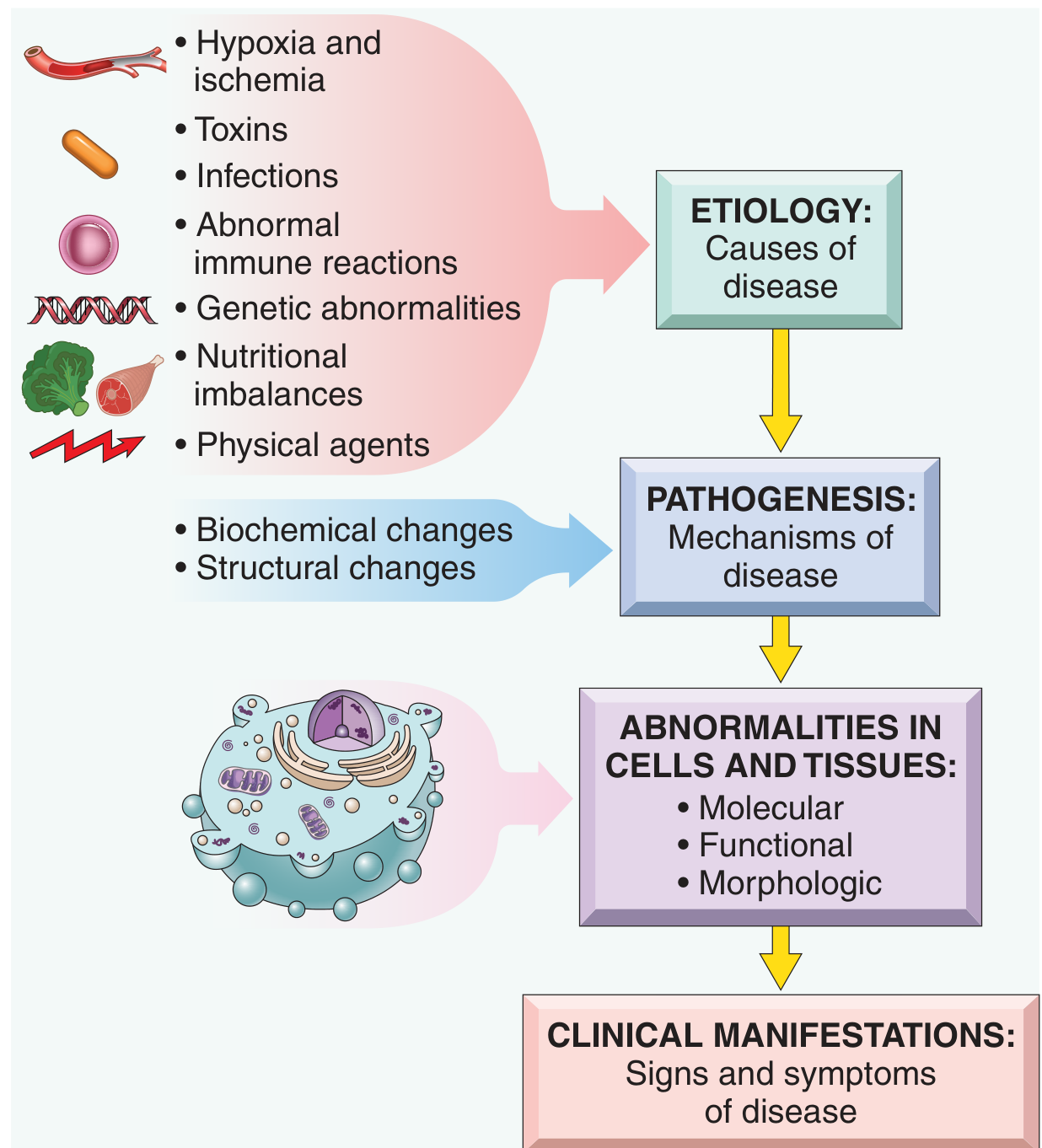
- Etiology of TB = Mycobacterium tuberculosis (the bacteria is the CAUSE)
- Pathogenesis of TB = bacteria enters lung → macrophages engulf it → granuloma forms → caseous necrosis → cavitation (this is the HOW)
- Clinical manifestations = cough, hemoptysis, weight loss, fever
- Etiology - cause
- Pathogenesis - mechanism of development
- Morphologic changes - structural changes in cells/tissues (what you see under microscope)
- Clinical significance - functional consequences (signs and symptoms)
PART 2 - OVERVIEW OF CELLULAR RESPONSES TO STRESS
Normal Cell
     |
     ↓ Stress/Stimulus
     |
     ├──── ADAPTATION (cell survives, adjusts)
     |         Examples: hypertrophy, hyperplasia, atrophy, metaplasia
     |
     ├──── REVERSIBLE INJURY (cell is damaged but can recover)
     |         If stress is removed → cell recovers
     |
     └──── IRREVERSIBLE INJURY → CELL DEATH
               Two main types:
               ├── NECROSIS (pathological death)
               └── APOPTOSIS (programmed death)
PART 3 - CAUSES OF CELL INJURY
| # | Cause | Examples |
|---|---|---|
| 1 | Hypoxia & Ischemia | Most common cause! Block in artery, anemia, lung disease |
| 2 | Toxins | Alcohol, carbon monoxide, drugs, insecticides, asbestos |
| 3 | Infectious agents | Bacteria (toxins), viruses, fungi, parasites |
| 4 | Immunologic reactions | Autoimmune, allergic, chronic immune responses |
| 5 | Genetic abnormalities | Down syndrome, sickle cell disease, enzyme deficiencies |
| 6 | Nutritional imbalances | Protein-calorie malnutrition, vitamin deficiencies, obesity |
| 7 | Physical agents | Trauma, heat, cold, radiation, electric shock |
Exam tip: "Hypoxia is the most common cause of cell injury." This line gets marks in every short answer.
- Hypoxia = oxygen deficiency only (supply of nutrients is maintained)
- Ischemia = reduced blood supply (so BOTH oxygen AND nutrients are lost)
- Therefore, ischemia causes injury faster and more severely than pure hypoxia
PART 4 - SEQUENCE OF EVENTS IN CELL INJURY
Step 1 - Reversible Cell Injury (the cell can still recover)
- Cellular swelling (most common and earliest change) - Na+/K+ pump fails → Na+ enters → water follows → cell swells
- Fatty change - lipids accumulate in cytoplasm (especially liver and heart cells)
- Under light microscope: cytoplasm looks pale and granular, vacuoles may appear
Step 2 - Point of No Return (Irreversible Injury)
- Severe mitochondrial damage (mitochondrial permeability transition pore opens) - no more ATP can be made
- Massive membrane damage - lysosomal contents leak out, digesting the cell from inside
PART 5 - THE 4 MAIN MECHANISMS OF CELL INJURY
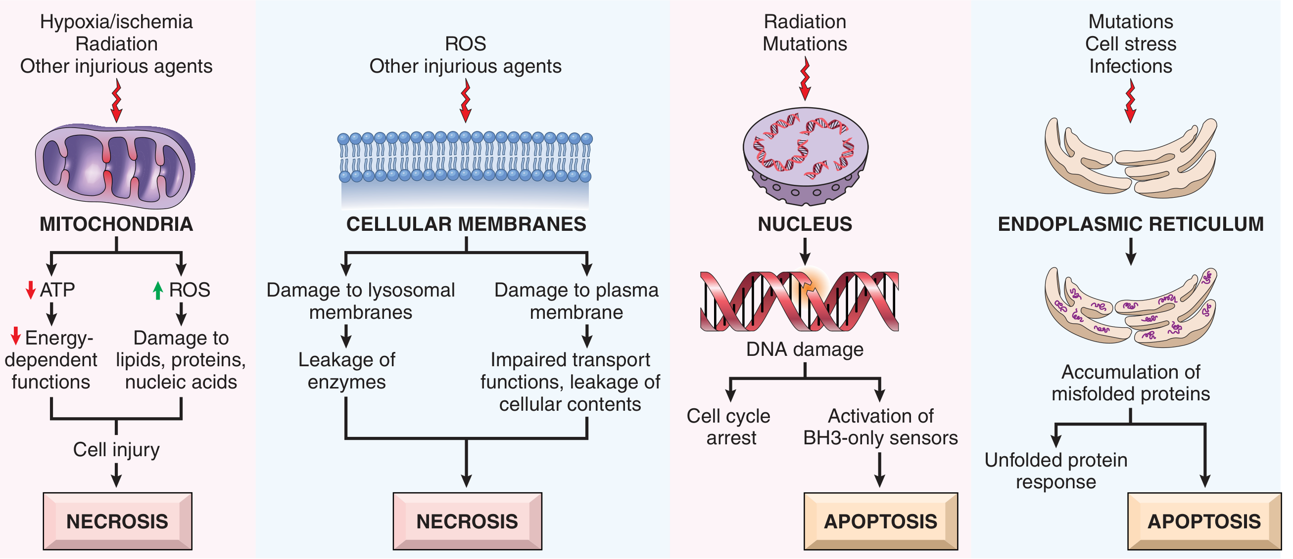
Mechanism 1 - Mitochondrial Dysfunction
- Injury → mitochondria damaged → ATP falls
- Without ATP: Na+/K+ pump fails → cell swells; Ca2+ builds up; cell membrane fails
- Also: damaged mitochondria generate Reactive Oxygen Species (ROS) - free radicals that damage everything
- Mitochondrial permeability transition pore opens → membrane potential lost → complete energy failure → NECROSIS
Mechanism 2 - Membrane Damage
- Damage to plasma membrane → cells cannot maintain ion gradients → swelling and death
- Damage to lysosomal membrane → lysosomal enzymes (acid hydrolases) leak → digest the cell from inside → NECROSIS
Mechanism 3 - DNA Damage
- Radiation, mutations, free radicals damage DNA
- Cell detects DNA damage → tries to repair → if too severe → apoptosis pathway activated
- Key molecule: p53 protein - the "guardian of the genome" - detects DNA damage and triggers apoptosis
Mechanism 4 - Endoplasmic Reticulum (ER) Stress
- Mutations, infections, cell stress → misfolded proteins accumulate in ER
- ER activates the unfolded protein response (UPR)
- If overwhelmed → triggers APOPTOSIS
PART 6 - REACTIVE OXYGEN SPECIES (ROS) / FREE RADICALS
O₂  +  incomplete reduction
         ↓
    Superoxide (O₂⁻)     ← most common initial ROS
         ↓  [superoxide dismutase = SOD]
    Hydrogen peroxide (H₂O₂)   ← can cross membranes
         ↓  [Fe²⁺ via Fenton reaction]
    Hydroxyl radical (•OH)    ← MOST DANGEROUS
| Antioxidant mechanism | What it does |
|---|---|
| Superoxide dismutase (SOD) | Converts O₂⁻ → H₂O₂ |
| Catalase | Converts H₂O₂ → H₂O + O₂ |
| Glutathione peroxidase | Destroys H₂O₂ and lipid peroxides |
| Vitamin C | Scavenges free radicals |
| Vitamin E | Protects lipid membranes from oxidation |
- Reperfusion injury (when blood returns after ischemia - paradoxically causes MORE damage)
- Inflammation (neutrophils deliberately make ROS to kill bacteria)
- Radiation
- Aging
- Toxins (CCl₄ - carbon tetrachloride, paracetamol overdose)
PART 7 - CALCIUM AND CELL INJURY
- Normal: cytosol Ca²⁺ is very LOW (kept in ER and mitochondria)
- Injury → Ca²⁺ floods cytosol
- Excess Ca²⁺ activates destructive enzymes:
- Phospholipases → destroy membranes
- Proteases → degrade proteins
- ATPases → deplete energy
- Endonucleases → cut DNA
QUICK SUMMARY - YOUR 5 KEY POINTS TO WRITE IN YOUR NOTEBOOK TONIGHT
- Pathology = Etiology (WHY) + Pathogenesis (HOW) + Morphology (WHAT) + Clinical significance (WHAT HAPPENS)
- Earliest change in reversible injury = Cellular swelling (due to Na+/K+-ATPase failure from ATP depletion)
- Point of no return = severe mitochondrial damage + massive membrane damage (lysosomal enzyme leak)
- 4 mechanisms of cell injury: Mitochondrial dysfunction, Membrane damage, DNA damage, ER stress
- Most common cause of cell injury = Hypoxia/Ischemia. Most dangerous ROS = Hydroxyl radical (•OH)
SELF-TEST QUESTIONS (do these after reading - close your notes and answer)
- What is the difference between etiology and pathogenesis? Give an example.
- What is the FIRST and EARLIEST morphologic change in reversible cell injury?
- Why does calcium accumulation lead to cell death? Name 4 enzymes activated.
- Name the 3 main ROS in order of formation. Which enzyme converts O₂⁻ to H₂O₂?
- Why does ischemia cause faster injury than pure hypoxia?
TOMORROW (Day 2) - CELL DEATH: Necrosis + Apoptosis
- The 6 types of necrosis with their classic examples
- What necrotic cells look like under the microscope (pyknosis, karyorrhexis, karyolysis)
- Apoptosis - what it is, when it happens, and how it differs from necrosis