
Give me notes on Endodontic emergencies and retreatment for 50 marks university exam like answer . Please refer articles . Give me answer like include the contents and also refernces
endodontic emergencies management
endodontic retreatment nonsurgical outcome
PMID: 34958512
PMID: 38280514
PMID: 37899987
PMID: 35762859
PMID: 38145805
endodontic flare-up interappointment pain incidence
endodontic retreatment indications causes failure
post-treatment disease endodontic failure microbiology
endodontic retreatment prognosis success factors
PMID: 40122230
endodontic pain management antibiotics prescribing
ENDODONTIC EMERGENCIES AND RETREATMENT
50-Mark University Examination Answer
CONTENTS
- Definition and Classification of Endodontic Emergencies
- Causes / Etiology
- Types of Endodontic Emergencies (Pre-treatment, Inter-appointment, Post-treatment)
- Clinical Diagnosis and Assessment - The 3D Approach
- Management of Each Emergency Type
- Flare-ups - Incidence, Predisposing Factors, Prevention and Management
- Endodontic Retreatment - Definition, Indications, Contraindications
- Causes of Endodontic Failure (Why Retreatment is Needed)
- Diagnosis of Failed Root Canal Treatment
- Procedure of Non-Surgical Retreatment (Orthograde Retreatment)
- Challenges in Retreatment - Separated Instruments, Ledges, Perforations, Posts
- Surgical Retreatment (Periradicular Surgery) vs. Non-Surgical Retreatment
- Prognosis and Outcome of Retreatment
- References
1. DEFINITION AND CLASSIFICATION OF ENDODONTIC EMERGENCIES
| Category | Timing |
|---|---|
| Pre-treatment emergency | Before any endodontic treatment has begun |
| Inter-appointment emergency (Flare-up) | Between appointments during active root canal treatment |
| Post-treatment emergency | After root canal treatment is completed (days, months, or years later) |
2. CAUSES / ETIOLOGY
- Dental caries extending to the pulp
- Traumatic injuries (fractures, luxation, avulsion)
- Failing or inadequate previous root canal treatment
- Periodontal disease (combined endo-perio lesions)
- Cracked tooth syndrome
- Resorption (internal/external)
- Iatrogenic causes (over-instrumentation, over-extension of filling, strip perforation)
- Missed canals
- Coronal leakage after treatment
3. TYPES OF ENDODONTIC EMERGENCIES
A. Pre-Treatment Emergencies
- Mild pulpal inflammation; pulp is vital and can recover
- Pain: sharp, provoked by thermal stimuli, disappears within seconds of stimulus removal
- Treatment: remove caries, apply sedative dressing (zinc oxide eugenol), re-evaluate; root canal treatment is NOT necessary at this stage
- Pulp inflammation beyond recovery; lingering pain (>30 seconds after stimulus)
- Spontaneous, radiating pain; may be worse at night
- Treatment: Pulpectomy (complete removal of pulp tissue) and root canal treatment
- Acute inflammatory pulpal hyperemia with exquisite sensitivity to percussion
- Treatment: Adjust occlusion immediately, proceed with root canal treatment
- Inflammation of the periapical tissues; tooth is extremely tender to percussion
- May be a consequence of irreversible pulpitis, over-instrumentation, or excess irrigant
- Radiograph: may be normal or show early periapical widening
- Treatment: Root canal treatment; establish drainage if abscess present; analgesics (NSAIDs)
- Collection of pus at the apex; severe, throbbing, spontaneous pain with swelling
- Tooth exquisitely tender to touch; patient may be febrile and systemically unwell
- May progress to cellulitis or Ludwig's angina (life-threatening)
- Treatment:
- Incision and drainage (I&D) of fluctuant swelling
- Pulp extirpation and establishment of drainage through the root canal
- Antibiotics ONLY if systemic signs present (fever >38°C, lymphadenopathy, trismus, cellulitis spreading to deep fascial spaces)
- Amoxicillin 500 mg TDS x 5 days (first line); Metronidazole 400 mg TDS added for anaerobes
- For penicillin allergy: Clindamycin or Azithromycin
- Diffuse spreading infection into fascial spaces; NOT fluctuant
- Signs: Diffuse swelling, induration, dysphagia/trismus indicates dangerous spread
- Requires urgent referral for IV antibiotics and possible surgical drainage
- Incomplete crack through the tooth; pain on biting/release; localized sharp pain
- Diagnosis: Bite test with Tooth Slooth; transillumination; methylene blue dye
- Treatment depends on depth of crack: splinting, crown, root canal, or extraction
B. Inter-Appointment Emergencies (Flare-Ups)
- Necrotic pulps with periapical pathology
- Single-visit treatment (controversial)
- Inadequate cleaning and shaping
- Extrusion of debris/irrigants beyond the apex
- Over-instrumentation beyond the working length
- Use of calcium hydroxide in infected canals (may push microorganisms apically)
- Patient factors: high anxiety, female gender, posterior teeth, pre-existing pain
- Accurate determination of working length (electronic apex locator + radiograph)
- Copious irrigation within the canal
- Avoid over-instrumentation
- Prescribe prophylactic NSAIDs (Ibuprofen 400 mg pre-operatively)
- Leave the tooth open only in exceptional cases (controversial, may worsen infection)
- Re-open tooth and re-irrigate canals with NaOCl (copious irrigation)
- Prescribe NSAIDs (Ibuprofen 400-600 mg QID) as primary analgesics
- Antibiotics ONLY if systemic involvement (swelling, fever, lymphadenopathy)
- Occlusal adjustment to relieve bite pressure
- Intracanal corticosteroid-antibiotic dressing (e.g., Ledermix paste) for severe cases
- Drain abscess if fluctuant swelling develops
C. Post-Treatment Emergencies
- Immediate post-operative pain (within 24-72 hours): Usually self-limiting; manage with NSAIDs
- Persistent apical periodontitis (weeks-months): Pain, tenderness, radiographic periapical lesion persisting - consider retreatment
- Cracked root: Vertical root fracture (VRF); deep isolated periodontal pocketing, sinus tract; confirmed by CBCT; usually requires extraction
- Coronal leakage: Bacterial re-invasion of root canal system through a defective restoration; retreatment indicated
- Symptomatic apical periodontitis after years: New infection following microbial re-colonization; retreatment required
- Overfilling/over-extension: Extruded sealer or gutta-percha causing foreign body reaction or nerve irritation; monitor or surgical removal
4. CLINICAL DIAGNOSIS: THE 3D APPROACH
D1 - Diagnosis
- Thorough history: onset, duration, character of pain (throbbing, sharp, dull), aggravating/relieving factors, history of trauma, previous dental treatment
- Clinical examination: Extra-oral (swelling, lymphadenopathy, trismus), intra-oral (caries, fracture, sinus tract, probing depths)
- Pulp sensibility tests: Cold test (Endo-Ice), EPT (electric pulp tester)
- Percussion test, palpation test
- Radiographic assessment: Periapical radiograph (PA), CBCT if complex anatomy or uncertain diagnosis
- Differentiate between: Inflammation vs. Infection (critical for antibiotic prescribing)
D2 - Definitive Dental Treatment
- This is the MOST important step; drugs alone are inadequate
- Treatment directed by diagnosis:
- Vital pulp: pulpectomy
- Necrotic + abscess: drainage (through canal and/or incision)
- Post-treatment disease: retreatment or surgical endodontics
- Root canal treatment relieves pain by removing the source of infection/inflammation
D3 - Drugs (Adjuncts Only)
- Analgesics: NSAIDs are first-line (Ibuprofen 400-600 mg QID); paracetamol as adjunct
- Opioids: rarely needed; avoid
- Antibiotics: ONLY when systemic infection present (fever, cellulitis, lymphadenopathy, trismus, rapid spread); NOT for localized pain or as a substitute for drainage
- Corticosteroids: dexamethasone 8 mg IM/IV in acute severe cases to reduce inflammation
- Antibiotic of choice: Amoxicillin 500 mg TDS; add Metronidazole 400 mg for anaerobes; Clindamycin if penicillin allergic
Important principle: Drugs should NEVER replace definitive dental treatment. Antibiotics for localized endodontic pain without systemic involvement contribute to antibiotic resistance and are NOT indicated. - Abbott PV, Int Endod J, 2022
5. ENDODONTIC RETREATMENT
Definition
6. CAUSES OF ENDODONTIC FAILURE (WHY RETREATMENT IS NEEDED)
A. Microbiological Causes (Primary)
- Persistent intraradicular infection: Enterococcus faecalis is the most common organism found in failed cases (highly resistant to calcium hydroxide and NaOCl)
- Extraradicular infection: Actinomyces israelii forming biofilm on root surface
- Reinfection through coronal leakage (most preventable cause)
B. Technical/Procedural Causes
| Cause | Details |
|---|---|
| Missed canals | MB2 of upper molar, second canal in lower anteriors |
| Inadequate cleaning/shaping | Untouched canal walls, inadequate irrigation |
| Short/inadequate obturation | Under-filling leaves space for bacterial recolonization |
| Ledge formation | Prevents proper instrumentation to working length |
| Apical transportation/zipping | Loss of original canal curvature |
| Separated instruments | Prevents adequate disinfection beyond the fragment |
| Perforations | Strip, furcal, or apical perforations |
| Over-extension | Extruded material causing foreign body reaction |
| Iatrogenic vertical root fracture | Caused by excessive lateral condensation forces |
C. Biological Causes
- Cyst formation (true radicular cyst - self-sustaining, does not heal without surgery)
- External apical root resorption
- Dense scar tissue formation
- Biofilm on root surfaces (extraradicular)
7. INDICATIONS FOR RETREATMENT
Non-Surgical Retreatment (Orthograde) Indications:
- Persistent/new periapical pathology (asymptomatic or symptomatic) on radiograph
- Symptomatic tooth with previously treated root canals
- Inadequate root canal filling detected on pre-prosthetic assessment
- Coronal leakage suspected with no signs of root fracture
- Procedural complications (ledge, short fill) accessible orthograde
- No contraindication to non-surgical approach
Surgical Retreatment Indications:
- Failed non-surgical retreatment (or technical impossibility orthograde)
- True radicular cyst confirmed
- Root apex anatomy abnormality (C-shaped canal, open apex that prevents orthograde seal)
- Separated instrument in apical third that cannot be bypassed
- Over-extended post that cannot be removed safely
- Biopsy required (suspicion of malignancy in persistent periapical lesion)
- Perforation in apical or furcal region inaccessible orthograde
Contraindications to Retreatment:
- Vertical root fracture (extraction indicated)
- Poor bone support with untreatable periodontal disease
- Non-restorable crown
- Systemic conditions that preclude surgery (uncontrolled diabetes, anticoagulation, bisphosphonate use)
- Poor strategic value of the tooth
8. DIAGNOSIS OF FAILED ROOT CANAL TREATMENT
Clinical Signs and Symptoms:
- Pain on percussion or biting
- Sinus tract / parulis present
- Swelling
- Tooth may be symptom-free (asymptomatic failure detected radiographically)
- Deep isolated periodontal pocket (suggests VRF)
Radiographic Assessment:
- Periapical radiograph: persistence or enlargement of periapical radiolucency after >4 years
- CBCT (Cone Beam CT): Gold standard for complex cases
- Detects missed canals, VRF, perforation, apical anatomy
- Zanza et al. (2023) state: "CBCT should be considered the first choice for retreatment diagnosis, since it can thoroughly affect the diagnosis and treatment plan" (PMID: 37899987)
- Brochado Martins et al. (2025) confirm CBCT reveals stricter periapical healing criteria - 36% strict vs. 87% loose criteria - highlighting limitations of 2D radiographs (PMID: 40122230)
AAE Criteria for Failure:
- Enlarging periapical lesion
- New periapical lesion where none existed
- Failure to reduce size of periapical lesion over 4 years
- Presence of clinical symptoms
9. PROCEDURE OF NON-SURGICAL (ORTHOGRADE) RETREATMENT
Step 1: Pre-Operative Assessment
- Full history, clinical and radiographic examination
- CBCT if indicated
- Inform consent: patient counselled about potential complications (instrument separation, perforation, inability to complete), alternative options (surgery, implant, extraction), and realistic prognosis
- Rubber dam isolation is mandatory
Step 2: Access Preparation
- Remove existing coronal restoration if compromised (amalgam, composite, crown)
- Crowns: remove or gain access through crown only if crown integrity is acceptable
- Post removal: ultrasonic vibration around post to dissolve cement; Gonon/Ruddle post removal systems; rotate counter-clockwise if threaded
Step 3: Locating Canal Orifices
- Use dental operating microscope (DOM) or surgical loupes
- Ultrasonic tips (e.g., ProUltra tips) for troughing around orifices
- CBCT-guided orifice location for missed canals
Step 4: Removal of Previous Root Canal Filling Material
- Rotary systems: ProTaper Retreatment files (D1, D2, D3), RaCe Retreatment, Mtwo Retreatment burs - most efficient technique
- Solvent-aided softening: Chloroform (most effective; limited use due to potential carcinogenicity), Eucalyptol, orange oil, Endosolv; used sparingly to soften coronal GP then mechanically remove
- Heat carriers: System B heat carrier to plasticize and remove thermoplastic GP
- Working length verification by apex locator and radiograph after GP removal
- Residual sealer (zinc oxide-eugenol, calcium silicate) removed by irrigation with NaOCl + EDTA + ultrasonic activation
- XP-endo Finisher or ultrasonic irrigation effectively removes Ca(OH)2 remnants
Step 5: Re-Instrumentation (Chemo-Mechanical Preparation)
- Re-establish working length with electronic apex locator
- Enlarge to larger apical size than original (minimum #30 apically for adequate disinfection)
- Irrigation protocol:
- 2.5-5.25% NaOCl (antibacterial, tissue dissolving)
- 17% EDTA (smear layer removal)
- Final rinse: NaOCl, then distilled water
- Activated irrigation: Passive Ultrasonic Irrigation (PUI), Sonic activation (EDDY), or XP-endo Finisher to improve debridement
Step 6: Intracanal Medicament
- Calcium hydroxide placed between appointments for cases with:
- Persisting exudate
- Large periapical lesion
- Symptomatic teeth
- Leave for 1-4 weeks
Step 7: Obturation
- Lateral condensation with GP + sealer OR Warm vertical compaction (System B + Obtura II) OR single cone with bioceramic sealer
- Olivieri et al. (2024) found retreatment success rates of 71-87% at 1-3 years; periapical lesion size significantly affects outcome - smaller lesions have better prognosis (PMID: 38145805)
Step 8: Coronal Restoration
- Immediate post-treatment coronal seal is essential
- Delayed restoration increases risk of coronal leakage and re-infection
- Full coverage crown recommended for posterior teeth
10. MANAGEMENT OF SPECIFIC RETREATMENT CHALLENGES
A. Separated Instrument Management
- Attempt removal as first option (Zanza et al., 2023)
- Techniques: ultrasonic vibration (IRS kit, Masserann kit), Instrument Removal System (IRS), microtube technique
- Factors determining management: location (coronal vs. apical third), canal curvature, length of instrument
- If unable to remove: bypass the instrument using pre-curved small K-files (#06, #08)
- If bypass impossible: apical surgery to remove instrument fragment
- If no periapical pathology associated: monitor
B. Ledge Management
- Ledge = artificially created step in the root canal wall due to inadequate curving of instruments
- Bypass with small hand file (#08-#10) pre-curved and rotated through the ledge
- Confirmed by working length change on radiograph
- Zanza et al. (2023) note: "The presence of ledges does not intrinsically reduce the success rate of root canal retreatments if properly managed"
C. Perforation Management
- Immediate management is critical - the longer the perforation is left untreated, the worse the prognosis
- Material of choice: Mineral Trioxide Aggregate (MTA) or other calcium silicate-based repair cements (BiodentineTM)
- Strip perforation (furcal): MTA placed under microscope guidance; CBCT to assess size
- Large perforations: may require surgical access for management
D. Post Removal
- Threaded posts: rotate counter-clockwise with pliers
- Cemented posts: ultrasonic vibration along shaft to disrupt cement (Masserann kit, Gonon extractor)
- Carbon fiber/ceramic posts: ultrasonic troughing; difficult to remove; may require sacrifice
- Caution: risk of root fracture increases with aggressive post removal
11. SURGICAL RETREATMENT (PERIRADICULAR SURGERY / APICOECTOMY)
Procedure:
- Flap design: Full-thickness mucoperiosteal flap (triangular or rectangular); preserve interdental papillae
- Osteotomy: Remove cortical bone overlying root apex using bur; expose apical 3mm of root
- Root resection (Apicoectomy): Resect 3 mm of root apex at 0-10° bevel (perpendicular preferred to reduce exposed dentinal tubules)
- Retrograde preparation: Use ultrasonic tips (CT tips) to prepare cavity 3 mm deep along the root axis (replaces the old contra-angle burs)
- Retrograde filling: MTA, Biodentine, IRM (Intermediate Restorative Material) - MTA is superior
- Curettage of granulation tissue: Send for histopathology to rule out pathology
- Flap repositioning and suturing
Outcomes - Surgical vs. Non-Surgical:
12. PROGNOSIS AND OUTCOME OF ENDODONTIC RETREATMENT
Success Rates (Key Evidence):
| Study | Type | Success Rate |
|---|---|---|
| Sabeti et al., 2024 (PMID: 38280514) | SR + Meta-analysis | 78% (strict) - 87% (loose criteria) |
| Olivieri et al., 2024 (PMID: 38145805) | SR + Meta-analysis | 71-87% (1-3 yr); 77% strict (4-5 yr) |
| Brochado Martins et al., 2025 (PMID: 40122230) | SR + Meta-analysis (CBCT) | 85% loose; 45% strict (CBCT assessment) |
Factors Improving Prognosis:
- Absence of periapical lesion or small lesion (<5 mm)
- Adequate root filling length (to within 0-2 mm of radiographic apex)
- Extended follow-up (long-term healing continues)
- Operator experience
- Proper coronal restoration post-treatment
- Absence of vertical root fracture
Factors Worsening Prognosis:
- Large periapical lesion (PAI score 4-5)
- Mandibular molars and multi-rooted teeth
- Multiple-visit retreatment (in some studies suggests greater complexity)
- Persistent infection / biofilm
- Separated instrument beyond foramen
- Perforation not adequately sealed
- Coronal leakage
13. ENDODONTIC EMERGENCIES IN SPECIAL SITUATIONS
Pregnancy:
- Radiographs: permissible with leaded apron; minimize exposure
- Analgesia: Paracetamol is safe; NSAIDs avoid in 3rd trimester; avoid aspirin
- Antibiotics: Amoxicillin is safe; Metronidazole use with caution (avoid 1st trimester); Clindamycin is safe; Tetracyclines are CONTRAINDICATED (tooth discoloration, teratogenic)
- Treatment: root canal treatment is preferred over extraction; avoid elective treatment in 1st trimester; 2nd trimester is safest
Immunocompromised Patients:
- Higher risk of rapid spread of odontogenic infection
- Lower threshold for antibiotics
- Coordinate with physician before invasive treatment
- Antifungal cover may be needed in severely immunocompromised
SUMMARY TABLE
| Emergency | Diagnosis | Primary Treatment | Drugs |
|---|---|---|---|
| Reversible Pulpitis | Provoked, brief pain, vital pulp | Sedative dressing, caries removal | Analgesics PRN |
| Irreversible Pulpitis | Lingering pain, spontaneous, vital | Pulpectomy, RCT | NSAIDs |
| Acute Apical Periodontitis | Percussion tender, PA widening | RCT, adjust occlusion | NSAIDs |
| Acute Apical Abscess (localized) | Swelling fluctuant, sinus tract | I&D, drainage through canal | Antibiotics only if systemic signs |
| Cellulitis | Diffuse non-fluctuant swelling | Urgent drainage, refer | IV antibiotics |
| Flare-up | Post-appointment pain/swelling | Re-irrigate, I&D if needed | NSAIDs; antibiotics if systemic |
REFERENCES
-
Abbott PV. Present status and future directions: Managing endodontic emergencies. International Endodontic Journal. 2022;55(5):406-445. PMID: 34958512 (Level 7 - Expert Review)
-
Sabeti M, Chung YJ, Aghamohammadi N, et al. Outcome of Contemporary Nonsurgical Endodontic Retreatment: A Systematic Review of Randomized Controlled Trials and Cohort Studies. Journal of Endodontics. 2024;50(4):436-449. PMID: 38280514 (Level 1 - Systematic Review + Meta-Analysis)
-
Olivieri JG, Encinas M, Nathani T, et al. Outcome of root canal retreatment filled with gutta-percha techniques: A systematic review and meta-analysis. Journal of Dentistry. 2024;142:104809. PMID: 38145805 (Level 1 - Systematic Review + Meta-Analysis)
-
Bucchi C, Rosen E, Taschieri S. Non-surgical root canal treatment and retreatment versus apical surgery in treating apical periodontitis: A systematic review. International Endodontic Journal. 2023;56(10):1076-1094. PMID: 35762859 (Level 1 - Systematic Review)
-
Zanza A, Reda R, Testarelli L. Endodontic Orthograde Retreatments: Challenges and Solutions. Clinical, Cosmetic and Investigational Dentistry. 2023;15:401-420. PMID: 37899987 (Level 7 - Comprehensive Review, PMC: PMC10612510)
-
Brochado Martins JF, Georgiou AC, Nunes PD, et al. CBCT-Assessed Outcomes and Prognostic Factors of Primary Endodontic Treatment and Retreatment: A Systematic Review and Meta-Analysis. Journal of Endodontics. 2025;51(6):678-692. PMID: 40122230 (Level 1 - Systematic Review + Meta-Analysis)
-
Aliabadi T, Saberi EA, Motameni Tabatabaei A. Antibiotic use in endodontic treatment during pregnancy: A narrative review. European Journal of Translational Myology. 2022;32(4). PMID: 36268928 (Level 7 - Narrative Review)
-
Sabeti MA, Karimpourtalebi N, Shahravan A, et al. Clinical and Radiographic Failure of Nonsurgical Endodontic Treatment and Retreatment Using Single-cone Technique With Calcium Silicate-based Sealers: A Systematic Review and Meta-analysis. Journal of Endodontics. 2024;50(6):736-749. PMID: 38548045 (Level 1 - Systematic Review + Meta-Analysis)
-
Hargreaves KM, Berman LH. Cohen's Pathways of the Pulp. 11th ed. Elsevier Mosby; 2016. (Standard Endodontic Textbook Reference)
-
Torabinejad M, Walton RE, Fouad AF. Endodontics: Principles and Practice. 5th ed. Elsevier Saunders; 2014. (Standard Endodontic Textbook Reference)
Exam Tip: For a 50-mark answer, expand on the 3D's concept with clinical examples, draw diagrams of periapical lesions, include the classification table of emergencies, and discuss at least 3-4 retreatment challenges with their solutions. Mention specific file systems (ProTaper Retreatment, Mtwo) and solvents (chloroform, eucalyptol) by name, and always quote success rates from systematic reviews to demonstrate evidence-based knowledge.
Can u provide illustrative images like hand drawn images . If u can or not ???
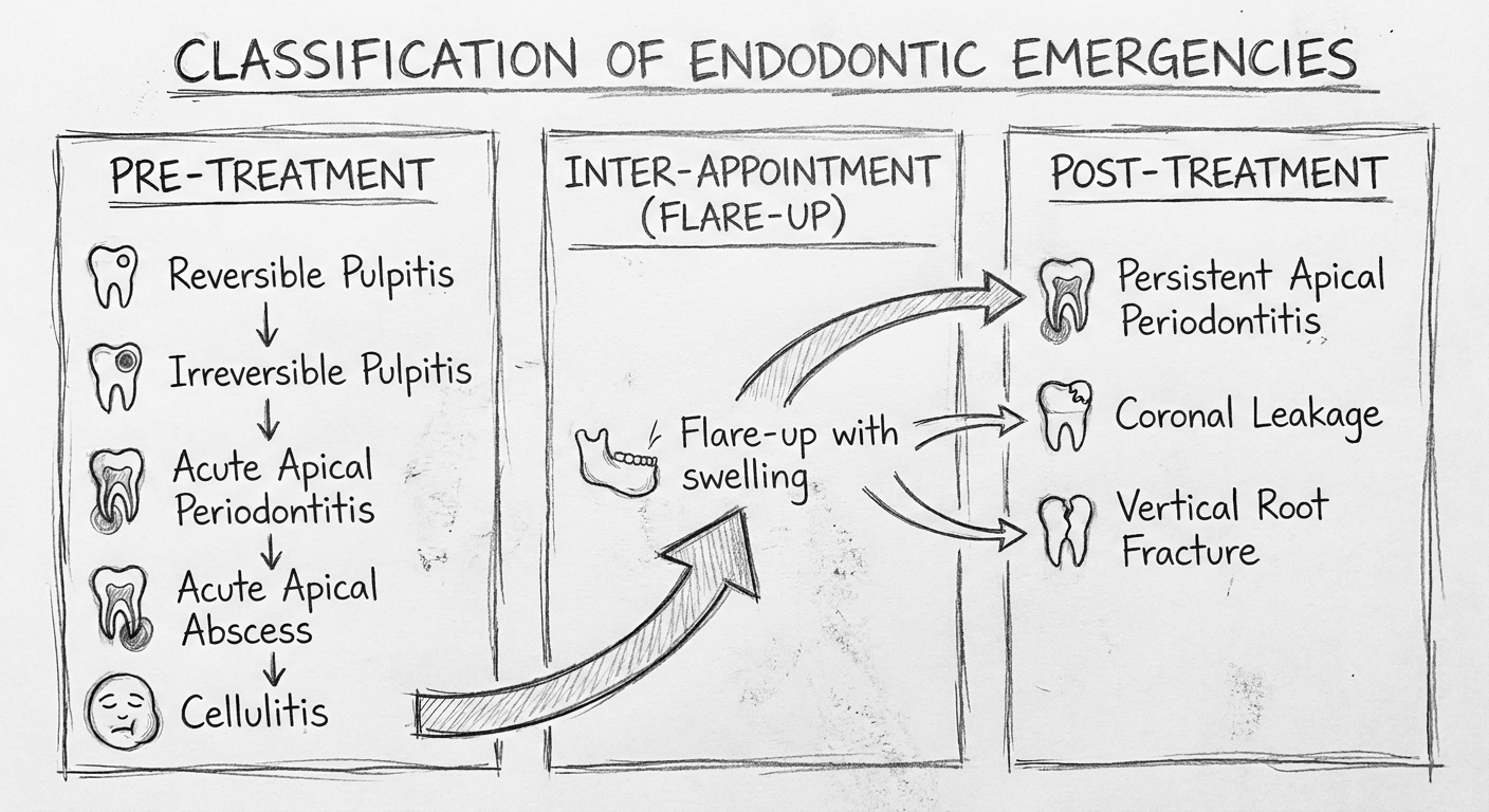
Hand-drawn pencil sketch style medical diagram showing classification of endodontic emergencies. Three columns labeled: "Pre-Treatment", "Inter-Appointment (Flare-up)", "Post-Treatment". Under Pre-Treatment: Reversible Pulpitis, Irreversible Pulpitis, Acute Apical Periodontitis, Acute Apical Abscess, Cellulitis. Under Inter-Appointment: Flare-up with swelling arrow. Under Post-Treatment: Persistent Apical Periodontitis, Coronal Leakage, Vertical Root Fracture. Arrows connecting stages showing progression. Simple hand-drawn look with light pencil lines, clean labels, educational diagram style on white background.
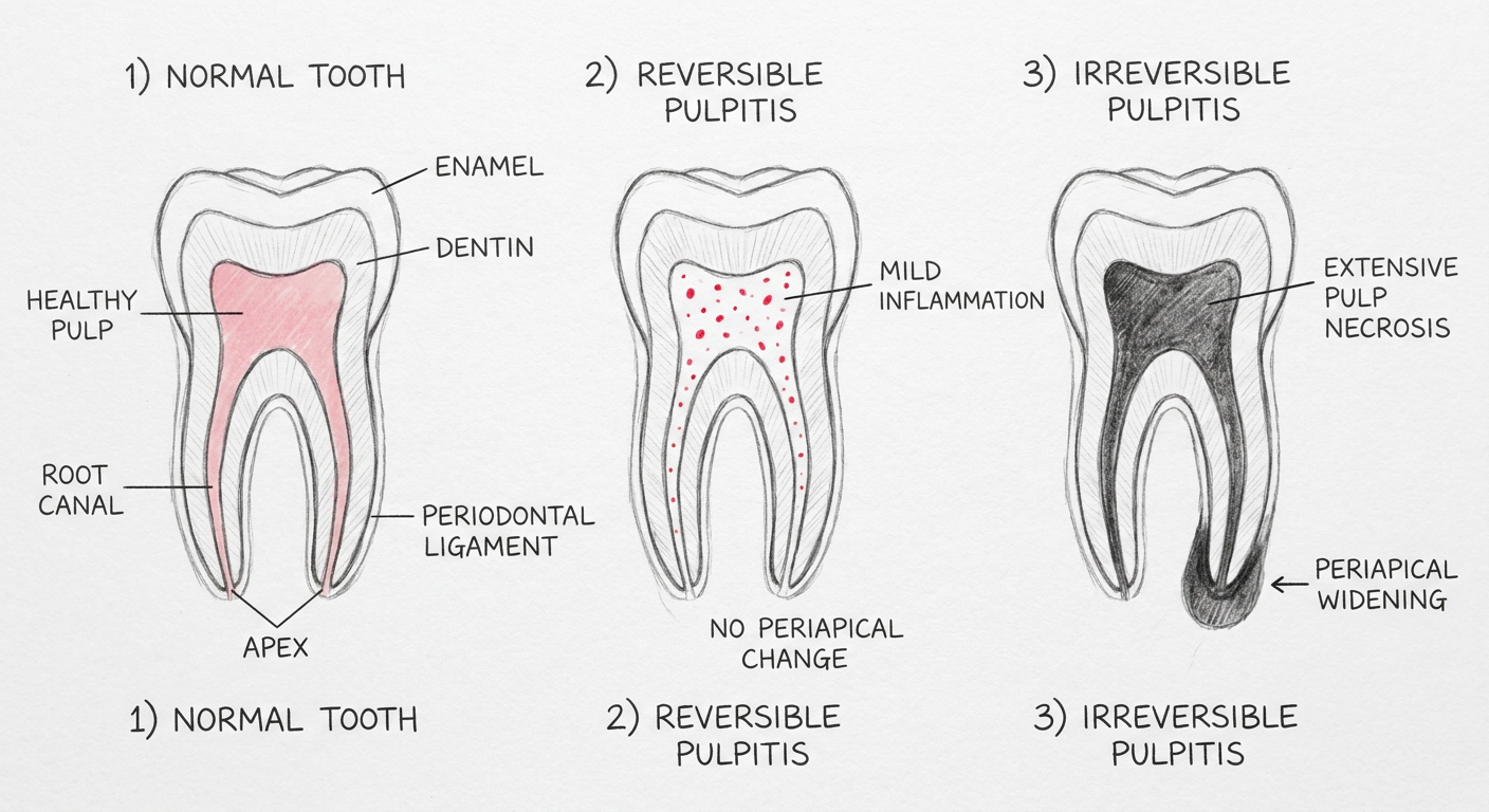
Hand-drawn pencil sketch medical diagram showing three tooth cross-sections side by side comparing: 1) Normal tooth with healthy pink pulp labeled, dentin, enamel, root canal, apex, periodontal ligament. 2) Reversible Pulpitis - mild inflammation shown in pulp chamber with small red dots, no periapical change. 3) Irreversible Pulpitis - extensive pulp necrosis shown dark, periapical widening at apex indicated with arrow. Labels for each stage. Educational dental anatomy sketch style, white background, clean pencil line art.
Hand-drawn pencil sketch medical dental diagram showing acute apical abscess spreading to cellulitis. Left side: cross-section of a lower molar tooth with necrotic pulp (dark), pus collection shown as dotted area at root apex labeled "Acute Apical Abscess", fluctuant swelling shown in gum. Right side: same jaw showing spreading cellulitis with diffuse swelling arrows radiating outward, labeled "Cellulitis - Diffuse spread", arrows pointing to fascial spaces labeled "submandibular space", "sublingual space", danger zone arrow labeled "Ludwig's Angina risk". Simple educational hand-drawn line art, white background, black pencil sketch style.
Hand-drawn flowchart diagram showing the "3D Approach" for endodontic emergency management. Top box: "ENDODONTIC EMERGENCY - Patient presents with pain/swelling". Three sequential boxes connected by large downward arrows: Box 1 "D1 - DIAGNOSIS" with bullet points: History, Clinical exam, Pulp tests (cold/EPT), Percussion, Radiograph/CBCT, Differentiate inflammation vs infection. Box 2 "D2 - DEFINITIVE DENTAL TREATMENT" with bullet points: Pulpectomy, Drainage (I&D), Root canal treatment, Retreatment. Box 3 "D3 - DRUGS (Adjunct Only)" with bullet points: NSAIDs - first line, Antibiotics - ONLY if systemic signs, Analgesics. Bottom note box: "IMPORTANT: Drugs NEVER replace treatment!". Hand-drawn style with pencil sketch boxes and arrows, educational diagram, white background.
Error calling model 'gemini-3-pro-image' (RESOURCE_EXHAUSTED): 429 RESOURCE_EXHAUSTED. {'error': {'code': 429, 'message': 'Resource has been exhausted (e.g. check quota).', 'status': 'RESOURCE_EXHAUSTED'}}
We encountered an error while processing your request. Please try again or reach out to us for help.
Images ??
📌 Diagram 1 — Classification of Endodontic Emergencies
📌 Diagram 2 — Pulp Stages: Normal → Reversible → Irreversible Pulpitis
- Wait a few minutes and ask me again: "Generate the remaining endodontic diagrams" - I'll retry the other 4
- Or ask for them one at a time
Yes visible
Hand-drawn pencil sketch medical dental diagram showing acute apical abscess spreading to cellulitis. Left side: cross-section of a lower molar tooth with necrotic pulp shown dark, pus collection as dotted area at root apex labeled "Acute Apical Abscess", fluctuant swelling in gum. Right side: same jaw showing spreading cellulitis with diffuse swelling arrows radiating outward labeled "Cellulitis - Diffuse spread", arrows pointing to fascial spaces labeled submandibular space, sublingual space, danger zone labeled "Ludwig's Angina risk". Simple educational hand-drawn line art, white background, black pencil sketch style.
Hand-drawn flowchart diagram showing the 3D Approach for endodontic emergency management. Top box: "ENDODONTIC EMERGENCY". Three sequential large boxes connected by downward arrows: Box 1 labeled "D1 - DIAGNOSIS" listing History, Clinical exam, Pulp vitality tests, Percussion test, Radiograph. Box 2 labeled "D2 - DEFINITIVE DENTAL TREATMENT" listing Pulpectomy, Drainage, Root canal treatment. Box 3 labeled "D3 - DRUGS (Adjunct Only)" listing NSAIDs first line, Antibiotics only if systemic signs, Analgesics. Warning note at bottom: "Drugs NEVER replace treatment". Clean hand-drawn pencil sketch style, educational flowchart, white background.
Error calling model 'gemini-3-pro-image' (RESOURCE_EXHAUSTED): 429 RESOURCE_EXHAUSTED. {'error': {'code': 429, 'message': 'Resource has been exhausted (e.g. check quota).', 'status': 'RESOURCE_EXHAUSTED'}}
We encountered an error while processing your request. Please try again or reach out to us for help.