
Parkinson’s and Parkinson plus syndrome and their psychiatric manifestation. Explain in a simplified easy to remember way
Parkinson disease Lewy body substantia nigra dopamine pathway diagram
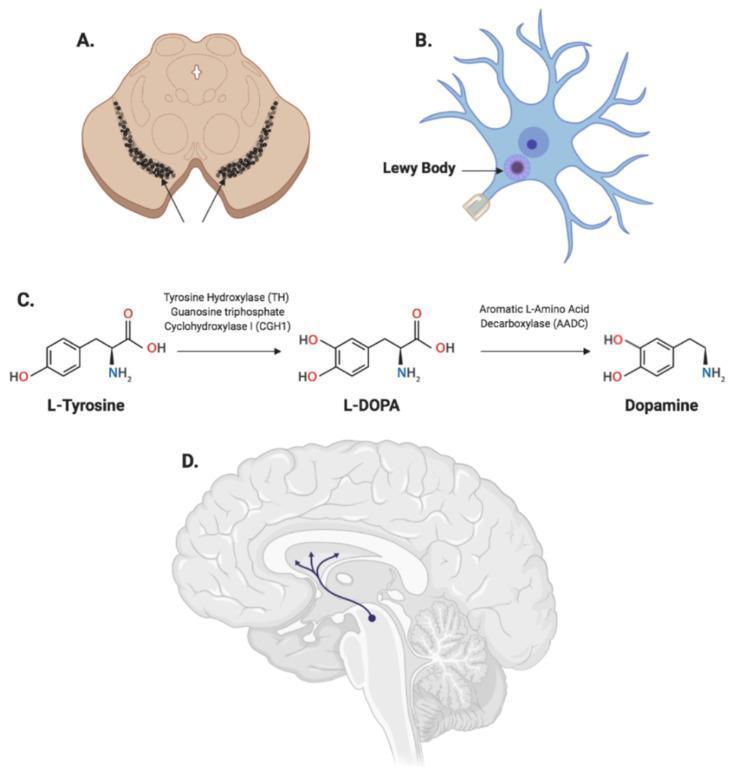
Educational infographic illustrating the neuro-pathophysiology of Parkinson's Disease (PD) through four distinct panels. Panel A displays a cross-sectional anatomical diagram of the midbrain, highlighting the bilateral, arc-shaped dark regions of the substantia nigra pars compacta. Panel B shows a schematic of a dopaminergic neuron containing a prominent intracellular Lewy Body, characterized as a dense, circular proteinaceous inclusion within the cell body. Panel C presents a biochemical flowchart of dopamine synthesis, detailing the enzymatic conversion of L-Tyrosine to L-DOPA via tyrosine hydroxylase (TH) and GCH1, followed by decarboxylation to Dopamine by aromatic L-amino acid decarboxylase (AADC). Panel D provides a sagittal view of the human brain, illustrating the nigrostriatal pathway where neurons from the substantia nigra project to the striatum within the basal ganglia. This combined visual explores the relationship between gross anatomical changes, cellular proteinopathy, neurotransmitter biochemistry, and neural circuit dysfunction in the progression of PD.
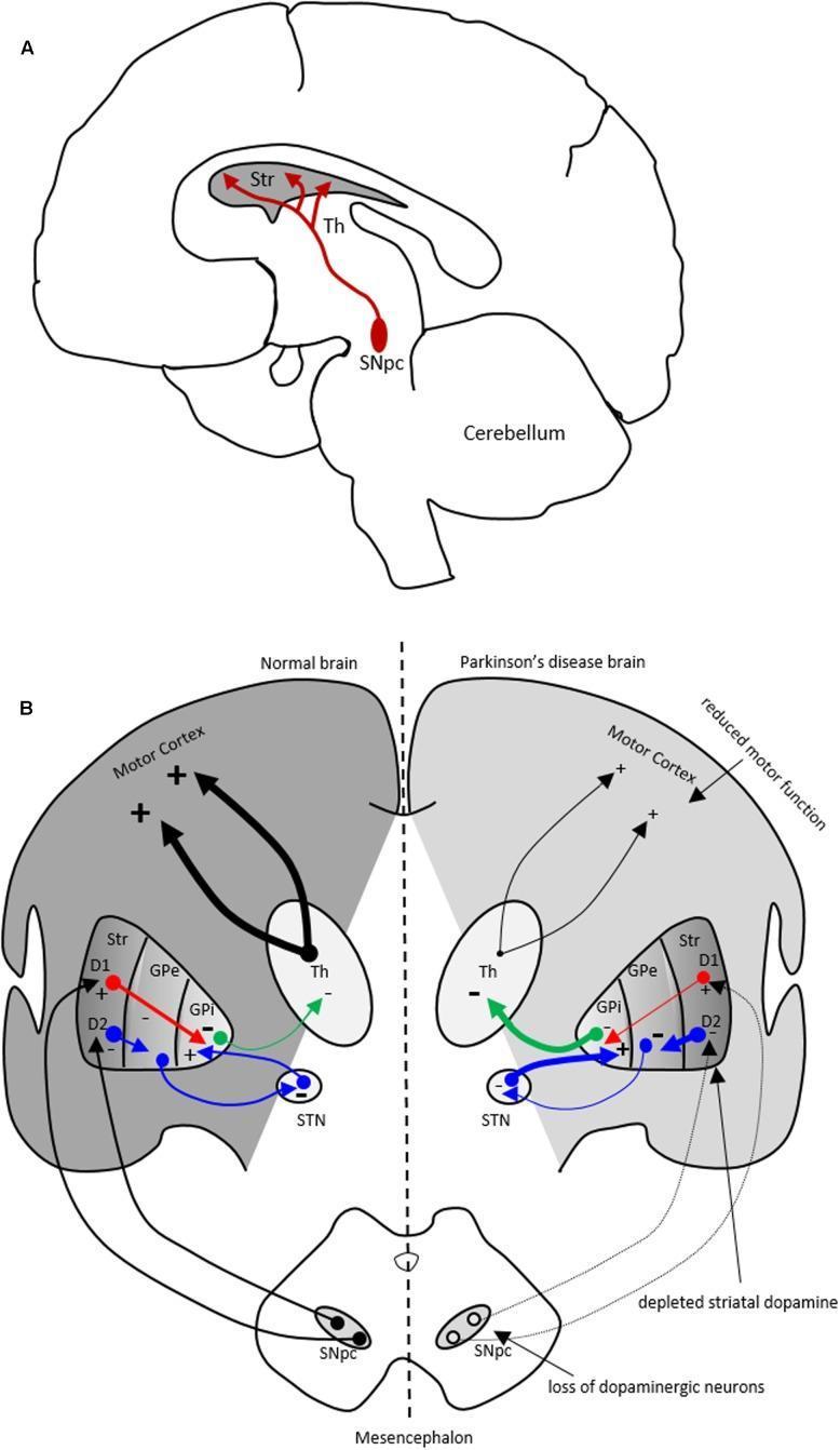
Educational diagram illustrating the pathophysiology of Parkinson's disease (PD) through neuroanatomical pathways and motor circuitry. Part A shows a sagittal anatomical diagram of the human brain identifying the nigrostriatal dopaminergic pathway. Red arrows represent dopamine projections originating from the substantia nigra pars compacta (SNpc) in the midbrain to the striatum (Str) and thalamus (Th). Part B provides a side-by-side comparison chart of motor basal ganglia circuitry in a 'Normal brain' versus a 'Parkinson's disease brain.' The schematic highlights the direct (D1) and indirect (D2) pathways involving the striatum (Str), globus pallidus externa (GPe), subthalamic nucleus (STN), and globus pallidus interna (GPi). In the PD brain, the diagram visually demonstrates the loss of dopaminergic neurons in the SNpc and depletion of striatal dopamine. Changes in line thickness indicate altered signaling intensities: decreased inhibitory input to the GPi and increased inhibitory output (green arrow) from the GPi to the thalamus. This increased inhibition leads to reduced thalamocortical excitation to the motor cortex, explaining the clinical manifestation of reduced motor function characteristic of PD.
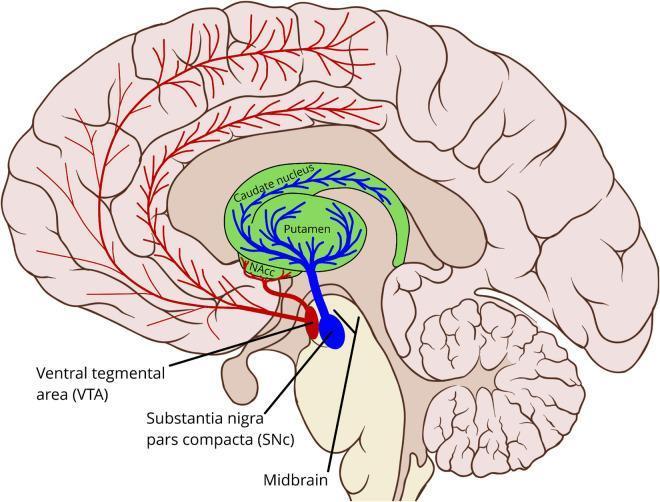
Anatomical diagram of the human brain in sagittal section illustrating the major dopaminergic pathways. The illustration highlights two primary midbrain sources: the Substantia Nigra pars compacta (SNc), color-coded in blue, and the Ventral Tegmental Area (VTA), color-coded in red. The nigrostriatal pathway is depicted as blue projections extending from the SNc to the striatum, which includes the green-shaded caudate nucleus and putamen. The mesolimbic and mesocortical pathways are represented by red lines originating from the VTA, projecting to the nucleus accumbens (NAcc) and extensively across the cerebral cortex, including prefrontal and motor regions. The diagram demonstrates the spatial relationship between the midbrain, the C-shaped basal ganglia structures, and the broader cortical distribution of dopamine. This resource is intended for neuroanatomical education regarding neurotransmitter systems and their clinical relevance to movement disorders (e.g., Parkinson's disease) and reward-based learning.
Parkinson plus syndromes PSP MSA corticobasal comparison features
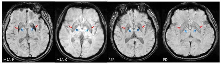
This diagnostic comparison contains four axial Susceptibility-Weighted Imaging (SWI) MRI slices of the brain at the level of the basal ganglia, showcasing differential iron accumulation patterns in Parkinsonian syndromes. The panels compare multiple system atrophy - parkinsonian variant (MSA-P), cerebellar variant (MSA-C), progressive supranuclear palsy (PSP), and Parkinson’s disease (PD). In the MSA-P case, distinctive posterolateral putaminal hypointensity is visible (indicated by closed red arrows), demonstrating a lateral-to-medial signal gradient and significant iron deposition in the lateral globus pallidus. In the PSP image, open blue arrows highlight increased iron-related signals localized to the anterior and medial aspects of the globus pallidus, which serves as a specific diagnostic marker. The MSA-C and PD slices provide a baseline for comparison, showing less pronounced or more diffuse signal changes. These images illustrate key neuroradiological hallmarks used in differentiating atypical parkinsonian syndromes (APS) based on localized mineral deposition in deep gray matter structures.
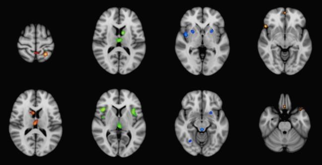
Diagnostic neuroimaging figure presenting a series of axial T1-weighted brain slices in MNI-152 template space. The visualization uses anatomic likelihood estimation (ALE) meta-analysis results to map significant gray matter atrophy patterns across four neurodegenerative movement disorders, distinguished by color-coded overlays. Corticobasal degeneration (CBD), shown in red, demonstrates atrophy primarily in the parietal lobes. Progressive supranuclear palsy (PSP), shown in green, highlights significant volume loss in the medial thalamus, insula, and midbrain. Multisystem atrophy Parkinson-type (MSA-P), represented in blue, shows localized atrophy within the putamen and brainstem structures. Idiopathic Parkinson's disease (IPD), indicated in gold/orange, shows scattered involvement including the frontal cortex. The comparison illustrates the differential spatial distribution of cortical and subcortical gray matter loss that characterizes these parkinsonian syndromes, providing educational value for differentiating atypical parkinsonian disorders from IPD based on structural MRI findings.
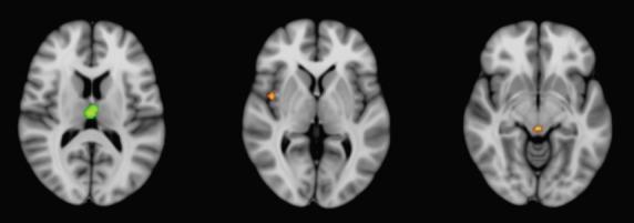
This diagnostic image displays three axial neuroimaging slices superimposed on the MNI-152 brain template, illustrating results from a conjunction analysis of gray matter atrophy in atypical parkinsonian syndromes (APS). The visualization identifies overlapping regions of volume loss between distinct disease groups. The leftmost slice shows a green-colored cluster in the superomedial thalamus, indicating shared atrophy common to corticobasal degeneration (CBD) and progressive supranuclear palsy (PSP). The middle slice reveals an orange cluster localized to the right insula (Brodmann Area 13), while the rightmost slice depicts an orange cluster in the midbrain; these orange regions signify shared gray matter reduction between progressive supranuclear palsy (PSP) and multisystem atrophy Parkinson-type (MSA-P). This neuroanatomical comparison serves as an educational tool for identifying common neurodegenerative patterns and distinguishing shared pathological involvement among APS variants, highlighting the thalamus, insula, and midbrain as critical areas of convergence for different parkinsonian disorders.
Parkinson's Disease & Parkinson-Plus Syndromes: Simplified Guide
The Big Picture First
Parkinson's Disease (PD) = the "original" - dopamine loss, responds well to L-DOPA, mainly motor symptoms + some psychiatric features. Parkinson-Plus syndromes = "Parkinson's with extras" - parkinsonism PLUS something more, poor L-DOPA response, more aggressive.
PART 1: Parkinson's Disease
Core Pathology (The "What's Broken?")
- What dies: Dopaminergic neurons in the substantia nigra pars compacta
- What accumulates: Lewy bodies (clumps of alpha-synuclein protein)
- Pathway affected: Nigrostriatal pathway → loss of dopamine → too much inhibition of motor output
Motor Symptoms (The Classic "TRAP" mnemonic)
- Tremor (resting, "pill-rolling")
- Rigidity (cogwheel)
- Akinesia/Bradykinesia (slowness)
- Postural instability
PART 2: Psychiatric Manifestations of PD
| Letter | Symptom | Key Details |
|---|---|---|
| D | Depression | Most common (up to 50%). Due to dopamine/serotonin depletion. Often precedes motor symptoms. Treat with SSRIs, SNRIs, dopamine agonists (pramipexole) |
| A | Anxiety | GAD, panic attacks. Often "off"-period anxiety (worsens when medication wears off). Poor evidence for treatment; benzodiazepines used cautiously |
| P | Psychosis | Hallucinations (45-65% of PD-dementia patients) - mostly visual (seeing people/animals). Delusions in 25-30%. Often medication-induced (L-DOPA, dopamine agonists). Treat with clozapine (best evidence) or quetiapine |
| H | Hallucinations | Visual > auditory. Can be drug-induced OR disease progression |
| H | Hypersexuality / Impulse control disorders | Dopamine agonists (pramipexole, ropinirole) cause gambling, hypersexuality, binge eating |
- Apathy (not the same as depression - no sad mood, just no motivation/initiative). Treat with dopamine agonists, stimulants.
- REM Sleep Behavior Disorder (RBD) - acting out dreams, often a very early prodromal sign (can precede PD by 10+ years)
- Dementia (PDD) - occurs in ~80% eventually. Develops >1 year after motor symptoms. Impairs attention, executive function, visuospatial, memory. Treat with acetylcholinesterase inhibitors (rivastigmine).
PART 3: Parkinson-Plus Syndromes
The "PLUS" means Parkinson's features + additional system involvement + poor L-DOPA response
1. PSP - Progressive Supranuclear Palsy
| Feature | Detail |
|---|---|
| Pathology | Tauopathy (tau inclusions in brainstem + basal ganglia) |
| Key sign | Vertical gaze palsy (cannot look up/down) - hallmark |
| Posture | Axial rigidity (neck extension, falls backward - "plank falls") |
| Psychiatric | Pseudobulbar affect (uncontrollable laughing/crying), apathy, progressive dementia, disinhibition |
| Timing | Falls EARLY (within 1 year of onset) |
| L-DOPA | Minimal/no response |
| Prognosis | Fatal in 5-7 years |
2. CBD - Corticobasal Degeneration
| Feature | Detail |
|---|---|
| Pathology | Tauopathy (same tau variants as PSP, more cortical) |
| Key sign | Alien limb phenomenon (one hand moves on its own) + asymmetric rigidity |
| Motor | Apraxia (can't plan movements), dystonia, myoclonus, action tremor |
| Psychiatric | Dementia, apathy, behavioral changes (FTD-like), frontal release signs |
| L-DOPA | Poor response |
3. MSA - Multiple System Atrophy
| Feature | Detail |
|---|---|
| Pathology | Synucleinopathy (alpha-synuclein in OLIGODENDROCYTES - glial cytoplasmic inclusions) |
| Three circuits fail | Striatonigral (parkinsonism), Olivopontocerebellar (ataxia), Autonomic (orthostatic hypotension, incontinence) |
| Subtypes | MSA-P (parkinsonism dominant) vs MSA-C (cerebellar dominant) |
| Psychiatric | Relatively LESS psychiatric than others. Depression, anxiety. Notably - cognitive decline is less prominent than PDD/PSP |
| L-DOPA | ~30% partial response |
| Distinguishing clue | Severe autonomic failure early (faintness on standing, can't urinate) |
4. DLB - Dementia with Lewy Bodies
| Feature | Detail |
|---|---|
| Pathology | Synucleinopathy (cortical + subcortical Lewy bodies) |
| Core features | Fluctuating cognition, vivid visual hallucinations, parkinsonism, REM sleep behavior disorder |
| "1-year rule" | Dementia appears within 1 year of parkinsonism → DLB. >1 year delay → PDD |
| Psychiatric | Hallucinations (very prominent and early), delusions, depression, extreme neuroleptic sensitivity (antipsychotics can be FATAL) |
| Key warning | AVOID typical antipsychotics and even risperidone - severe sensitivity reaction |
| Treatment | Rivastigmine (best evidence for cognition + hallucinations) |
PART 4: Quick Comparison Table
| Feature | PD | PSP | CBD | MSA | DLB |
|---|---|---|---|---|---|
| Pathology | Synuclein | Tau | Tau | Synuclein (oligodendrocytes) | Synuclein |
| Tremor | Resting (pill-roll) | Rare | Action/minimal | Postural | Variable |
| L-DOPA | Excellent | None | None | 30% partial | Variable |
| Key sign | Tremor | Vertical gaze palsy | Alien limb | Autonomic failure | Visual hallucinations |
| Symmetry | Asymmetric onset | Symmetric, axial | Asymmetric | Symmetric | Variable |
| Dementia | Late (>1yr) | Moderate | Moderate | Mild | Early (within 1yr) |
| Falls | Late | Early | Late | Moderate | Variable |
| Psychiatric hallmark | Depression + drug psychosis | Pseudobulbar + apathy | FTD-like behavior | Mild depression | Hallucinations + neuroleptic sensitivity |
PART 5: Memory Hooks Summary
PD     = Dopamine down → TRAP motor + DAPHH psychiatric
PSP    = TAU + stares poorly + falls backward + pseudobulbar
CBD    = TAU + alien limb + asymmetric + apraxia
MSA    = SYNUCLEIN in glia + Motor + Sphincter + Ataxia
DLB    = SYNUCLEIN cortical + Dementia FIRST + vivid hallucinations
- Dementia before or within 1 year of parkinsonism = DLB
- Dementia more than 1 year after parkinsonism = PDD (Parkinson's disease dementia)