
glandular componentof breast and its lining epithelium
Reading File
The breast is a modified sweat (apocrine) gland. Its glandular component consists of lobules and ducts lined by two epithelial layers. The inner layer is secretory epithelium (luminal cells), and the outer is a myoepithelial cell layer. Let me pull the detailed textbook content now.
Searching sources...
Finding Sources
Finding Sources
Finding Sources
Finding Sources
Reading File
Reading File
Reading File
Reading File
Reading File
Loading Image
Loading Image
Now I have rich content from three major textbooks. Let me also fetch one more useful image and look at the histology images.
Loading Image
Loading Image
I now have all the content needed for a thorough answer. Here is the complete response:
Glandular Component of the Breast and Its Lining Epithelium
Overview
The mature breast consists of three principal tissue types: (1) glandular epithelium, (2) fibrous stroma, and (3) adipose tissue. The glandular apparatus is a branching duct system organized radially from the nipple-areolar complex (NAC), comprising 15-20 lobes, each with a lactiferous duct opening at the nipple.
- Sabiston Textbook of Surgery, p. 1369
- Robbins & Cotran Pathologic Basis of Disease, p. 961
Gross Anatomy of the Glandular System
Cutaway diagram of the mature resting breast:
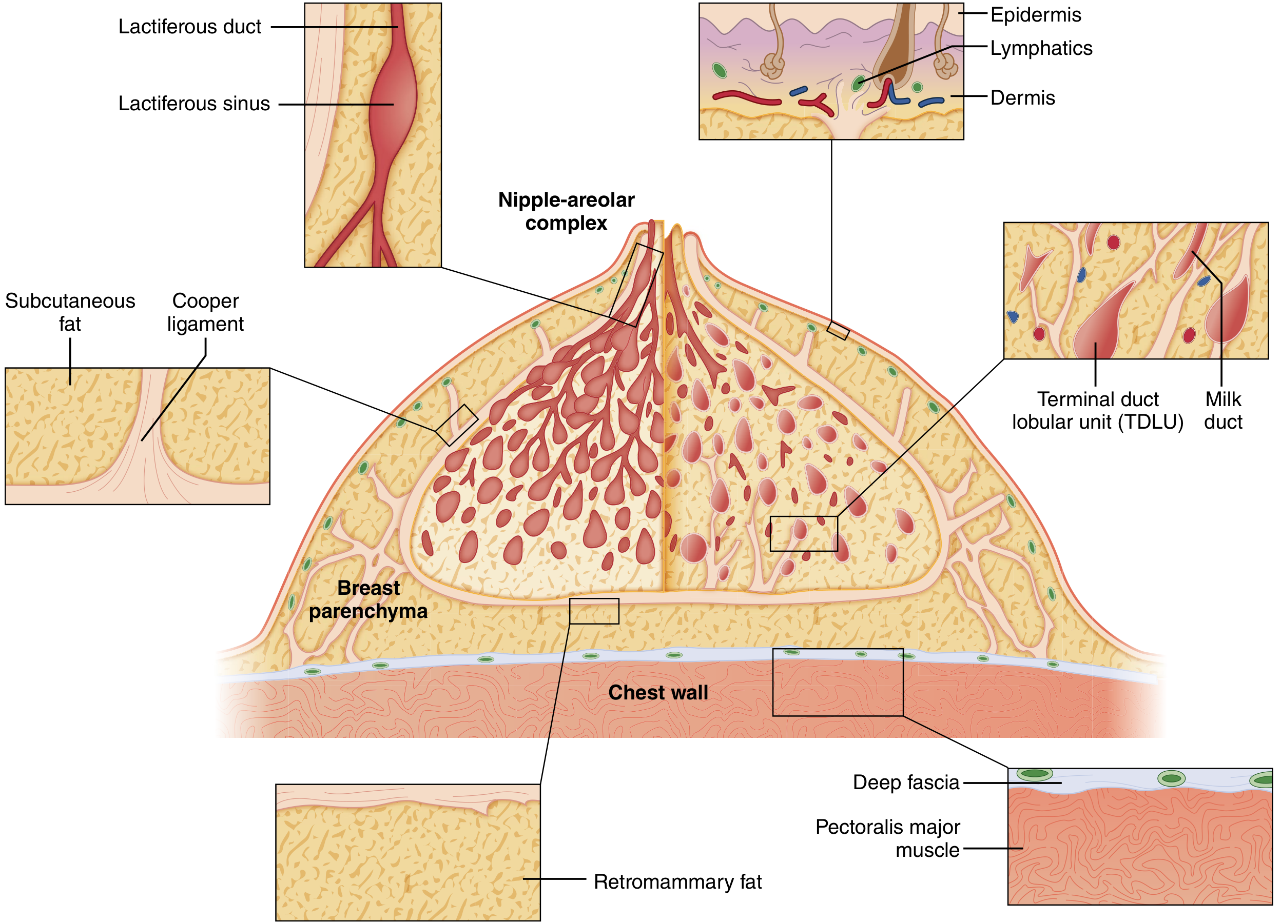
The ductal-lobular system is arranged like an inverted tree:
| Level | Structure | Notes |
|---|---|---|
| Nipple | Major lactiferous ducts (15-20) | Open independently at the nipple |
| Subareolar | Lactiferous sinuses | Dilated ampullary segments |
| Mid-breast | Segmental and subsegmental ducts | Progressive branching |
| Periphery | Terminal ducts | Lead into lobules |
| Secretory unit | Acini (alveoli) | Milk-forming glands |
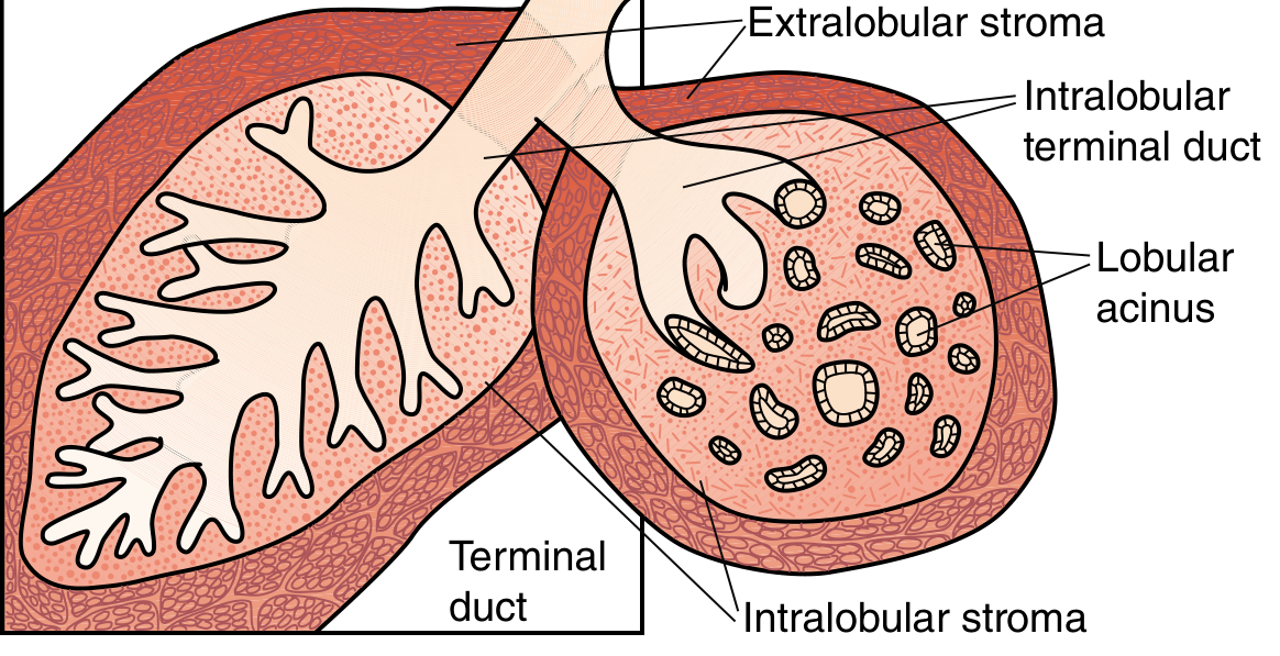
The terminal duct + its lobule = Terminal Duct Lobular Unit (TDLU) - the structural and functional unit of the breast. Each lobule is composed of grape-like clusters of small acini. TDLUs are surrounded by a specialized loose intralobular connective tissue containing capillaries, lymphocytes, and mononuclear cells - distinct from the denser interlobular stroma outside.
Diagram: TDLU Structure
Lining Epithelium - Region by Region
The entire ductal-lobular system is lined by two distinct epithelial cell layers:
- Inner luminal (secretory) cells - columnar to cuboidal, face the lumen
- Outer myoepithelial cells - contractile, propel milk toward the nipple; located between the luminal cells and the basement membrane
| Region | Epithelial Lining |
|---|---|
| Nipple (superficial major ducts) | Keratinizing squamous epithelium - abruptly transitions to double-layered epithelium below |
| Lactiferous sinuses (subareolar) | Stratified cuboidal epithelium |
| Lactiferous ducts and terminal ducts | Simple cuboidal epithelium + numerous myoepithelial cells |
| Acini/alveoli (lobules) | Simple cuboidal/low columnar epithelium with myoepithelial cells |
"Within the nipple, superficial portions of the major ducts are lined by keratinizing squamous epithelium that abruptly transitions to a double-layered epithelium with inner luminal and outer myoepithelial cell layers that lines the remainder of the ductal-lobular system."
- Robbins & Cotran, p. 961
The Basement Membrane
Outside both epithelial layers (luminal + myoepithelial), a continuous basement membrane containing laminin, type IV collagen, and proteoglycans surrounds the ductal-lobular system. This is a critical anatomical boundary:
- Intact basement membrane = in situ carcinoma (e.g., DCIS - no invasion)
- Breached basement membrane = invasive carcinoma (tumor gains access to lymphatics and blood vessels - metastatic potential)
Cell Types in the Breast (Summary Diagram)
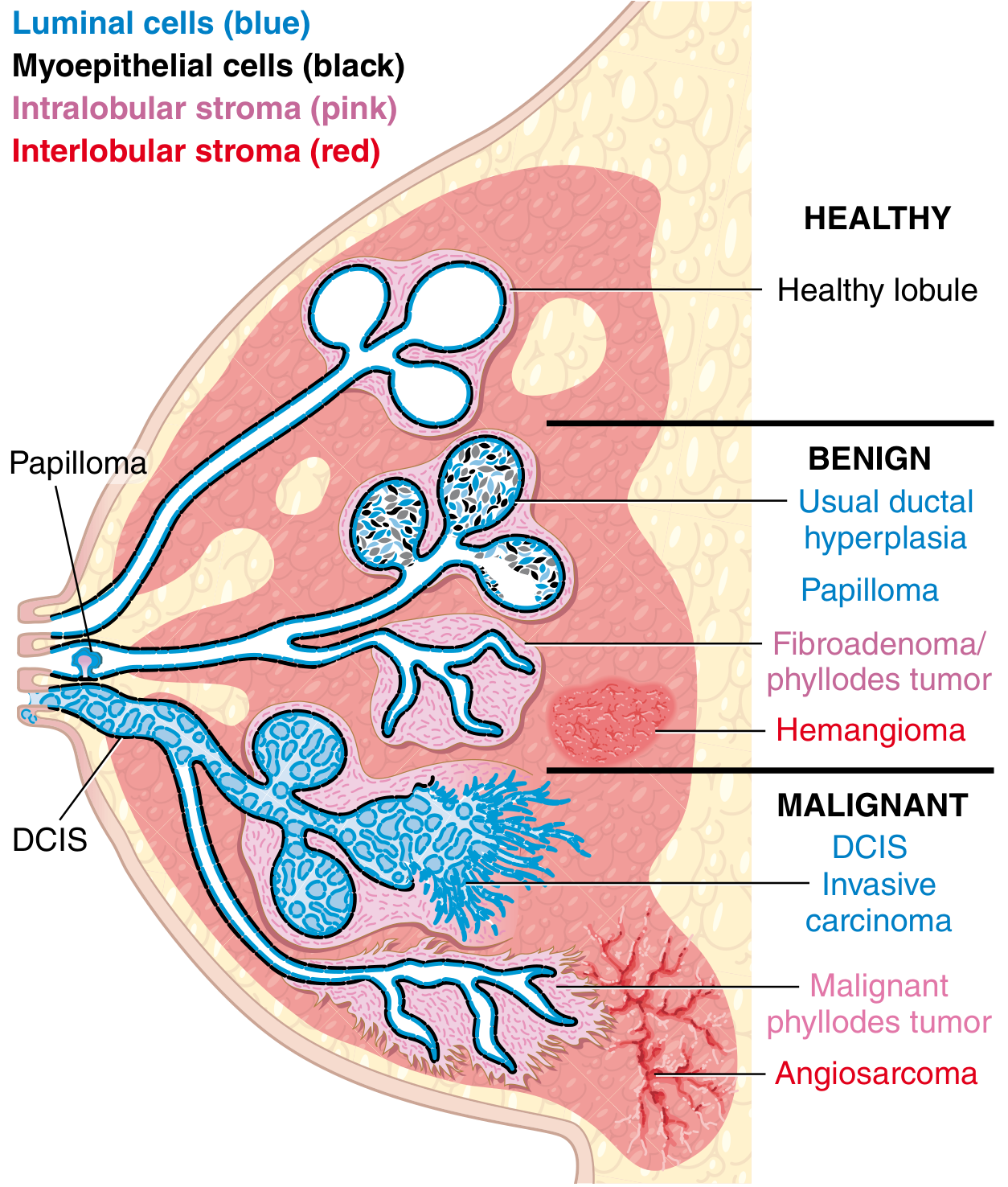
The cellular constituents of the breast include:
- Two structural units: ducts and lobules
- Two epithelial cell types: luminal cells and myoepithelial cells
- Two stromal compartments: intralobular (loose, specialized) and interlobular (dense, fibrous)
Each of these is a source of both benign and malignant disease. - Robbins & Cotran, p. 961
Histology During Different Physiological States
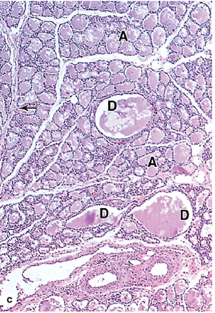
Lactating breast (H&E, x60) - Alveoli (A) filled with milk, excretory ducts (D) prominent, intralobular stroma sparse:
| State | Glandular Appearance |
|---|---|
| Pre-pubertal | Only lactiferous sinuses near nipple; rudimentary ducts |
| Puberty (non-pregnant) | Duct elongation; TDLUs small; acini rudimentary; mostly stroma + fat |
| Pregnancy | Massive lobular proliferation under estrogen, progesterone, prolactin, placental lactogen; alveoli grow extensively; cuboidal secretory epithelium |
| Lactation | Alveoli maximally dilated and filled with milk; stroma minimal |
| Post-weaning | Epithelial apoptosis, lobular regression (partial) |
| Post-menopause | Glandular involution; largely replaced by adipose tissue |
During the menstrual cycle, the lobules also cycle: relatively quiescent in the follicular phase, then secretory activity and epithelial proliferation under estrogen + progesterone in the luteal phase, followed by partial regression at menstruation.
Key Clinical Points
- Most breast carcinomas originate from the TDLU epithelium (not large ducts, despite traditional "ductal" terminology)
- Myoepithelial cells are absent in invasive carcinoma - their loss on immunohistochemistry (p63, SMA, calponin) helps distinguish invasive from in situ disease
- Accessory breast tissue (ducts/lobules extending into axilla or chest wall subcutaneous tissue) can give rise to carcinoma outside grossly evident breast tissue
Sources: Sabiston Textbook of Surgery, p. 1369-1370; Robbins & Cotran Pathologic Basis of Disease, p. 961; Junqueira's Basic Histology, p. 1156
This is a shared conversation. Sign in to Orris to start your own chat.