
Circulatory disorders explain in detail along with artries and veins disorders explain them in detail
Reading File
Now let me search the medical library for comprehensive content on circulatory disorders, arterial disorders, and venous disorders in parallel.
Finding Sources
Finding Sources
Finding Sources
Finding Sources
Finding Sources
Reading File
Finding Sources
Reading File
Finding Sources
Finding Sources
Finding Sources
Finding Sources
Now let me read the detailed content from key textbooks on atherosclerosis and venous disease:
Reading File
Reading File
Reading File
Reading File
Reading File
Reading File
Reading File
Reading File
Reading File
I now have excellent content from multiple authoritative textbooks. Let me fetch the key diagnostic images to include in the answer.
Loading Image
Loading Image
Loading Image
I now have comprehensive material from multiple authoritative sources. Let me compile the detailed answer.
Circulatory Disorders: A Detailed Overview
Circulatory disorders encompass any condition that impairs the normal flow of blood through the cardiovascular system. They are broadly divided into arterial disorders (affecting arteries that carry oxygenated blood away from the heart) and venous disorders (affecting veins that return deoxygenated blood to the heart). Both categories involve pathologic changes to vessel walls, hemodynamics, or the blood itself.
PART 1: OVERVIEW OF THE CIRCULATORY SYSTEM
The circulatory system consists of:
- Arteries - thick-walled, elastic/muscular vessels with three layers: tunica intima (inner endothelium), tunica media (smooth muscle and elastic tissue), and tunica adventitia (outer connective tissue)
- Veins - thinner-walled vessels containing valves to prevent backflow; high-capacitance, low-pressure system
- Capillaries - microscopic exchange vessels connecting arterioles to venules
Blood flow depends on the pressure gradient, vessel caliber, and blood viscosity. Disruptions to any of these factors produce circulatory disorders.
PART 2: ARTERIAL DISORDERS
1. Atherosclerosis (The Most Common Arterial Disease)
Atherosclerosis is a chronic inflammatory disease of large- and medium-sized muscular arteries. The fundamental lesion is the atheroma (fibrofatty plaque) - a raised focal lesion within the intima consisting of a lipid core covered by a fibrous cap.
Pathogenesis - Stage by Stage:
Stage 1 - Fatty Streak
The earliest lesion can appear in children as young as 10-14 years. A yellow streak runs along major arteries, composed of smooth muscle cells filled with cholesterol and foam cells (lipid-laden macrophages). It is not yet flow-limiting.
Stage 2 - Fibrous Plaque
Large numbers of smooth muscle cells, foam cells, and leukocytes accumulate. As the plaque grows, it projects into the vessel lumen, causing narrowing that can lead to ischemia or infarction.
Stage 3 - Complicated Lesion
Rupture of the fibrous plaque activates the coagulation cascade and thrombus forms. The end result is a calcified, ulcerated plaque with areas of hemorrhage and thrombus.
Morphology of an Atherosclerotic Plaque:
Plaques have three principal components:
- Cells: smooth muscle cells (SMCs), macrophages, and T cells
- Extracellular matrix (ECM): collagen, elastic fibers, proteoglycans
- Intracellular and extracellular lipids (cholesterol crystals - "cholesterol clefts")
The plaque has a superficial fibrous cap (SMCs + dense collagen), a shoulder region (macrophages + T cells), and a deep necrotic core (lipid, necrotic debris, foam cells, fibrin, organized thrombus).
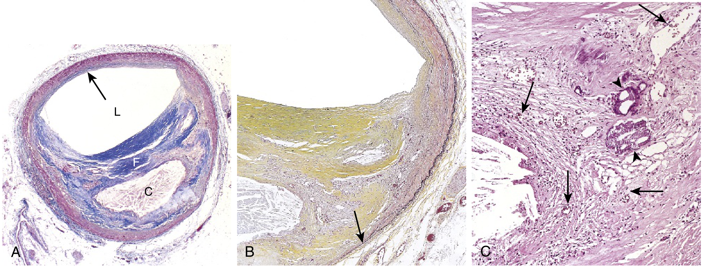
Atherosclerotic plaque in a coronary artery. F = fibrous cap, C = necrotic core, L = narrowed lumen (Robbins & Kumar Basic Pathology)
Distribution of Lesions (in descending order of severity):
- Infrarenal abdominal aorta
- Coronary arteries
- Popliteal arteries
- Internal carotid arteries
- Circle of Willis
The upper extremities and mesenteric/renal arteries (except ostia) are typically spared.
Risk Factors:
- Smoking
- Diabetes mellitus
- Hypertension
- Hyperlipidemia
- Male gender and advancing age
- Family history
- Obesity and reduced physical activity
Consequences of Atherosclerosis:
a) Atherosclerotic Stenosis
As plaques enlarge, the vessel lumen narrows. Critical stenosis occurs when the vessel is approximately 70% occluded. In the coronary artery, this produces stable angina - chest pain on exertion that is relieved by rest. In other vascular beds, stenosis causes:
- Bowel ischemia
- Ischemic encephalopathy
- Intermittent claudication (ischemic leg pain on walking)
- Chronic ischemic heart disease (IHD)
b) Acute Plaque Change (Most Dangerous)
Three types of plaque change occur:
- Rupture/ulceration/erosion - exposes highly thrombogenic substances, induces thrombus formation
- Hemorrhage into a plaque - rupture of the fibrous cap or neovessels causes intraplaque hematoma, rapid plaque expansion, or rupture
- Atheroembolism - ruptured plaque discharges debris producing microemboli (cholesterol, calcium, platelet aggregates)
Stable vs. Vulnerable Plaques:
- Stable plaques: thick fibrous cap, small lipid core, minimal inflammation - low rupture risk
- Vulnerable plaques: thin fibrous cap, large lipid core, marked inflammation - high rupture risk
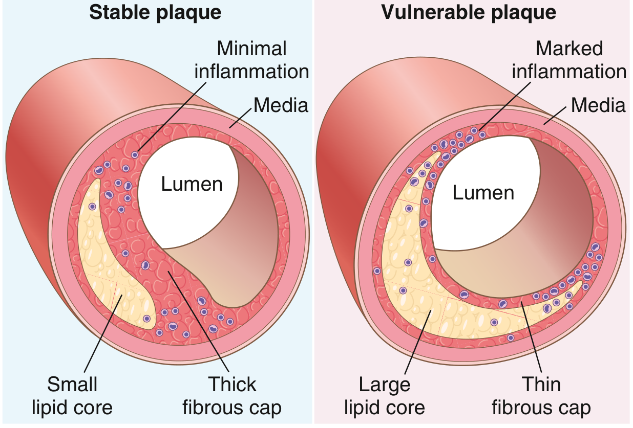
Stable and vulnerable atherosclerotic plaques (Robbins & Kumar Basic Pathology)
Major Clinical Outcomes of Atherosclerosis:
| Affected Vessel | Clinical Outcome |
|---|---|
| Coronary arteries | Myocardial infarction, angina |
| Carotid/cerebral arteries | Stroke, TIA |
| Aorta | Aortic aneurysm |
| Peripheral arteries | Peripheral arterial disease, gangrene |
| Renal arteries | Renovascular hypertension |
2. Arterial Embolism
An embolus is a foreign body (most commonly a detached blood clot) carried by the blood to a site distant from its origin, where it lodges and causes occlusion.
Sources:
- 85% originate from the heart (intracardiac thrombi)
- Left ventricular thrombus from MI: accounts for 60-70% of arterial emboli
- Atrial thrombi (mitral stenosis, rheumatic heart disease): 5-10%
- Atrial fibrillation is co-present in 60-75% of patients with peripheral embolic events
Mechanism of Damage:
After acute obstruction, the embolus can:
- Propagate proximally or distally
- Fragment and embolize to further distal vessels
- Precipitate venous thrombosis by local inflammatory reaction
Embolic occlusion occurs most often at major arterial bifurcations (because vessel diameters change abruptly there). The common femoral artery bifurcation is the most frequent site (35-50% of cases).
Myonephropathic-Metabolic Syndrome: Revascularization after ischemia releases potassium, lactic acid, and myoglobin - causing life-threatening hyperkalemia, metabolic acidosis, and myoglobinuria. This accounts for ~1/3 of deaths after arterial embolism revascularization.
Atheroembolism: Microemboli of cholesterol, calcium, and platelet aggregates dislodged from proximal plaques lodge in distal end arteries.
3. Aneurysms
An aneurysm is a localized permanent dilation of a blood vessel. It results from weakening of the vessel wall.
Types:
- True aneurysm: involves all three layers of the vessel wall (intima, media, adventitia)
- False (pseudo) aneurysm: a contained rupture - blood leaks through a defect into surrounding tissue, forming a hematoma with a fibrous wall
- Fusiform aneurysm: symmetrical, circumferential dilation
- Saccular aneurysm: spherical outpouching on one side
Pathogenesis:
In severe atherosclerosis, pressure atrophy of the tunica media and loss of elastic tissue weakens the wall sufficiently to allow aneurysmal dilation. Calcification of atheromas makes vessels hard and brittle.
Common Sites:
- Abdominal aortic aneurysm (AAA): most common, usually infrarenal; risk of rupture (catastrophic internal hemorrhage)
- Thoracic aortic aneurysm: associated with hypertension, Marfan syndrome
- Intracranial ("berry") aneurysm: at cerebral artery bifurcations; rupture causes subarachnoid hemorrhage
- Popliteal artery aneurysm: most common peripheral arterial aneurysm
4. Arterial Dissection
An aortic dissection occurs when blood enters the medial layer of the aortic wall through a tear in the intima, creating a false lumen that propagates along the vessel.
Causes:
- Hypertension (most common)
- Connective tissue disorders (Marfan, Ehlers-Danlos syndrome)
- Trauma
- Cystic medial necrosis
Classification (Stanford):
- Type A: involves the ascending aorta - surgical emergency
- Type B: involves the descending aorta only - often managed medically
Clinical Features: Sudden, severe "tearing" or "ripping" chest/back pain; pulse deficits; differential blood pressures between arms.
5. Vasospastic Disorders (Peripheral Arterial Spasm)
These disorders produce an abnormal vasomotor response in distal small arteries. The arterial wall itself is normal - symptoms arise from episodic vasoconstriction.
Examples:
- Raynaud disease: idiopathic episodic vasospasm of digits triggered by cold or emotion; color sequence: white (ischemia) → blue (cyanosis) → red (reperfusion)
- Raynaud phenomenon: secondary to underlying disease (scleroderma, lupus, etc.)
- Livedo reticularis: net-like mottled skin discoloration due to cutaneous vascular spasm
- Acrocyanosis: persistent cyanosis of hands/feet
- Erythromelalgia: episodic burning pain + redness of extremities due to abnormal vasodilation
6. Peripheral Arterial Disease (PAD)
PAD refers to stenotic or occlusive disease of the non-coronary arteries (usually lower limbs) due to atherosclerosis.
Clinical Stages (Fontaine Classification):
| Stage | Symptoms |
|---|---|
| I | Asymptomatic |
| IIa | Mild claudication (walking >200m) |
| IIb | Moderate-severe claudication (<200m) |
| III | Ischemic rest pain |
| IV | Ulceration or gangrene |
Investigations:
- Ankle-Brachial Index (ABI): normal 0.9-1.3; <0.9 indicates PAD; <0.4 = critical limb ischemia
- Duplex ultrasound
- CT angiography
7. Arteriovenous Fistula
An abnormal direct communication between an artery and a vein, bypassing the capillary bed. Causes: congenital defects, penetrating injuries, arterial aneurysm rupture into adjacent vein, or inflammatory necrosis.
Effects:
- Proximal artery and distal veins become distended, tortuous, and aneurysmal
- Chronic venous hypertension causes dermatitis and skin ulceration
- Large fistulas cause increased cardiac output, tachycardia, widened pulse pressure, or high-output cardiac failure
- ~60% are associated with a false aneurysm
8. Arterial Trauma
Partial lacerations continue to bleed (intact portion prevents arterial retraction). Complete transection usually bleeds less due to arterial spasm and thrombus formation - but delayed hemorrhage can occur when spasm resolves or thrombus liquefies.
Blunt injury disrupts the intima, leading to dissection, progressive obstruction, and thrombosis. Vasospasm can accompany adjacent trauma and spontaneously resolves in the absence of structural disruption.
PART 3: VENOUS DISORDERS
1. Varicose Veins
Varicose veins are dilated, tortuous, elongated superficial veins resulting from incompetent venous valves. Prevalence: 14-46% in women, 11-29% in men.
Pathogenesis:
- Primary: intrinsic structural/functional abnormality in the vein wall or venous valves, leading to valvular reflux and venous hypertension
- Secondary: deep-vein thrombosis (DVT) damages valves, causing post-thrombotic syndrome
Most Commonly Affected: Great saphenous vein and small saphenous vein in the lower limbs.
Risk Factors: Female sex, advancing age, prolonged standing, obesity, pregnancy, family history.
Symptoms:
- Dull ache, throbbing, heaviness, or pressure after prolonged standing (relieved by elevation)
- Cramping, burning, pruritus, swelling
- Cosmetic concern
Complications:
- Superficial thrombophlebitis
- Hemorrhage from ruptured varicosity
- Chronic venous insufficiency with skin changes
2. Chronic Venous Insufficiency (CVI)
CVI results from venous hypertension due to incompetent valves and/or venous obstruction, with extravasation of fluid and blood elements into tissue.
Causes:
- Primary: intrinsic valve incompetence
- Secondary: post-DVT syndrome (most common cause of advanced CVI)
- After DVT, valve leaflets become thickened and contracted, preventing retrograde flow
- Large proximal veins may remain occluded after thrombosis
- Secondary incompetence develops distally due to high pressures
Other Causes:
- May-Thurner syndrome: left iliac vein occluded by overlying right common iliac artery
- Extrinsic compression by tumor or retroperitoneal fibrosis
- Arteriovenous fistulas
- Congenital deep-vein agenesis/hypoplasia
- Klippel-Trenaunay and Parkes-Weber syndromes
Clinical Features:
- Pitting edema (ankles → knees → thighs with increasing severity)
- Stasis dermatitis: hyperpigmentation, erythema, eczema
- Lipodermatosclerosis: induration + hemosiderin deposition + inflammation, just above the ankle
- Atrophie blanche: white scar tissue with telangiectasias near the medial malleolus
- Venous ulcers: near the medial malleolus (the most advanced complication)
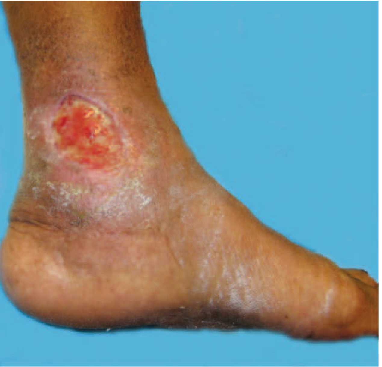
Venous insufficiency with active venous ulcer near the medial malleolus (Harrison's Principles of Internal Medicine)
CEAP Classification of Chronic Venous Disease:
| Class | Description |
|---|---|
| C0 | No visible or palpable signs |
| C1 | Telangiectasia / reticular veins |
| C2 | Varicose veins |
| C3 | Edema |
| C4 | Skin changes (hyperpigmentation, eczema, lipodermatosclerosis) |
| C5 | Healed venous ulcer |
| C6 | Active venous ulcer |
Diagnosis:
- Venous duplex ultrasonography (primary test): detects obstruction (absence of flow, echogenic thrombus, non-compressible vein) and venous reflux (prolonged reversal of flow during Valsalva maneuver)
- Color-assisted Doppler ultrasound
- CT venography for proximal/central veins
Bedside Tests:
- Brodie-Trendelenburg test: distinguishes primary from secondary varicose veins
- Perthes test: assesses patency of the deep venous system
3. Deep Vein Thrombosis (DVT)
DVT is the formation of a blood clot within a deep vein, most commonly in the lower extremities (calf, popliteal, femoral, or iliac veins).
Virchow's Triad (three predisposing factors):
- Stasis: immobility, long flights, heart failure
- Endothelial injury: surgery, trauma, catheters
- Hypercoagulability: malignancy, inherited thrombophilias (factor V Leiden, prothrombin mutation), OCP use, pregnancy
Clinical Features:
- Unilateral leg swelling, warmth, erythema
- Calf tenderness (Homan's sign - dorsiflexion pain, though non-specific)
- Many are clinically silent
Most Feared Complication: Pulmonary Embolism (PE)
- Thrombus fragments and travels to the pulmonary vasculature
- Ranges from asymptomatic to massive, fatal PE
- Presents with dyspnea, pleuritic chest pain, tachycardia, hypoxia
Other Complications:
- Post-thrombotic syndrome (PTS): chronic venous insufficiency + persistent leg pain, swelling, skin changes, ulceration
- Phlegmasia cerulea dolens: massive DVT causing severe obstruction, cyanosis, and limb-threatening ischemia
Diagnosis:
- Wells Score for pre-test probability
- D-dimer (high sensitivity, low specificity)
- Venous compression ultrasound (gold standard for lower limb DVT)
- CT pulmonary angiography (for PE)
Treatment:
- Anticoagulation: direct oral anticoagulants (DOACs) such as rivaroxaban, apixaban are first line
- Low molecular weight heparin (LMWH) - especially in cancer-associated VTE
- Thrombolysis for massive, limb-threatening DVT
- Inferior vena cava (IVC) filter when anticoagulation is contraindicated
4. Superficial Thrombophlebitis
Thrombosis + inflammation of a superficial vein. Common in varicose veins. Presents as a tender, red, palpable cord along a vein. Usually treated with NSAIDs and compression; anticoagulation may be added if near the saphenofemoral junction to prevent extension into the deep system.
5. Portal Vein Thrombosis
Occlusion of the extrahepatic portal vein. Can be catastrophic or produce only vague symptoms.
Causes:
- Cirrhosis (portal vein thrombosis in 25% of cirrhotics)
- Hypercoagulable states (polycythemia vera, factor V Leiden, antiphospholipid antibody syndrome)
- Inflammatory processes (pancreatitis, intraabdominal sepsis)
- Idiopathic (~1/3 of cases)
Clinical Effects:
- Abdominal pain
- Portal hypertension - esophageal varices prone to rupture
- Ascites (often massive and intractable when present)
6. Superior Vena Cava (SVC) Syndrome
Obstruction of the superior vena cava (most often by malignancy, especially lung cancer or lymphoma, or by thrombosis from central venous catheters).
Features: Facial/neck swelling, venous distension of neck and chest wall, arm edema, headache, blurred vision. A true oncological emergency.
7. Hepatic Vein Obstruction (Budd-Chiari Syndrome)
Obstruction of hepatic vein outflow at any level from hepatic venules to the right atrium.
Causes: Hypercoagulable states, myeloproliferative disorders, tumor invasion, webs.
Features: Hepatomegaly, ascites, abdominal pain, and eventually cirrhosis and liver failure. Produces the classic "nutmeg liver" gross appearance (centrilobular congestion).
PART 4: GENERAL COMPARISON - ARTERIAL vs. VENOUS DISORDERS
| Feature | Arterial Disorders | Venous Disorders |
|---|---|---|
| Wall thickness | Thick (three layers) | Thin |
| Blood pressure | High | Low |
| Pathology type | Occlusion, atherosclerosis, embolism, aneurysm | Thrombosis, insufficiency, varices |
| Pain character | Ischemic, claudication, severe at rest (late) | Dull ache, heaviness, worse with standing |
| Skin changes | Pale, cold, atrophic, no hair | Pigmented, warm, edematous, eczema |
| Ulcer location | Toes, heel, between toes | Medial/lateral malleolus |
| Pulses | Reduced or absent | Normal |
| Edema | Absent or minimal | Present, pitting |
| Key diagnostic test | ABI, CT angiography | Duplex ultrasound |
Sources
- Robbins & Kumar Basic Pathology - Chapters 8 (Atherosclerosis, p. 319-322) and pathologic basis of vascular disease
- Harrison's Principles of Internal Medicine 22E - Chapters on Venous Disease and Chronic Venous Insufficiency, pp. 2223-2224
- ROSEN's Emergency Medicine - Arterial Embolism and PAD, pp. 1177-1180
- Bailey and Love's Short Practice of Surgery 28th Edition - Atherosclerosis pathophysiology and IHD, p. 969
This is a shared conversation. Sign in to Orris to start your own chat.