
Abruptio placenta rule out
Reading File
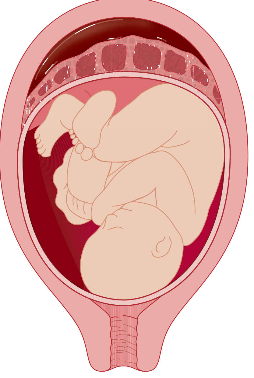
Abruptio placentae (placental abruption) is the premature separation of a normally implanted placenta from the uterine wall before delivery of the fetus. It is a major cause of antepartum hemorrhage and can be life-threatening for both mother and fetus.
Searching sources...
Finding Sources
Finding Sources
Searching PubMed
"placental abruption"[MeSH Terms] AND management
Reading File
Reading File
Reading File
Reading File
Reading File
Reading File
Loading Image
Loading Image
Reading File
Here is a comprehensive clinical overview of Abruptio Placentae (Placental Abruption) - Rule Out, drawn from authoritative medical textbooks.
Abruptio Placentae - Rule Out
Definition
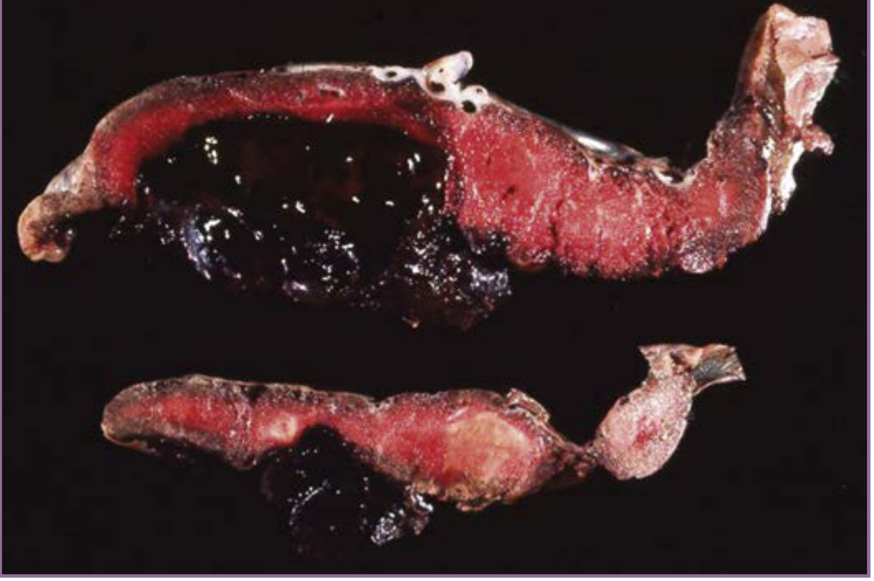
Abruptio placentae is the premature separation of a normally implanted placenta from the uterine wall after 20 weeks' gestation, before delivery of the fetus. It is a major cause of antepartum hemorrhage with significant maternal and fetal morbidity and mortality.
Epidemiology
- Complicates approximately 1% of all pregnancies (clinically recognized)
- Incidence peaks between 24-32 weeks of gestation
- If placentas are routinely examined at delivery, subclinical evidence is found in ~4% of cases
- Accounts for approximately 10% of all preterm births
- Abruption sufficient to cause fetal death occurs in ~1 of every 420 deliveries
- Disproportionate increase seen among Black women in the United States
Pathogenesis
Bleeding occurs between the decidua basalis and the placenta, dissecting the decidua apart. This leads to:
- Loss of placental surface area for gaseous exchange and fetal nutrition
- Hematoma expansion causing further dissection
- Revealed hemorrhage if dissection reaches the placental edge and tracks through the cervix
- Concealed hemorrhage with circumferential dissection and near-total separation
The underlying mechanism in many cases is vasospasm of abnormal maternal arterioles. Some cases result from venous hemorrhage into areas of necrotic decidua. With ongoing bleeding:
- Fetal hypoxia and possible fetal death
- Activation of the coagulation cascade -> DIC
- Maternal hypovolemia -> hemorrhagic shock
- Bleeding into myometrium -> Couvelaire uterus (uterine atony, increased hemorrhage risk)
Risk Factors
| Category | Specific Risk Factors |
|---|---|
| Strongest | Prior abruption (up to 20-fold increase; 25% recurrence with 2 prior events) |
| Hypertension | Chronic HTN (5x risk), superimposed preeclampsia (8x risk), preeclampsia alone |
| Substance use | Cigarette smoking (2.5x risk, dose-response); cocaine/crack use (up to 10%) |
| Trauma | Motor vehicle accidents (most common traumatic cause), intimate partner violence |
| Obstetric | Multiparity, oligohydramnios, pPROM, chorioamnionitis, uterine fibroids (2x risk) |
| Demographics | Maternal age <20 or >35 years |
| Other | Thrombophilia/hyperhomocysteinemia, PAPP-A <5th percentile on 1st trimester screen |
Clinical note: After trauma, evidence of abruption may not be apparent for up to 24 hours. Women with vaginal bleeding or contractions post-MVA should be observed for at least 24 hours; asymptomatic patients can be discharged after 6 hours of monitoring.
Clinical Features
Spectrum of Severity
| Feature | Mild Abruption | Severe Abruption (~15% of cases) |
|---|---|---|
| Vaginal bleeding | Slight or absent | Heavy or absent (concealed) |
| Uterine tone | Mild tenderness | Tetanic contraction, board-like rigidity |
| Maternal vitals | Normal | Hypotension, shock |
| Fetal status | No distress | Fetal distress or death |
| Fibrinogen | Normal (>300 mg/dL) | <150 mg/dL |
| Coagulopathy | Absent | DIC present |
Classic Triad
- Painful, dark vaginal bleeding (70% of patients)
- Uterine tenderness/pain (~2/3 of patients)
- Uterine contractions/irritability (~1/3 of patients)
Note: About 10% of women present only with occult bleeding (concealed abruption - no external blood loss). Always consider abruption in a patient with uterine pain, fetal distress, or hemodynamic instability even without visible bleeding.
Diagnostic Approach
Key Points
- Abruption remains a clinical diagnosis - imaging is primarily useful to exclude placenta previa
- Ultrasonography is insensitive - fresh blood is isoechoic to placenta; a normal USS does NOT rule out abruption
- Electronic fetal monitoring (CTG) has 100% negative predictive value for adverse outcomes when reassuring
- MRI is diagnostic but impractical in the acute setting
Investigations
| Investigation | Purpose | Finding in Abruption |
|---|---|---|
| CBC | Blood loss assessment | Falling Hb/Hct |
| Fibrinogen | Most important coag test | Decreased (normal in pregnancy: 400-650 mg/dL) |
| FDP (fibrin degradation products) | Most sensitive test for coagulopathy | Almost always elevated |
| Platelet count | Coagulopathy | Usually decreased |
| PT / aPTT | Coagulopathy | Normal to prolonged |
| Thrombin time | Parallels fibrinogen fall | Good marker of severity |
| Type & cross-match | Transfusion preparation | - |
| Kleihauer-Betke test | Fetal-maternal hemorrhage | Positive in fetal cells in maternal blood |
| Ultrasound | Exclude placenta previa; look for retroplacental clot | May be normal even with significant abruption |
| CTG (cardiotocography) | Fetal wellbeing | Fetal distress; most sensitive for identifying abruption impact |
Differential Diagnosis
When ruling out abruptio placentae, consider:
| Condition | Key Distinguishing Feature |
|---|---|
| Placenta previa | Painless, bright red bleeding; confirmed/excluded by USS |
| Uterine rupture | Catastrophic hypotension, cessation of contractions, abnormal fetal lie |
| Amniotic fluid embolism | Sudden cardiovascular collapse, hypoxia |
| Preeclampsia complications (HELLP) | Epigastric pain, elevated LFTs, thrombocytopenia |
| Pyelonephritis | CVA tenderness, fever, urinary symptoms |
| Appendicitis / cholecystitis | Localized RLQ/RUQ pain, fever |
| Ovarian torsion | Sudden onset, periumbilical or adnexal pain |
| Early labor | Uterine contractions without significant tenderness or hemorrhage |
Management
Immediate Stabilization (All Cases)
- Two large-bore IV access - fluid resuscitation
- Continuous fetal monitoring (CTG)
- Emergency obstetric consultation
- Labs: CBC, metabolic panel, coagulation panel (fibrinogen, FDP), type and cross-match
- RhoGAM (anti-D Ig 300 μg IM) if mother is Rh-negative
- Transfuse as needed: PRBCs, FFP, cryoprecipitate (for DIC)
Delivery Decisions
| Scenario | Management |
|---|---|
| Severe abruption (any gestational age) | Immediate delivery (vaginal or C-section based on obstetric status) |
| Abruption >34 weeks, stable mother and fetus | Vaginal delivery preferred; amniotomy + oxytocin if needed |
| Abruption with fetal death | Vaginal delivery preferred (unless hemodynamically unstable or obstetric contraindication) |
| Mild abruption, preterm (<34 weeks), stable | Conservative management with close monitoring; tocolytics controversial |
DIC Management
- Coagulopathy develops in ~10% of abruptions, strongly associated with fetal demise or massive hemorrhage
- Use component therapy: FFP (raises fibrinogen ~25 mg/dL per unit), cryoprecipitate (fibrinogen + factors VIII, XIII), platelets
- Target fibrinogen >200 mg/dL
Maternal and Fetal Complications
| Complication | Notes |
|---|---|
| DIC | ~10% of cases; most severe with fetal death |
| Hemorrhagic shock / exsanguination | Leading cause of maternal death |
| Couvelaire uterus | Myometrial infiltration by blood; leads to uterine atony |
| Acute kidney injury / renal cortical necrosis | From shock and DIC |
| Amniotic fluid embolism | Abruption is a predisposing factor |
| Fetal distress and death | ~15% of patients; risk proportional to % of placenta separated |
| Preterm birth / IUGR | Major cause of perinatal morbidity |
| Cerebral palsy / IVH / PVL | Perinatal asphyxia consequences |
Prevention
- Treat chronic hypertension adequately
- Smoking cessation (dose-response relationship)
- Avoidance of cocaine and substance abuse
- Treat hyperhomocysteinemia with folate supplementation
- Counsel high-risk patients (prior abruption = up to 20-fold recurrence risk) about warning signs
Key Clinical Pearls for "Rule Out"
"Abruption remains a clinical diagnosis - imaging is useful only to exclude placenta previa." - Creasy & Resnik's Maternal-Fetal Medicine
- A normal ultrasound does NOT rule out abruption - fresh blood is isoechoic to placenta
- Concealed abruption can occur with no visible bleeding but significant hemodynamic compromise
- CTG is the most sensitive tool - 100% NPV for adverse outcomes when reassuring
- Always consider abruption after trauma, even minor trauma, and observe for at least 6-24 hours
- Fibrinogen <150 mg/dL indicates severe abruption with active DIC
- Fetal death occurs in ~15% - risk increases with percentage of placental surface involved
Sources: ROSEN's Emergency Medicine, 9e; Tintinalli's Emergency Medicine, 9e; Creasy & Resnik's Maternal-Fetal Medicine, 9e
This is a shared conversation. Sign in to Orris to start your own chat.