
Pathology slide- hyaline degeneration
Reading File
Now let me search the medical library for hyaline degeneration content and images in parallel.
Finding Sources
Finding Sources
Let me search for the core pathology of hyaline degeneration and look for pathology slide images specifically.
Finding Sources
Finding Sources
Excellent! Found a dedicated "Hyaline Change" section in Robbins. Let me read that section and also look for pathology slide images.
Reading File
Finding Sources
Now let me look for images related to hyaline in the textbook block and also search for arteriolar hyalinosis slide images.
Finding Sources
Finding Sources
Now let me get the Robbins Mallory hyaline morphology content and search for slide images in the blocks.
Reading File
Searching the Web
hyaline degeneration pathology slide histology H&E staining types microscopy
Let me now look for images in the Robbins textbook blocks for hyaline-related content, and also check for the arteriolar hyalinosis image from the kidney textbook.
Finding Sources
Reading File
Reading File
I now have an arteriolar hyalinosis slide image. Let me fetch it and also fetch the Mallory hyaline image from Robbins.
Loading Image
Finding Sources
Loading Image
Reading File
I now have excellent images and textbook content. Let me also quickly fetch the Mallory hyaline image from primary biliary cholangitis for a clearer example.
Loading Image
I now have rich textbook content and pathology slide images. Here is the comprehensive response:
Hyaline Degeneration - Pathology
Definition
"Hyaline" refers to an alteration - within cells or in the extracellular space - that produces a homogeneous, glassy, pink (eosinophilic) appearance on routine H&E staining. It is a descriptive histological term, not a marker of a single disease process. The change is produced by a variety of molecular alterations and does not represent one specific pattern of cell injury.
- Robbins, Cotran & Kumar Pathologic Basis of Disease (Hyaline Change section)
Classification: Intracellular vs Extracellular
A. Intracellular Hyaline
| Type | Location | Composition | Associated Condition |
|---|---|---|---|
| Reabsorption droplets | Renal proximal tubule cytoplasm | Reabsorbed plasma proteins | Nephrotic syndrome, proteinuria |
| Russell bodies | Plasma cells | Immunoglobulin (Ig) accumulation | Chronic inflammation, rhinoscleroma |
| Mallory hyaline (Mallory bodies) | Hepatocyte cytoplasm | Keratin 8 & 18 intermediate filaments (ubiquitinated) | Alcoholic hepatitis, NASH, PBC |
B. Extracellular Hyaline
| Type | Location | Mechanism | Associated Condition |
|---|---|---|---|
| Hyalinized collagen | Old scars, fibrous tissue | Collagen fusion/cross-linking | Old infarcts, fibrosis |
| Arteriolar hyalinosis | Arteriole walls (esp. kidney) | Extravasated plasma proteins + basement membrane deposition | Hypertension, Diabetes mellitus |
| Fibrin caps / capsular drops | Glomerular capillaries / Bowman's capsule | Hyaline deposition | Diabetic kidney disease |
Pathology Slides
1. Mallory Hyaline - Alcoholic Liver Disease (H&E, x200)
The slide below shows perivenular hepatocytes containing Mallory hyaline (rope-like eosinophilic cytoplasmic inclusions) with a neutrophilic reaction surrounding them - a hallmark of alcohol-associated hepatitis:
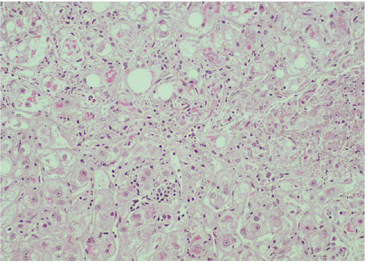
Fig. Perivenular hepatocytes containing Mallory hyaline with neutrophilic reaction (H&E x200). - Yamada's Textbook of Gastroenterology
2. Mallory Hyaline - Primary Biliary Cholangitis (H&E, x200)
Mallory hyaline is not exclusive to alcohol. Here it appears in periportal hepatocytes in a patient with primary biliary cholangitis, alongside steatosis:
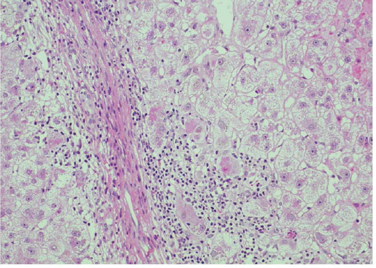
Fig. Periportal hepatocyte containing Mallory hyaline in primary biliary cholangitis (H&E x200). - Yamada's Textbook of Gastroenterology
3. Arteriolar Hyalinosis - Diabetic Kidney Disease (Jones Silver Stain)
This is exracellular hyaline. The three-panel image shows classic exudative lesions of diabetic kidney disease using Jones methenamine silver stain:
- Panel A - Concomitant hyalinosis of both afferent AND efferent arterioles (arrows) - characteristic of DKD (unlike hypertension, which affects only afferent)
- Panel B - Fibrin/hyaline caps in glomerular capillaries (wavy arrows)
- Panel C - Capsular drop (asterisk) - hyaline under parietal epithelium of Bowman's capsule
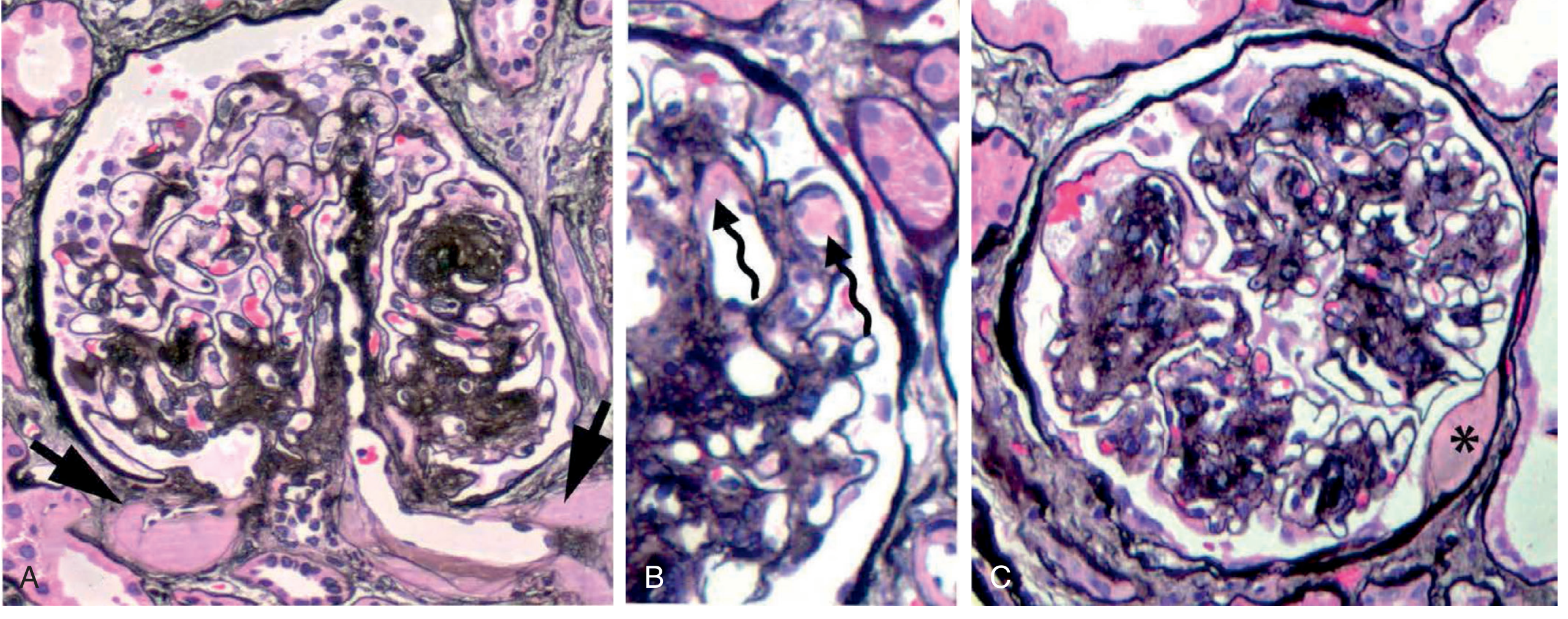
Fig. 39.9 Exudative lesions of diabetic kidney disease - Brenner and Rector's The Kidney
Key H&E Microscopy Features to Recognize
| Feature | Appearance |
|---|---|
| Color | Bright pink/eosinophilic - homogeneous, glassy |
| Texture | Structureless, no cellular detail |
| Mallory bodies | Rope-like or web-like intracytoplasmic inclusions |
| Arteriolar hyalinosis | Vessel wall thickened, lumen narrowed, smooth muscle replaced by pink glassy material |
| Russell bodies | Round, glassy pink inclusions in plasma cells |
Clinical Correlates Summary
| Condition | Type of Hyaline | Key Histological Feature |
|---|---|---|
| Alcoholic hepatitis | Mallory hyaline (intracellular) | Ballooned hepatocytes + neutrophils |
| NASH / metabolic steatohepatitis | Mallory hyaline | Ballooned hepatocytes (zone 3) |
| Hypertension (benign) | Arteriolar hyalinosis | Afferent arteriole only |
| Diabetes mellitus | Arteriolar hyalinosis | Both afferent AND efferent arterioles |
| Nephrotic syndrome | Protein reabsorption droplets | Proximal tubule pink granules |
| Chronic inflammation | Russell bodies | Plasma cells with pink globular inclusions |
| Old scar / infarct | Hyalinized collagen | Acellular pink fibrous tissue |
Key exam point: Concomitant efferent + afferent arteriolar hyalinosis is pathognomonic of diabetic nephropathy. Benign hypertensive nephrosclerosis only affects the afferent arteriole. The biochemical basis of extracellular collagen hyalinization remains incompletely understood, but arteriolar hyalinosis results from extravasated plasma proteins and deposited basement membrane material.
This is a shared conversation. Sign in to Orris to start your own chat.