
Ultrasound padhkar samjhne ka sabse asan tarika
Ultrasound seekhne ka sabse aasan tarika hai - pehle basics samjho, phir systematically practice karo. Main abhi medical library aur resources check karta hoon.
Searching sources...
Reading File
Searching the Web
ultrasound basics easy learning guide Hindi medical students
Finding Sources
Finding Sources
Finding Sources
Reading File
Finding Sources
Reading File
Loading Image
Finding Sources
Reading File
Ab mujhe poori picture mil gayi hai. Bahut accha material hai - ab ek comprehensive guide likhta hoon Hindi mein.
Ultrasound Padhna - Sabse Aasan Tarika 🔊
1. Pehle Samjho - Ultrasound Kaam Kaise Karta Hai?
Probe ek transducer hoti hai jo sound waves emit karti hai. Ye waves tissue se guzarti hain aur wapas aati hain. Jo wave wapas aati hai uska signal monitor pe brightness ke roop mein dikhta hai.
Simple rule: Jitna dense tissue, utna zyada reflection, utna zyada white
2. 3 Sabse Zaroori Words - Image Padhne Ka Aadhar
Ye 3 terms yaad kar lo, aur ultrasound ki 80% report samajh aayegi:
| Term | Screen pe colour | Example |
|---|---|---|
| Anechoic (An-echo) | Bilkul kala (Black) | Pani, bile, cyst, urine in bladder |
| Hypoechoic | Dhundla grey (Dark grey) | Muscles, fluid-filled structures |
| Hyperechoic | Chamkila white (Bright white) | Bone, gallstone, fat, gas |
| Isoechoic | Aas-paas ke tissue jaisa | Normal liver vs spleen comparison |
Trick yaad karne ka:
- "Hypo" = kam = dark grey
- "Hyper" = zyada = bright white
- "An" = bilkul nahi = pure kala
3. Probes (Transducers) - Kaunsa Kab Use Hota Hai?
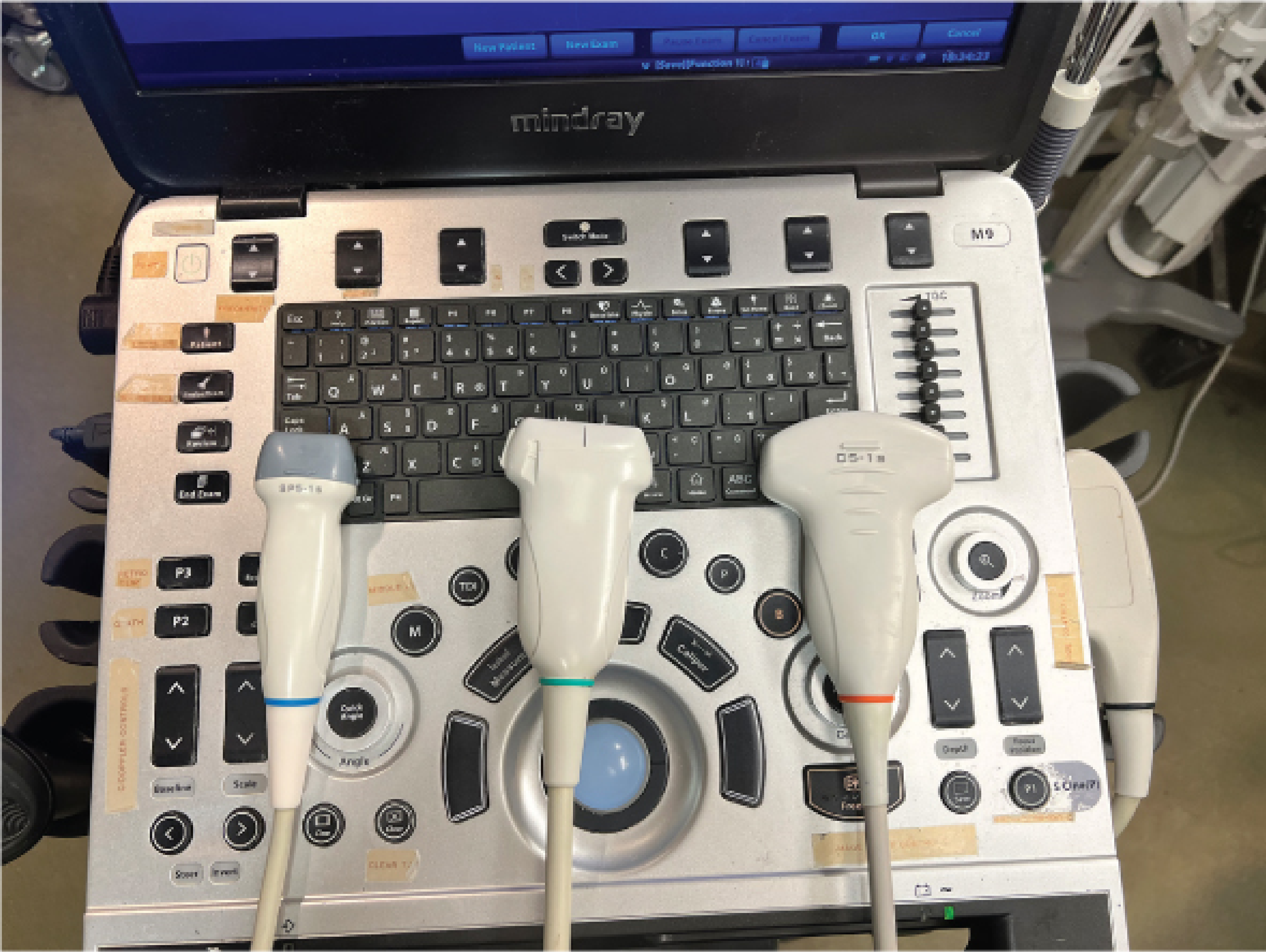
| Probe | Frequency | Depth | Use kahan? |
|---|---|---|---|
| Curvilinear (Convex) | 3-5 MHz | Deep | Abdomen, pelvis, kidney, liver |
| Phased Array (Cardiac) | 3-5 MHz | Deep | Heart (ribs ke beech fit ho jati) |
| Linear (Vascular) | 7-12 MHz | Shallow (<4 cm) | Veins, nerves, superficial structures |
Rule of thumb:
- Deep dekha = Low frequency probe (3-5 MHz)
- Shallow / superficial dekha = High frequency probe (7-12 MHz)
- Frequency aur depth ka ulta relation hai - high frequency = acchi resolution lekin kam depth
4. 2 Important Artifacts - Inhe Pehchanna Zaroori Hai
Posterior Acoustic Shadowing
- Gallstone ya bone ke peeche kala area dikhta hai
- Kyun? Kyunki hard structure ne saari waves absorb/reflect kar li, peeche kuch nahi pahuncha
- Gallstone ki pehchan isi se hoti hai
Posterior Acoustic Enhancement
- Cyst ya fluid ke peeche zyada bright area dikhta hai
- Kyun? Fluid ne waves ko attentuate nahi kiya, to peeche zyada signal aaya
- Simple cyst ka hallmark hai
5. Step-by-Step Learning Plan
Phase 1 - Theory (1-2 hafte)
- Physics samjho (waves, reflection, impedance)
- Probe types yaad karo
- Echoicity terms seekho (anechoic, hypo, hyper)
Phase 2 - Normal Anatomy (2-4 hafte)
Organs ka normal appearance pehchano:
- Liver - uniform medium-grey (isoechoic)
- Kidney - cortex + medulla alag alag
- Gallbladder - anechoic (kali)
- Bladder - anechoic (kali)
- Aorta - pulsating tubular structure
Phase 3 - Common Pathologies
- Gallstones (hyperechoic + shadowing)
- Kidney stones (hyperechoic + shadowing)
- Cysts (anechoic + enhancement)
- Ascites (free fluid - anechoic)
- Pleural effusion
6. Best Free Resources (Practice ke liye)
YouTube Videos:
https://www.youtube.com/watch?v=4kWZcIYA1YI
Online Resources:
- Geeky Medics Ultrasound Guide - free, clear diagrams
- POCUS 101 - detailed knobology + probes
7. Sabse Bada Tip - "WIFA Rule"
Ultrasound report padhte waqt hamesha 4 cheezein dekho:
- W - What structure hai? (Organ identify karo)
- I - Is it isoechoic/hypo/hyper? (Texture)
- F - Fluid hai? Anechoic area hai? (Fluid check)
- A - Any artifact? (Shadow ya enhancement)
Summary Table - Quick Revision
| Kya Dikhta Hai | Matlab |
|---|---|
| Kala area (no echo) | Fluid / cyst / bile |
| Bright white spot | Stone / bone / gas / fat |
| Dark grey | Muscles / liver disease |
| White spot + kala shadow neeche | Gallstone / kidney stone |
| Kala area + extra bright neeche | Simple cyst (benign) |
Source: Fischer's Mastery of Surgery (8th ed.), Yamada's Textbook of Gastroenterology (7th ed.) - Physics of Ultrasound chapters
This is a shared conversation. Sign in to Orris to start your own chat.