
surgical anatomy of breast Surgery exam.
Reading File
Now let me search the medical library for surgical anatomy of the breast.
Finding Sources
Finding Sources
Excellent - found the dedicated section. Let me read it along with the Fischer's Mastery surgical anatomy content in parallel.
Reading File
Reading File
Let me continue reading the Mulholland section and get the Fischer's section on surgical anatomy of the breast.
Reading File
Finding Sources
Reading File
Reading File
Now let me fetch the key anatomical images and also read the arterial supply and nerve sections from Mulholland.
Loading Image
Loading Image
Loading Image
Now let me read the arterial/venous supply and nerve sections from Mulholland, and also fetch the Fischer's lymphatic/blood supply diagram.
Reading File
Loading Image
Now let me read the arterial supply and nerve content, and also get the Fischer's lymphatic/blood supply figure.
Reading File
Finding Sources
I now have comprehensive content from two authoritative surgical textbooks. Let me get the arterial supply paragraph from Mulholland that was truncated, and also the nerves of the axilla content (for surgical importance).
Reading File
Finding Sources
Finding Sources
I now have all the content needed for a comprehensive answer. Let me compile it.
Surgical Anatomy of the Breast
Sources: Mulholland & Greenfield's Surgery 7e (Ch. 74); Fischer's Mastery of Surgery 8e (Ch. 51); Current Surgical Therapy 14e
1. Surface Boundaries
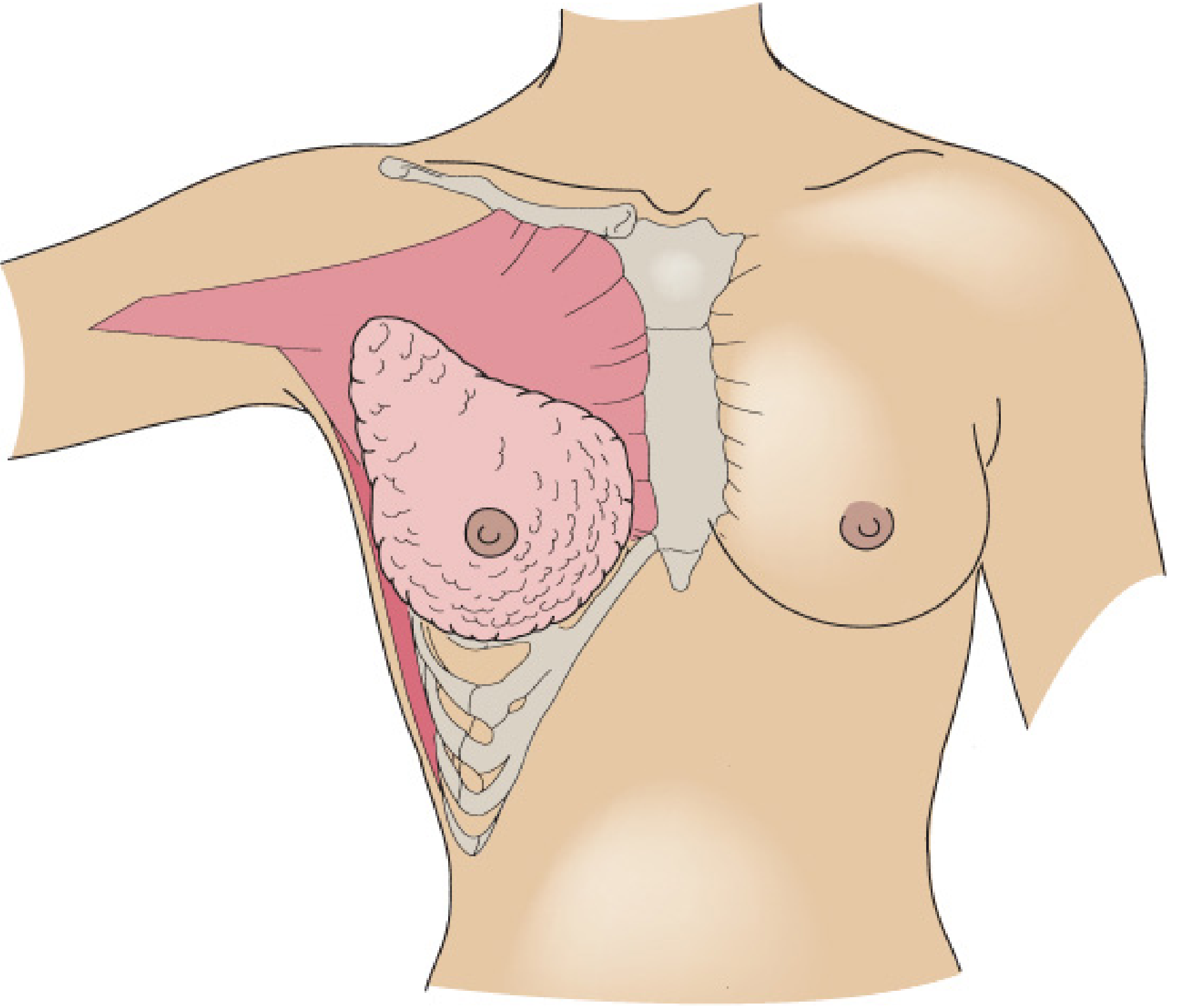
The adult breast occupies the anterior thoracic wall within the superficial fascia. Its boundaries are:
| Border | Landmark |
|---|---|
| Superior | 2nd rib |
| Inferior | 6th rib |
| Medial | Lateral sternal edge |
| Lateral | Midaxillary line |
Breast tissue also extends superolaterally into the axilla as the Axillary Tail of Spence, making the upper outer quadrant the largest quadrant and the most common site of both benign and malignant disease.
2. Layers and Deep Relations
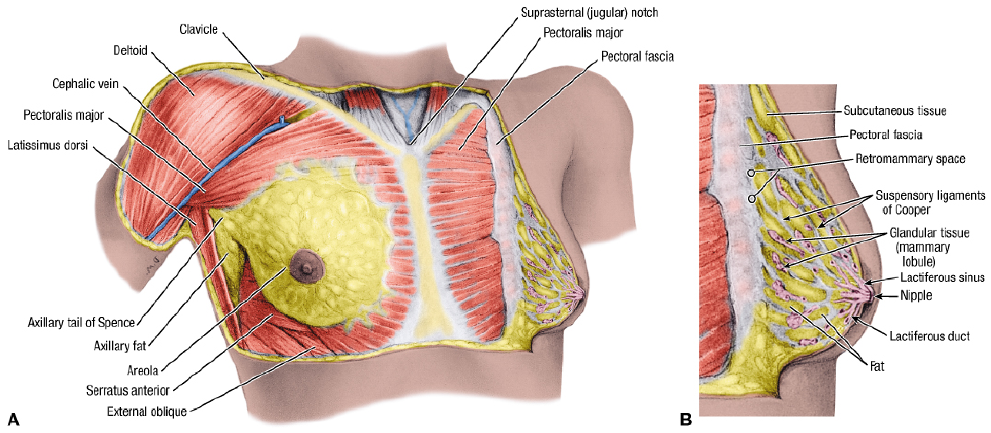
Posteriorly, the breast lies on the deep investing fasciae of:
- Pectoralis major (upper/medial)
- Serratus anterior (inferolateral)
- External abdominal oblique (inferior)
- Upper rectus sheath (inferomedial)
Between the breast and the pectoral fascia lies the retromammary (submammary) space - a relatively avascular plane used in mastectomy and augmentation. The breast is entirely contained within the superficial fascia, which provides an easy dissection plane.
3. Internal Structure
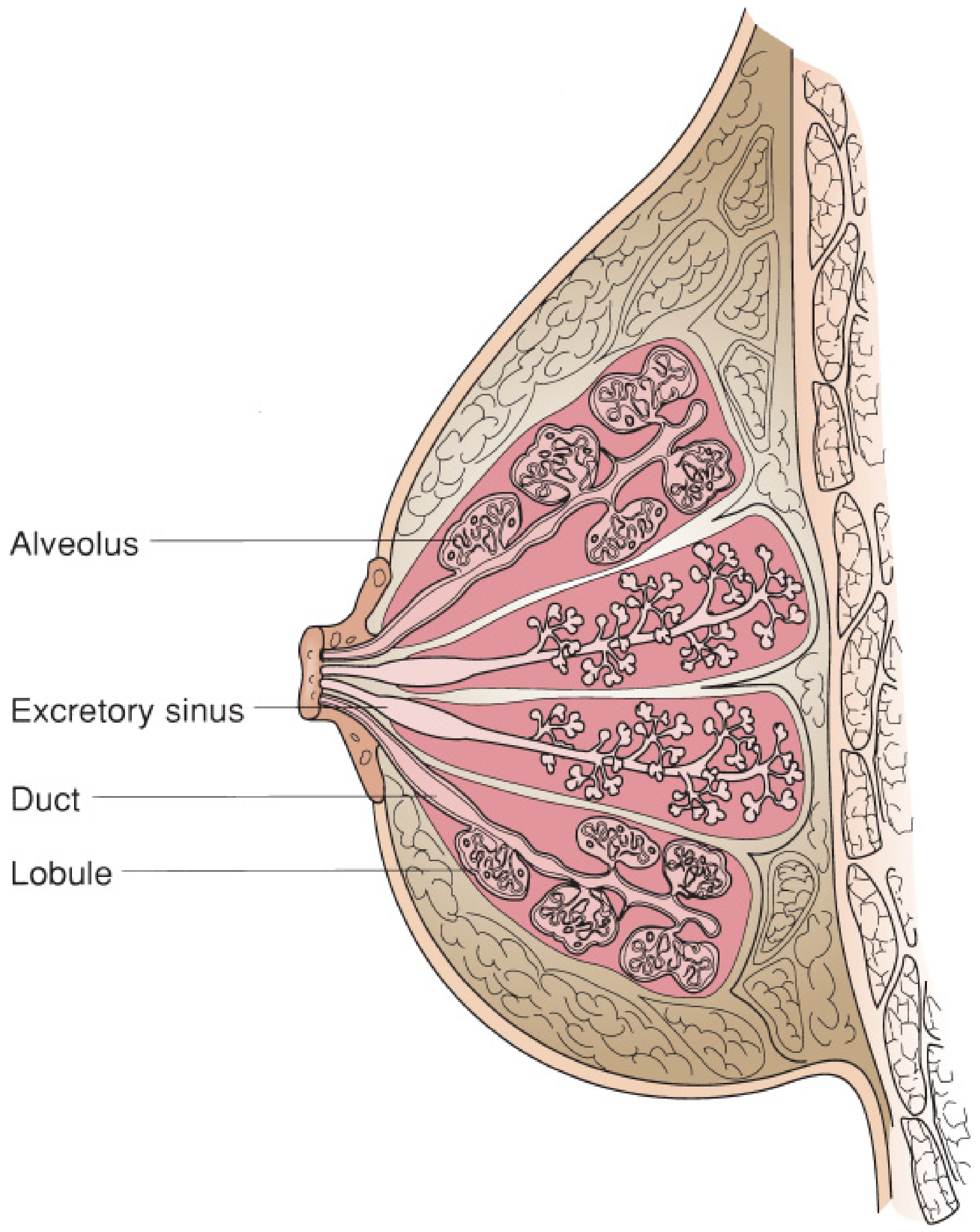
The breast parenchyma consists of:
- 15-20 lobes of glandular tissue, separated by connective tissue and fat
- Each lobe is composed of 20-40 lobules
- Each lobule has 10-100 alveoli
- Each lobe terminates in a lactiferous duct → dilates into a lactiferous sinus → opens at the nipple via a constricted orifice
- Glandular tissue is located more centrally; fat is more peripheral
Suspensory Ligaments of Cooper: Fibrous bands running from the deep fascia to the dermis - they provide support and mobility. More pronounced superiorly than inferiorly. Their tethering to skin causes skin dimpling in breast carcinoma.
Average weight: 150-225 g (nonlactating); can exceed 500 g when lactating. Left breast is commonly slightly larger than the right.
4. Nipple-Areolar Complex
- Skin is highly pigmented stratified squamous epithelium
- Deep to the nipple and areola are bundles of smooth muscle arranged radially, circumferentially, and longitudinally (allow erection of the nipple)
- Montgomery glands (modified sebaceous glands) on the areolar margin open as nodular elevations called Tubercles of Morgagni
- Nipple contains free sensory nerve endings + Meissner corpuscles
- Areola contains Ruffini-like endings + Krause end-bulbs
5. Blood Supply
Arterial Supply
Three main sources (principal supply enters superolaterally and superomedially):
| Source | Branches |
|---|---|
| Internal mammary (thoracic) artery | 2nd, 3rd, 4th perforating branches (2nd perforator is the largest) |
| Axillary artery | Superior thoracic artery; lateral thoracic artery; pectoral branches of thoracoacromial artery |
| Posterior intercostal arteries | 2nd, 3rd, 4th lateral branches |
The plane deep to the breast is typically avascular - key for dissection.
Venous Drainage
Parallels the arterial supply:
- Internal mammary veins
- Posterior intercostal veins
- Axillary vein tributaries
(Batson's plexus - the valveless vertebral venous plexus - provides a route for haematogenous spread to spine in breast cancer)
6. Lymphatic Drainage
Lymphatic flow is unidirectional through thin-walled, valveless vessels. Three interconnected plexuses:
- Interlobular plexus - primary, within the gland along lactiferous ducts
- Subareolar plexus (of Sappey) - drains skin, nipple/areola, and central gland
- Deep (posterior) plexus - on the deep surface of breast, communicates with fascia
Drainage Routes
| Route | % |
|---|---|
| Axillary lymph nodes | >75% |
| Internal mammary (parasternal) nodes | ~25% |
| Occasional: subclavicular, subperitoneal, inferior phrenic, rectus sheath | minority |
Key point: The flow to axillary vs. parasternal nodes is independent of the quadrant of origin.
7. Axillary Lymph Nodes - Levels
The axilla is a pyramidal space with:
- Anterior wall: Pectoralis major, pectoralis minor, pectoral fascia
- Posterior wall: Subscapularis, teres major, latissimus dorsi
- Medial wall: Serratus anterior + upper 4-5 ribs
- Lateral wall: Humerus (bicipital groove)
- Apex: Cervicoaxillary canal (bounded by clavicle anteriorly, scapula posteriorly, 1st rib medially)
- Base: Axillary fascia + skin
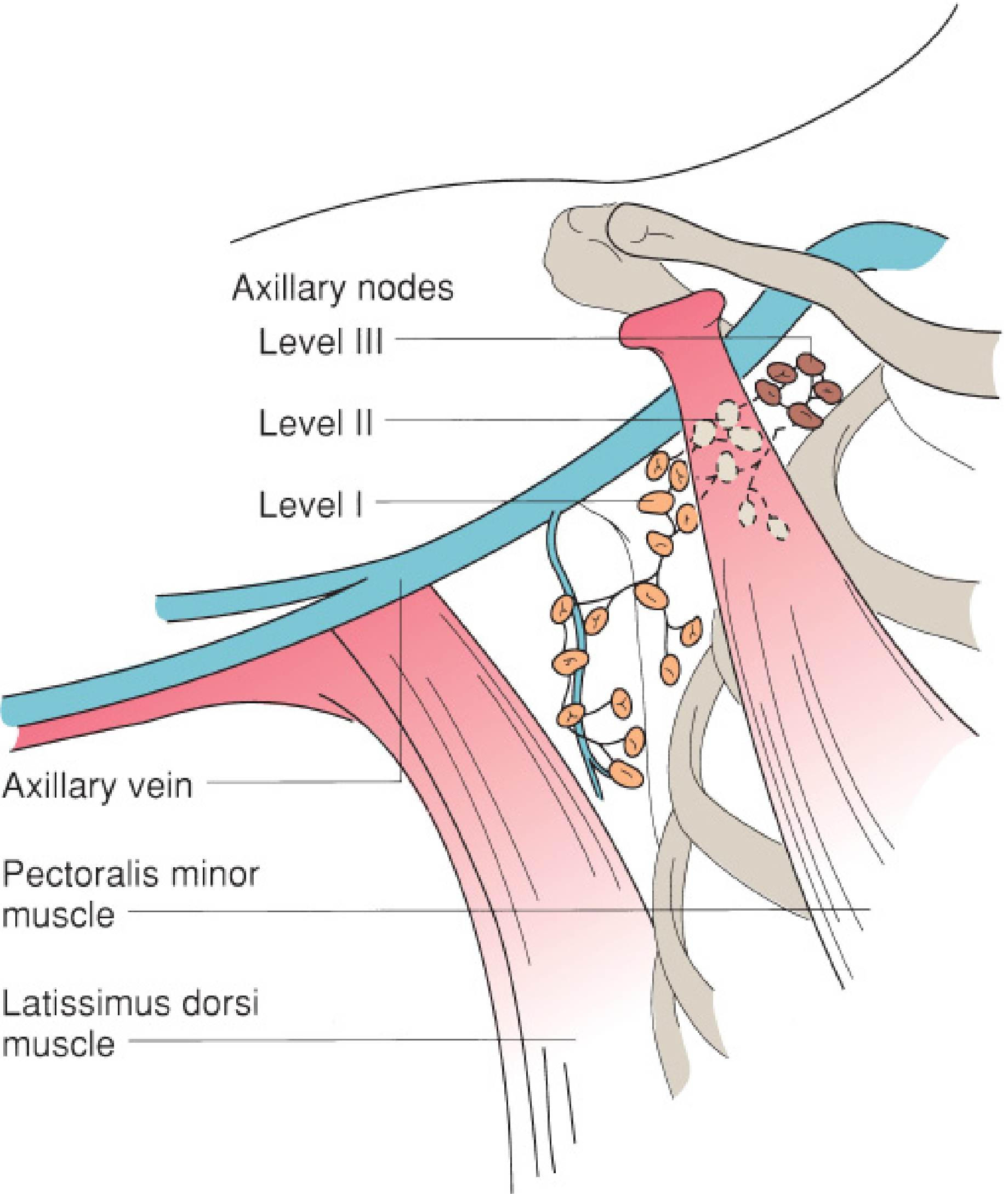
Level Classification (relative to pectoralis minor)
| Level | Location | Node Groups |
|---|---|---|
| Level I | Inferior & lateral to pec. minor | External mammary (anterior/pectoral), axillary vein, subscapular (scapular) nodes |
| Level II | Behind pec. minor | Central nodes, some subclavicular nodes |
| Level III | Medial to pec. minor | Subclavicular (apical/infraclavicular) nodes |
Rotter's (Interpectoral) Nodes: Located between pectoralis major and minor along the lateral pectoral nerve - surgically important in modified radical mastectomy.
Internal mammary nodes: Located in the 1st-6th intercostal spaces within 3 cm of the sternum.
Supraclavicular nodes: Contiguous with apex of axilla - their involvement indicates advanced disease (N3c in TNM staging).
8. Nerves - Surgically Important
During axillary surgery, the following nerves must be identified and preserved:
| Nerve | Origin | Action | If Damaged |
|---|---|---|---|
| Long thoracic nerve (of Bell) | C5, C6, C7 | Serratus anterior | Winged scapula |
| Thoracodorsal nerve | Posterior cord | Latissimus dorsi | Weakness of internal rotation/extension of arm |
| Medial pectoral nerve | Medial cord | Pectoralis major (lower) & minor | Chest wall deformity |
| Lateral pectoral nerve | Lateral cord | Pectoralis major (upper) | Atrophy of pec major upper head |
| Intercostobrachial nerve | Lateral cutaneous branch of T2 | Sensation - medial upper arm | Numbness/dysesthesia of inner arm (most commonly sacrificed) |
The boundaries of axillary lymph node dissection (ALND):
- Superior: Axillary vein
- Medial: Serratus anterior muscle + long thoracic nerve
- Lateral: Latissimus dorsi muscle + thoracodorsal nerve/bundle
9. Sensory Innervation of the Breast
- Main supply: lateral and anterior cutaneous branches of the 2nd through 6th intercostal nerves
- Small region of upper breast skin: anterior/medial branches of the supraclavicular nerve (from cervical plexus)
- The 4th intercostal nerve (lateral cutaneous branch) provides the most important innervation to the nipple-areolar complex - at risk in reconstructive and cosmetic procedures
10. Surgical Significance Summary
| Structure | Clinical Relevance |
|---|---|
| Suspensory ligaments of Cooper | Skin dimpling in carcinoma; traction dimple sign |
| Axillary tail of Spence | Must be included in mastectomy; site of missed tumors |
| Retromammary space | Avascular plane for mastectomy and implant placement |
| Subareolar plexus | Basis of sentinel lymph node biopsy (SLNB) |
| Level I-III nodes | Staging; axillary dissection extent |
| Long thoracic nerve | Winged scapula if damaged during ALND |
| Thoracodorsal nerve + vessels | Pedicle for latissimus dorsi flap reconstruction |
| Internal mammary nodes | Important in medial quadrant tumors; target in radiation |
| Rotter's nodes | Must clear in complete ALND |
Mulholland & Greenfield's Surgery, Scientific Principles and Practice - 7e, p. 3866-3870 (Ch. 74) | Fischer's Mastery of Surgery 8e, Ch. 51 (Mastery Keys: Surgical Anatomy of Breast)
This is a shared conversation. Sign in to Orris to start your own chat.