# TRANSITION STAGE OF CARBOHYDRATE METABOLISM: OXIDATIVE DECARBOXYLATION OF PYRUVATE
## THE BRIDGE BETWEEN GLYCOLYSIS AND THE TCA CYCLE
---
## 1. OVERVIEW AND SIGNIFICANCE
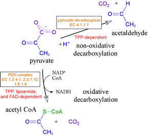
The transition stage (also called the **pyruvate dehydrogenase reaction** or the **link reaction**) represents the critical irreversible step that connects glycolysis (occurring in the cytoplasm) to the Krebs cycle (occurring in the mitochondrial matrix). This reaction involves the **oxidative decarboxylation of pyruvate to acetyl-CoA**.
### The Overall Reaction:
**Pyruvate + CoA-SH + NAD⁺ → Acetyl-CoA + CO₂ + NADH + H⁺**
- **ΔG°' = −33.4 kJ/mol (−8.0 kcal/mol)** — This makes the reaction **thermodynamically irreversible** under physiological conditions.
- This is why **fatty acids (which yield acetyl-CoA) cannot be converted back to glucose** in animals — there is no mechanism to reverse this step.
### Key Points:
- **Location:** Mitochondrial matrix
- **Substrate:** Pyruvate (3-carbon)
- **Product:** Acetyl-CoA (2-carbon acetyl group attached to Coenzyme A) + 1 CO₂
- Since glycolysis produces **2 pyruvate molecules per glucose**, this reaction occurs **twice per glucose molecule**
- Produces **2 NADH per glucose** (yielding ~5 ATP via oxidative phosphorylation if using the malate-aspartate shuttle)
---
## 2. TRANSPORT OF PYRUVATE INTO MITOCHONDRIA
Before the PDH reaction can occur, pyruvate must cross the mitochondrial membranes:
### Outer Mitochondrial Membrane:
- Freely permeable to pyruvate through **porins (voltage-dependent anion channels, VDACs)**
### Inner Mitochondrial Membrane:
- Pyruvate is transported by the **Mitochondrial Pyruvate Carrier (MPC)**, also known as the **pyruvate translocase**
- This is a **symporter** — it co-transports pyruvate with a **proton (H⁺)** (electroneutral symport)
- MPC consists of two subunits: **MPC1** and **MPC2**, which form a heterodimeric complex
- The driving force is the **proton gradient** generated by the electron transport chain
### 🏥 CLINICAL CORRELATION: MPC Deficiency
- Mutations in MPC1 or MPC2 cause impaired pyruvate transport
- Results in **lactic acidosis** because pyruvate accumulates in the cytoplasm and is converted to lactate
- Presents with neurological dysfunction, developmental delay
- Some studies link MPC dysfunction to cancer metabolism (Warburg effect) — cancer cells often downregulate MPC to maintain glycolytic flux
---
## 3. THE PYRUVATE DEHYDROGENASE COMPLEX (PDC/PDH COMPLEX)
### 3.1 General Structure
The PDH complex is one of the **largest known multienzyme complexes** in the cell:
- **Molecular weight:** ~7–10 million Daltons (in eukaryotes)
- **Diameter:** ~50 nm — visible under electron microscopy
- Located in the **mitochondrial matrix**
- Organized as a **multienzyme complex** for efficient substrate channeling
The complex contains **three catalytic enzymes**, **five coenzymes**, and **two regulatory enzymes**.
---
### 3.2 The Three Component Enzymes
| Component | Enzyme Name | EC Number | Abbreviation | Number of Subunits (in E. coli / Mammals) |
|-----------|------------|-----------|-------------|------------------------------------------|
| **E1** | Pyruvate Dehydrogenase (Pyruvate Decarboxylase) | EC 1.2.4.1 | PDH | 24 (E. coli) / 30 (mammals) |
| **E2** | Dihydrolipoyl Transacetylase | EC 2.3.1.12 | DLAT | 24 (both) |
| **E3** | Dihydrolipoyl Dehydrogenase | EC 1.8.1.4 | DLD | 12 (E. coli) / 12 (mammals) |
#### Architectural Organization:
- **E2 forms the structural core** of the complex
- In **E. coli**: E2 has **24 subunits** arranged in a **cube** (octahedral symmetry)
- In **mammals**: E2 has **60 subunits** arranged in a **pentagonal dodecahedron** (icosahedral symmetry — like a soccer ball)
- **E1** and **E3** are attached to the outer surface of the E2 core
- In mammals, an additional protein **E3-binding protein (E3BP or protein X)** anchors E3 to the E2 core
#### E2 Subunit — The Most Complex:
Each E2 subunit has multiple domains connected by flexible linkers:
1. **Lipoyl domain(s):**
- 1 lipoyl domain in *E. coli*
- **2 lipoyl domains** in mammals (L1 and L2 — though some sources report 1-3 depending on species)
- Each domain carries a **lipoamide** prosthetic group covalently attached to a specific **lysine** residue
- The lipoyl-lysine arm acts as a **swinging arm** (~14 Å long) that carries intermediates between the three active sites
2. **E1-binding domain (peripheral subunit-binding domain)**
3. **E3-binding domain** (via E3BP in mammals)
4. **Catalytic (acyltransferase) domain** at the C-terminus
---
### 3.3 The Five Coenzymes (Cofactors)
This is classically remembered by the mnemonic: **"TLC For Nerds"** or **"Tender Loving Care For Nancy"**
| Coenzyme | Vitamin Precursor | Associated Enzyme | Role | Tightly/Loosely Bound |
|----------|------------------|-------------------|------|----------------------|
| **Thiamine Pyrophosphate (TPP)** | Vitamin B₁ (Thiamine) | E1 | Decarboxylation of pyruvate | Tightly bound (prosthetic group) |
| **Lipoamide (Lipoic acid)** | Lipoic acid (not a classic vitamin) | E2 | Accepts acetyl group, transfer agent | Covalently bound (prosthetic group) |
| **Coenzyme A (CoA-SH)** | Vitamin B₅ (Pantothenic acid) | E2 | Accepts acetyl group to form acetyl-CoA | Substrate (loosely bound) |
| **FAD (Flavin Adenine Dinucleotide)** | Vitamin B₂ (Riboflavin) | E3 | Reoxidation of dihydrolipoamide | Tightly bound (prosthetic group) |
| **NAD⁺ (Nicotinamide Adenine Dinucleotide)** | Vitamin B₃ (Niacin) | E3 | Final electron acceptor | Substrate (loosely bound) |
#### Important Notes on Coenzymes:
- **Three are prosthetic groups** (TPP, lipoamide, FAD) — remain permanently attached
- **Two are co-substrates** (CoA-SH, NAD⁺) — bind, participate, and leave as products
- **Lipoic acid** is unique — it is attached via an **amide bond** to the **ε-amino group of a lysine residue** on E2, forming **lipoamide** (also called lipoyllysine)
- The lipoyl-lysine arm can extend ~14 Å, allowing it to swing between active sites (**substrate channeling**)
### 🏥 CLINICAL CORRELATION: B-Vitamin Deficiencies
**Thiamine (B₁) Deficiency:**
- **Beriberi** (wet — cardiac; dry — neurological)
- **Wernicke-Korsakoff Syndrome** — seen in chronic alcoholics
- Wernicke's encephalopathy: Confusion, ataxia, ophthalmoplegia (classic triad)
- Korsakoff's psychosis: Anterograde amnesia, confabulation
- Mechanism: Without TPP, PDH cannot function → impaired aerobic metabolism → reliance on anaerobic glycolysis → lactic acidosis; neurons are particularly vulnerable
- **Pyruvate and lactate levels increase** in blood
- Treatment: **IV thiamine** (must be given BEFORE glucose to avoid worsening)
**Riboflavin (B₂) Deficiency:**
- Ariboflavinosis: cheilosis, glossitis, angular stomatitis, corneal vascularization
- Affects multiple FAD-dependent enzymes including PDH complex
**Niacin (B₃) Deficiency:**
- **Pellagra:** 4 D's — Dermatitis, Diarrhea, Dementia, Death
- Affects NAD⁺-dependent reactions
**Pantothenic Acid (B₅) Deficiency:**
- Rare (pantothenic acid is "everywhere" — from Greek *pantos*)
- Dermatitis, enteritis, alopecia, adrenal insufficiency
- "Burning feet syndrome"
**Lipoic Acid Deficiency:**
- Not a classic vitamin deficiency in humans
- Used as a supplement (α-lipoic acid) — acts as an antioxidant
- Mutations in lipoic acid synthesis (LIAS gene) cause neonatal epilepsy and lactic acidosis
---
## 4. THE REACTION MECHANISM — STEP BY STEP
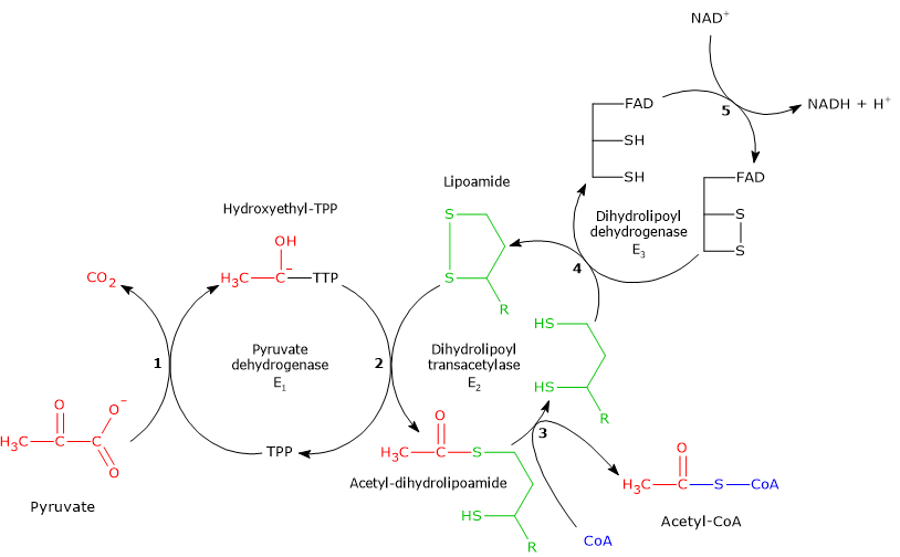
The five-step mechanism involves all three enzymes and all five coenzymes:
---
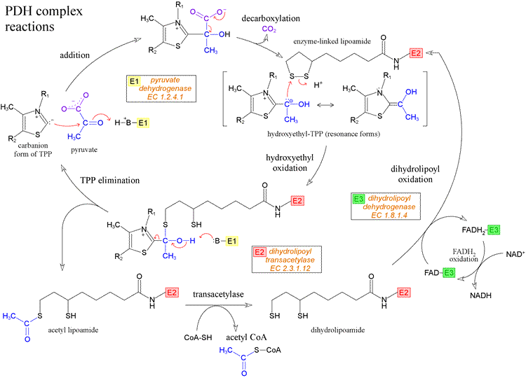
### STEP 1: Decarboxylation (by E1 — Pyruvate Dehydrogenase)
**Pyruvate + TPP → Hydroxyethyl-TPP (HETPP) + CO₂**
- Pyruvate binds to the **thiazolium ring** of TPP
- The **C-2 carbanion** of the thiazolium ring attacks the **carbonyl carbon (C-2)** of pyruvate
- This forms a covalent adduct: **lactyl-TPP**
- **Decarboxylation** occurs — CO₂ is released (this is the carbon from the carboxyl group, i.e., C-1 of pyruvate)
- The remaining 2-carbon fragment stays bound to TPP as **hydroxyethyl-TPP (HETPP)** (also called α-hydroxyethylidene-TPP or "active acetaldehyde")
**This is the rate-limiting step of the entire PDH complex**
Key Details:
- TPP requires **Mg²⁺** as a cofactor for binding to E1
- The reaction is analogous to the mechanism in other TPP-dependent enzymes (transketolase, α-ketoglutarate dehydrogenase, branched-chain α-keto acid dehydrogenase)
---
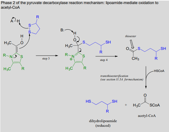
### STEP 2: Oxidative Transfer (by E1 and E2)
**Hydroxyethyl-TPP + Lipoamide (oxidized) → Acetyl-lipoamide + TPP**
- The hydroxyethyl group on TPP is **oxidized** and simultaneously **transferred** to the **lipoamide** on E2
- The disulfide bond (S-S) of lipoamide is **reductively cleaved**
- The acetyl group attaches to one of the sulfur atoms of lipoamide via a **thioester bond**, forming **acetyl-dihydrolipoamide** (also called 6-S-acetyldihydrolipoamide)
- TPP is regenerated on E1
This is where the **oxidation** occurs — the hydroxyethyl group (aldehyde oxidation state) is oxidized to an acetyl group (carboxylic acid oxidation state), with the electrons reducing the disulfide bond of lipoamide.
The long, flexible **lipoyl-lysine arm** of E2 reaches into the active site of E1 to accept the acetyl group — this is the essence of **substrate channeling**.
---
### STEP 3: Transacetylation (by E2 — Dihydrolipoyl Transacetylase)
**Acetyl-dihydrolipoamide + CoA-SH → Acetyl-CoA + Dihydrolipoamide**
- The acetyl group is transferred from lipoamide to **Coenzyme A**
- A new **thioester bond** is formed (between the acetyl group and the -SH of CoA)
- This produces **acetyl-CoA** — the final product
- Lipoamide is left in its **reduced form (dihydrolipoamide)** with two free -SH groups
**Acetyl-CoA** is now released and enters the **TCA cycle** (or is used for fatty acid synthesis, ketone body synthesis, cholesterol synthesis, etc.)
The **thioester bond** in acetyl-CoA is a **high-energy bond** (ΔG°' of hydrolysis ≈ −31.4 kJ/mol), which preserves the free energy of oxidation for use in subsequent reactions.
---
### STEP 4: Regeneration of Lipoamide (by E3 — Dihydrolipoyl Dehydrogenase)
**Dihydrolipoamide + FAD → Lipoamide (oxidized) + FADH₂**
- E3 **reoxidizes** dihydrolipoamide back to its oxidized (disulfide) form
- The electrons are transferred to **FAD** (which is tightly bound to E3)
- FAD is reduced to **FADH₂**
- E3 contains a **reactive disulfide** in its active site (between two cysteine residues), which participates in the electron transfer
This step is essential because without regeneration of oxidized lipoamide, the complex would be stuck after one catalytic cycle.
---
### STEP 5: Regeneration of FAD (by E3)
**FADH₂ + NAD⁺ → FAD + NADH + H⁺**
- FADH₂ transfers its electrons to **NAD⁺**
- NAD⁺ is reduced to **NADH + H⁺**
- FAD is regenerated
This is the **final step**, and the **NADH** produced carries the electrons to the **electron transport chain (Complex I)**, where it generates **~2.5 ATP** per NADH (via oxidative phosphorylation).
**Note:** This is one of the few reactions where **electrons flow from FADH₂ to NAD⁺** (thermodynamically "uphill" in isolation, but driven by the overall favorable energetics of the complex). This is possible because the FAD in E3 has a **higher reduction potential** than free FAD, and the overall reaction is exergonic.
---
### Summary of the Five Steps:
| Step | Enzyme | Coenzyme | Reaction Type | What Happens |
|------|--------|----------|--------------|-------------|
| 1 | E1 | TPP | Decarboxylation | Pyruvate loses CO₂ → HETPP |
| 2 | E1→E2 | Lipoamide | Oxidative transfer | HETPP → acetyl group transferred to lipoamide |
| 3 | E2 | CoA-SH | Transacetylation | Acetyl-lipoamide + CoA → Acetyl-CoA |
| 4 | E3 | FAD | Oxidation | Dihydrolipoamide reoxidized; FAD → FADH₂ |
| 5 | E3 | NAD⁺ | Oxidation | FADH₂ reoxidized; NAD⁺ → NADH |
---
## 5. SUBSTRATE CHANNELING
The PDH complex is a masterpiece of **substrate channeling**:
- Intermediates never leave the complex — they are passed directly from one active site to the next
- The **lipoyl-lysine swinging arm** physically carries intermediates between E1, E2, and E3 active sites
- This provides several advantages:
1. **Increased reaction rate** — no diffusion delay
2. **Prevention of side reactions** — reactive intermediates are protected
3. **Coordinated regulation** — all steps can be regulated as a unit
4. **No dilution** of intermediates in the matrix
---
## 6. ENERGETICS
### Per pyruvate molecule:
- **1 CO₂** released
- **1 NADH** produced (~2.5 ATP via ETC)
- **1 Acetyl-CoA** produced (yields 10 ATP when completely oxidized in TCA + ETC)
### Per glucose molecule (2 pyruvates):
- **2 CO₂** released
- **2 NADH** produced (~5 ATP)
- **2 Acetyl-CoA** produced
---
## 7. REGULATION OF THE PDH COMPLEX
The PDH complex is one of the most elaborately regulated enzymes. It is regulated at **two levels**:
### 7.1 Allosteric Regulation (Product Inhibition)
The products of the reaction directly inhibit the complex:
| Inhibitors (Products) | Activators (Substrates) |
|----------------------|----------------------|
| **Acetyl-CoA** (inhibits E2) | **CoA-SH** |
| **NADH** (inhibits E3) | **NAD⁺** |
| **ATP** (signals energy sufficiency) | **AMP, ADP** |
- **Acetyl-CoA/CoA-SH ratio:** High ratio inhibits; low ratio activates
- **NADH/NAD⁺ ratio:** High ratio inhibits; low ratio activates
---
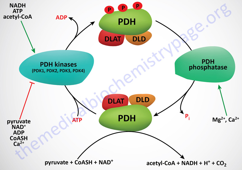
### 7.2 Covalent Modification (Phosphorylation/Dephosphorylation)
This is the **primary regulatory mechanism** for the PDH complex in mammals.
#### The Two Regulatory Enzymes:
| Enzyme | Full Name | Action | Effect on PDH |
|--------|-----------|--------|--------------|
| **PDH Kinase (PDK)** | Pyruvate Dehydrogenase Kinase | **Phosphorylates** E1 (on serine residues) | **INACTIVATES** PDH |
| **PDH Phosphatase (PDP)** | Pyruvate Dehydrogenase Phosphatase | **Dephosphorylates** E1 | **ACTIVATES** PDH |
#### Phosphorylation Details:
- PDK phosphorylates **3 specific serine residues** on the **E1α subunit**:
- **Site 1 (Ser-264):** Most important; phosphorylation here alone causes ~99% inactivation
- **Site 2 (Ser-271):** Additional inactivation
- **Site 3 (Ser-203):** Slowest to be phosphorylated
- **Only Site 1 phosphorylation is sufficient for complete inactivation**
- PDP removes phosphate groups to reactivate the complex
#### PDK Isoforms:
There are **4 isoforms** of PDK in humans (PDK1, PDK2, PDK3, PDK4):
- **PDK1:** Heart, skeletal muscle
- **PDK2:** Most widely distributed (all tissues)
- **PDK3:** Testis, kidney, brain
- **PDK4:** Heart, skeletal muscle — **upregulated during starvation, diabetes, and by glucocorticoids**
#### PDP Isoforms:
Two isoforms: **PDP1** and **PDP2**
- PDP1: Widely distributed; activated by **Ca²⁺**
- PDP2: Primarily in liver and adipose tissue; activated by **insulin** (via reduced phosphorylation of PDP2 itself)
---
#### Regulation of PDK (the kinase):
| **PDK Activators** (→ PDH Inactive) | **PDK Inhibitors** (→ PDH Active) |
|--------------------------------------|--------------------------------------|
| Acetyl-CoA | CoA-SH |
| NADH | NAD⁺ |
| ATP | ADP |
| Fatty acids (via acetyl-CoA, NADH) | **Pyruvate** (direct inhibitor of PDK) |
| Ketone bodies | |
| Starvation (↑PDK4 expression) | |
#### Regulation of PDP (the phosphatase):
| **PDP Activators** (→ PDH Active) | **PDP Inhibitors** (→ PDH Inactive) |
|--------------------------------------|--------------------------------------|
| **Ca²⁺** | NADH |
| **Mg²⁺** | |
| **Insulin** (especially in adipose tissue) | |
---
### 7.3 Tissue-Specific Regulation
#### In **Cardiac Muscle** during exercise:
- ↑Ca²⁺ from muscle contraction → activates PDP → activates PDH
- ↑ADP (energy demand) → inhibits PDK → keeps PDH active
- This ensures adequate acetyl-CoA production for the TCA cycle during increased energy demand
#### In **Liver** during fasting/starvation:
- ↑Fatty acid oxidation → ↑Acetyl-CoA, ↑NADH → activates PDK → inactivates PDH
- This **spares pyruvate for gluconeogenesis** (since pyruvate → oxaloacetate → glucose)
- PDK4 expression is **upregulated** by PPAR-α (activated by fatty acids) and glucocorticoids
#### In **Fed state/After a carbohydrate-rich meal:**
- **Insulin** activates PDP → activates PDH → promotes glucose oxidation
- ↑Pyruvate (from glycolysis) directly inhibits PDK → PDH stays active
#### In **Adipose tissue:**
- Insulin activates PDH → increases acetyl-CoA → promotes fatty acid synthesis (lipogenesis)
---
### 🏥 CLINICAL CORRELATIONS — REGULATION
**Dichloroacetate (DCA):**
- A structural analog of pyruvate
- **Inhibits PDK** → keeps PDH in the active (dephosphorylated) state
- Used experimentally to treat **lactic acidosis** (by activating PDH and promoting aerobic metabolism of pyruvate)
- Also investigated as an **anticancer agent** (shifts cancer cell metabolism from glycolysis to oxidative phosphorylation, promoting apoptosis)
---
## 8. CLINICAL CORRELATIONS — COMPREHENSIVE
---
### 8.1 PYRUVATE DEHYDROGENASE COMPLEX DEFICIENCY
This is the **most common defined cause of congenital lactic acidosis**.
#### Genetics:
- Most commonly due to mutations in the **PDHA1 gene** (encoding the **E1α subunit**)
- PDHA1 is located on the **X chromosome (Xp22.12)** — so it is **X-linked**
- Despite X-linkage, **both males and females** are affected:
- Males: More severely affected (hemizygous)
- Females: Variable severity due to **X-inactivation (lyonization)** — can range from severe to mild
- Less commonly, autosomal recessive mutations in:
- PDHB (E1β)
- DLAT (E2)
- DLD (E3) — also affects α-ketoglutarate dehydrogenase and branched-chain α-keto acid dehydrogenase (since they share E3)
- PDHX (E3-binding protein)
- Genes for PDK or PDP (very rare)
- Genes for lipoic acid synthesis (LIAS, LIPT1, LIPT2, BOLA3, NFU1)
#### Biochemistry:
- Impaired conversion of pyruvate to acetyl-CoA
- **Pyruvate accumulates** → converted to:
- **Lactate** (by LDH) → **Lactic acidosis**
- **Alanine** (by transamination) → **↑Alanine** in blood
- **Lactate/Pyruvate ratio** is typically **normal** (because the problem is not in the redox state; it's in PDH itself) — This distinguishes it from disorders of oxidative phosphorylation where L/P ratio is elevated
- Reduced acetyl-CoA → impaired TCA cycle → reduced ATP
- Brain is particularly affected because it relies heavily on glucose oxidation
#### Clinical Manifestations:
- **Severe neonatal form:**
- Congenital lactic acidosis
- Hypotonia
- Seizures
- Dysmorphic features (facial features similar to fetal alcohol syndrome)
- Agenesis of corpus callosum, structural brain abnormalities
- Often fatal in infancy
- **Moderate form (Leigh syndrome):**
- Subacute necrotizing encephalomyelopathy
- Progressive neurological deterioration
- Symmetric necrotic lesions in **basal ganglia, thalamus, brainstem**
- Psychomotor regression, dystonia, ataxia
- Characteristic MRI findings
- **Mild form:**
- Intermittent ataxia
- Peripheral neuropathy
- Exercise intolerance
#### Diagnosis:
- **↑Lactate, ↑pyruvate, ↑alanine** in blood and CSF
- Normal or low L/P ratio
- Enzyme assay on fibroblasts, lymphocytes, or muscle biopsy
- Genetic testing (PDHA1 mutation analysis)
#### Treatment:
- **Ketogenic diet** — provides acetyl-CoA via fatty acid oxidation, bypassing the PDH block
- High fat, low carbohydrate
- Acetyl-CoA is produced from β-oxidation without needing PDH
- **Thiamine supplementation** (high doses: 100-600 mg/day) — some mutations are "thiamine-responsive" (E1 has increased Km for TPP; high thiamine can partially overcome this)
- **Dichloroacetate (DCA)** — inhibits PDK, maximizes residual PDH activity
- Limited clinical benefit; causes peripheral neuropathy with chronic use
- **Avoid excessive carbohydrate intake** (would increase pyruvate → worsening lactic acidosis)
- **Citrate/bicarbonate** for acute acidosis management
- Prognosis is generally poor for severe forms
---
### 8.2 ARSENIC POISONING
#### Mechanism:
- **Arsenite (As³⁺)** binds to the **vicinal (adjacent) sulfhydryl groups** of **dihydrolipoamide** on E2
- Forms a stable **cyclic dithioarsine complex**
- This prevents the reoxidation of dihydrolipoamide → the complex is "stuck"
- Inhibits not only PDH but also **α-ketoglutarate dehydrogenase** (which uses the same mechanism)
- Also inhibits other lipoamide-containing enzyme complexes
#### Biochemical Consequences:
- Accumulation of pyruvate and α-ketoglutarate
- Lactic acidosis
- Impaired energy production (blocked TCA cycle)
- Also inhibits enzymes with essential sulfhydryl groups
#### Clinical Presentation:
- **Acute arsenic poisoning:**
- Garlic-like breath odor
- Severe GI symptoms (rice-water diarrhea, vomiting)
- Cardiovascular collapse (QT prolongation)
- Multi-organ failure
- Death
- **Chronic arsenic poisoning (arsenicosis):**
- Mees' lines (white transverse lines on nails)
- Skin hyperpigmentation and keratoses ("raindrops on a dusty road")
- Peripheral neuropathy
- Increased cancer risk (skin, bladder, lung)
#### Treatment:
- **Chelation therapy:**
- **Dimercaprol (BAL — British Anti-Lewisite)** — provides competing dithiol groups
- **Succimer (DMSA)** — oral chelation
- **Unithiol (DMPS)**
- These chelators have adjacent -SH groups that compete with lipoamide for arsenic binding
---
### 8.3 MERCURY AND LEAD POISONING
- **Mercury (organic mercury, Hg²⁺):** Also binds to sulfhydryl groups of lipoamide and other enzymes
- Can inhibit PDH complex
- Causes neurological damage (Minamata disease)
- **Lead (Pb²⁺):**
- Interferes with sulfhydryl-dependent enzymes
- Also inhibits PDH complex activity
- Primary toxicity is on heme synthesis (ALA dehydratase, ferrochelatase) and neurological function
---
### 8.4 LEIGH SYNDROME (Subacute Necrotizing Encephalomyelopathy)
- Can be caused by PDH complex deficiency (mutations in E1α, E2, E3BP)
- Also caused by defects in Complex I, Complex IV, Complex V, and other mitochondrial defects
- **Symmetric bilateral lesions** in basal ganglia and brainstem on MRI
- Progressive neurological decline
- Usually fatal in childhood
---
### 8.5 E3 (DIHYDROLIPOYL DEHYDROGENASE) DEFICIENCY
- **Unique** because E3 is shared among **three multienzyme complexes:**
1. **Pyruvate dehydrogenase complex**
2. **α-Ketoglutarate dehydrogenase complex** (TCA cycle)
3. **Branched-chain α-keto acid dehydrogenase complex** (BCKDH — branched-chain amino acid catabolism)
4. Also part of the **glycine cleavage system** (glycine degradation)
- Therefore, E3 deficiency causes a **combined deficiency** of all these complexes
#### Clinical Features:
- Lactic acidosis (PDH and α-KGDH deficiency)
- **Maple syrup urine disease (MSUD)**-like features (BCKDH deficiency → accumulation of branched-chain amino acids and α-keto acids → sweet-smelling urine)
- **Nonketotic hyperglycinemia** (glycine cleavage system deficiency)
- This combined picture is sometimes called **"combined lipoamide dehydrogenase deficiency"** or **"multiple α-keto acid dehydrogenase deficiency"**
- Very poor prognosis
---
### 8.6 THIAMINE DEFICIENCY AND PDH — DETAILED
#### Wernicke-Korsakoff Syndrome (expanded):
- **Most common in chronic alcoholism:**
- Alcohol directly inhibits intestinal thiamine absorption
- Poor nutrition in alcoholics
- Alcohol reduces hepatic storage of thiamine
- Alcohol increases thiamine utilization
- **Wernicke's Encephalopathy (acute):**
1. **Confusion** (global confusional state)
2. **Ophthalmoplegia** (lateral rectus palsy → CN VI; nystagmus)
3. **Ataxia** (cerebellar/vestibular dysfunction)
- Hemorrhagic lesions in **mammillary bodies, periventricular regions, medial thalamus**
- **EMERGENCY** — can progress to Korsakoff's if untreated
- **Korsakoff's Psychosis (chronic):**
1. **Anterograde amnesia** (cannot form new memories)
2. **Retrograde amnesia**
3. **Confabulation** (fabricating memories to fill gaps)
- Due to damage to **mammillary bodies** and **medial thalamic nuclei**
- Often irreversible
- **Beriberi:**
- **Wet beriberi:** High-output cardiac failure, edema, dilated cardiomyopathy
- **Dry beriberi:** Peripheral neuropathy (symmetrical, ascending), muscle wasting
- **Infantile beriberi:** In breastfed infants of thiamine-deficient mothers; cardiac failure, aphonia
- **Lactic acidosis** in thiamine deficiency:
- PDH inhibited → pyruvate cannot enter TCA cycle
- Pyruvate → lactate (via LDH)
- Brain especially vulnerable (high glucose dependence)
#### Why give thiamine BEFORE glucose in alcoholics:
- Administering glucose increases glycolysis → ↑pyruvate production
- Without functioning PDH (due to low thiamine), pyruvate → lactate
- Can precipitate or worsen Wernicke's encephalopathy
- **Always give thiamine first!** (or simultaneously with glucose)
---
### 8.7 PDH AND DIABETES MELLITUS
- In **Type 1 and uncontrolled Type 2 diabetes:**
- ↑Fatty acid oxidation → ↑Acetyl-CoA, ↑NADH
- These activate PDK → PDH is inactivated
- **PDK4 is strongly upregulated** (via PPAR-α activation by fatty acids, and glucocorticoid-related signaling)
- Pyruvate is diverted to **gluconeogenesis** → contributes to hyperglycemia
- This creates a vicious cycle in diabetes
- **Insulin** normally activates **PDP** → activates PDH
- In insulin-deficient/resistant states, PDH remains inactive
---
### 8.8 PDH AND CANCER (WARBURG EFFECT)
- Many cancer cells exhibit the **Warburg effect**: preferential use of glycolysis even in the presence of oxygen (aerobic glycolysis)
- Mechanisms involving PDH:
- **Upregulation of PDK1** (by HIF-1α — hypoxia-inducible factor) → inactivates PDH
- Pyruvate is diverted to lactate (by LDH-A, also upregulated)
- This maintains the glycolytic flux that cancer cells prefer for biosynthesis
- **DCA (dichloroacetate)** has been investigated as an anticancer agent:
- Inhibits PDK → activates PDH → shifts metabolism from glycolysis to OXPHOS
- Restores mitochondrial function → promotes apoptosis in cancer cells
- Clinical trials ongoing
---
### 8.9 PRIMARY BILIARY CHOLANGITIS (PBC) — formerly Primary Biliary Cirrhosis
- **Autoimmune disease** targeting the **E2 component (DLAT)** of PDH
- Patients develop **anti-mitochondrial antibodies (AMA)**, specifically **anti-PDC-E2 antibodies**
- AMA target the **lipoyl domain** of E2
- Found in **>90-95% of PBC patients** — highly sensitive and specific
- The E2 subunit is exposed on the surface of biliary epithelial cells (cholangiocytes), possibly after apoptosis or modification
- **Clinical Features:**
- Chronic progressive cholestatic liver disease
- Pruritus (itching — often the first symptom)
- Fatigue
- Jaundice (late feature)
- Hepatomegaly
- Xanthomas and xanthelasma (due to hypercholesterolemia)
- Eventually → cirrhosis, portal hypertension, liver failure
- **Diagnosis:**
- AMA positivity (anti-PDC-E2)
- Elevated alkaline phosphatase (ALP) and γ-GT
- Liver biopsy: granulomatous destruction of bile ducts ("florid duct lesion")
- **Treatment:**
- **Ursodeoxycholic acid (UDCA)** — first-line; improves bile flow
- **Obeticholic acid** — second-line
- Liver transplantation for end-stage disease
---
### 8.10 LIPOIC ACID SYNTHESIS DEFECTS
Mutations in genes involved in lipoic acid biosynthesis:
| Gene | Function | Clinical Features |
|------|----------|-------------------|
| **LIAS** | Lipoic acid synthase | Neonatal epilepsy, lactic acidosis, developmental delay |
| **LIPT1** | Lipoyltransferase 1 | Leigh-like syndrome, lactic acidosis |
| **LIPT2** | Lipoyltransferase 2 | Similar to above |
| **BOLA3** | Iron-sulfur cluster assembly | Lactic acidosis, hyperglycinemia, encephalopathy |
| **NFU1** | Iron-sulfur cluster biogenesis | Fatal encephalopathy, pulmonary hypertension |
These affect all lipoamide-dependent enzymes (PDH, α-KGDH, BCKDH, glycine cleavage system).
---
## 9. ANALOGOUS ENZYME COMPLEXES
The PDH complex mechanism is shared by two other α-keto acid dehydrogenase complexes:
| Complex | Substrate | Product | Location in Metabolism |
|---------|-----------|---------|----------------------|
| **Pyruvate dehydrogenase** | Pyruvate | Acetyl-CoA | Transition (glycolysis → TCA) |
| **α-Ketoglutarate dehydrogenase** | α-Ketoglutarate | Succinyl-CoA | TCA cycle |
| **Branched-chain α-keto acid dehydrogenase** | Branched-chain α-keto acids | Branched-chain acyl-CoAs | Amino acid catabolism |
All three:
- Have E1, E2, E3 structure
- Use the same 5 coenzymes (TPP, lipoamide, CoA, FAD, NAD⁺)
- **Share the same E3 subunit** (dihydrolipoyl dehydrogenase)
- Are regulated by phosphorylation/dephosphorylation (except α-KGDH in mammals)
- Are inhibited by arsenic (vicinal dithiol mechanism)
---
## 10. IRREVERSIBILITY AND METABOLIC SIGNIFICANCE
### Why is this reaction irreversible?
1. **Highly negative ΔG** (−33.4 kJ/mol) — thermodynamically very favorable
2. **CO₂ is released** — a gas that escapes/is rapidly removed
3. **NADH formation** — pulled forward by ETC
4. **Thioester bond** in acetyl-CoA is relatively stable
### Metabolic Consequence — The Irreversibility Barrier:
**Animals CANNOT convert fatty acids to glucose** because:
- Fatty acid β-oxidation → Acetyl-CoA
- Acetyl-CoA CANNOT be converted back to pyruvate (PDH is irreversible)
- Therefore, acetyl-CoA cannot be used for gluconeogenesis
- The 2 carbons of acetyl-CoA that enter the TCA cycle are lost as 2 CO₂
- (Exception: Plants and some microorganisms have the **glyoxylate cycle** which bypasses the CO₂-releasing steps of the TCA cycle, allowing net conversion of acetyl-CoA to oxaloacetate → glucose)
---
## 11. FATE OF ACETYL-CoA
Acetyl-CoA produced by PDH can enter multiple pathways:
1. **TCA cycle** (primary fate — energy production)
2. **Fatty acid synthesis** (lipogenesis — in liver, adipose, lactating breast)
3. **Cholesterol and steroid synthesis** (via HMG-CoA → mevalonate pathway)
4. **Ketone body synthesis** (in liver, during fasting/diabetes)
5. **Amino acid synthesis** (limited)
6. **Protein acetylation** (histone acetylation — epigenetic regulation)
---
## 12. SUMMARY TABLE — COMPLETE OVERVIEW
| Feature | Detail |
|---------|--------|
| **Reaction** | Pyruvate + CoA + NAD⁺ → Acetyl-CoA + CO₂ + NADH |
| **Location** | Mitochondrial matrix |
| **Enzyme complex** | Pyruvate Dehydrogenase Complex (PDC) |
| **Component enzymes** | E1 (PDH), E2 (DLAT), E3 (DLD) |
| **Regulatory enzymes** | PDH Kinase (PDK) — inactivates; PDH Phosphatase (PDP) — activates |
| **Coenzymes** | TPP (B₁), Lipoamide, CoA (B₅), FAD (B₂), NAD⁺ (B₃) |
| **Products per glucose** | 2 Acetyl-CoA, 2 CO₂, 2 NADH |
| **ATP yield per glucose** | 2 × 2.5 = 5 ATP (from NADH via ETC) |
| **Reversibility** | IRREVERSIBLE |
| **Rate-limiting step** | E1 (decarboxylation) |
| **Inhibitors** | Acetyl-CoA, NADH, ATP, arsenic, mercury |
| **Activators** | CoA-SH, NAD⁺, ADP, Ca²⁺, insulin, pyruvate (inhibits PDK) |
| **Key clinical disorders** | PDH deficiency, arsenic poisoning, Wernicke-Korsakoff, PBC, Leigh syndrome, E3 deficiency, cancer (Warburg effect) |
---
## 13. HIGH-YIELD MNEMONICS
### **Coenzymes of PDH — "The Lovely Coenzymes For Nerds":**
- **T** = TPP (Thiamine, B₁)
- **L** = Lipoic acid (Lipoamide)
- **C** = CoA (Pantothenic acid, B₅)
- **F** = FAD (Riboflavin, B₂)
- **N** = NAD⁺ (Niacin, B₃)
### **Vitamins: "B₁, B₂, B₃, B₅ + Lipoic acid":**
- Remember: Vitamins B₁, B₂, B₃, B₅ → all participate
### **Arsenic attacks "L":**
- Arsenic binds **L**ipoamide (dihydrolipoamide's -SH groups)
### **Phosphorylation = Inactivation for PDH:**
- "PDH is Put Down by PHosphorylation"
- PDK = Kinase = Kills (inactivates) PDH
- PDP = Phosphatase = Powers up PDH
---
This completes the comprehensive coverage of the transition stage of carbohydrate metabolism, including all enzymatic details, mechanisms, regulation, and clinical correlations.
Rewrite the whole content as it is plus add maximum number of high yield clear images along the content from your side