
Metastatic lung disease radiology and for 10 mark precise and for radiology pg exam
Metastatic Lung Disease - Radiology (10 Marks)
Definition & Routes of Spread
- Haematogenous - most common; tumour cells lodge in pulmonary capillaries and grow as discrete nodules
- Lymphatic - gives lymphangitic carcinomatosis
- Direct extension - from hilar nodes, pleura, or a primary bronchial carcinoma into adjacent tissue
- Endobronchial - rare; submucosal deposits cause airway obstruction
Radiological Patterns
1. Discrete Pulmonary Nodules (Haematogenous Metastases)
| Feature | Detail |
|---|---|
| Number | Multiple (occasionally solitary, seen in 2-3% of all series) |
| Distribution | Peripheral and basal - most evident on CT |
| Shape | Usually spherical and well-defined |
| Edge | Irregular in adenocarcinoma ("spiculated" or corona radiata sign) |
| Calcification | Rare, except in osteosarcoma and chondrosarcoma |
| Cavitation | Seen in squamous cell carcinoma metastases (e.g. from head and neck primary) |
| Growth rate | Explosive doubling in choriocarcinoma/osteosarcoma (<30 days); very slow in thyroid carcinoma |
- With multiple non-calcified nodules on plain CXR: probability of metastases >90% (near 100% in non-endemic areas).
- For CT-only visible nodules <1 cm: probability may fall to 50%, depending on community prevalence of infectious granulomas.
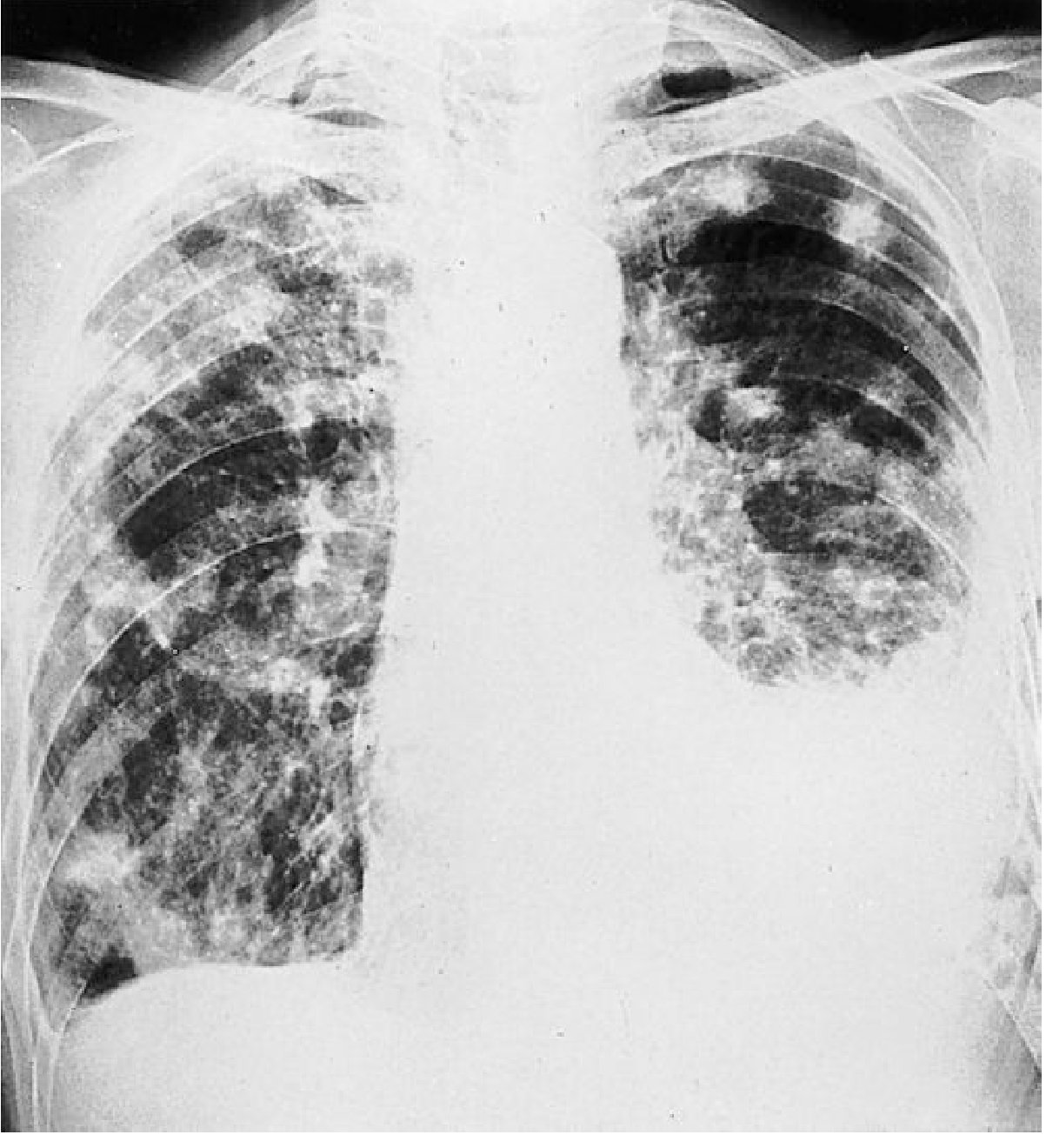
2. Cannon-ball Metastases
- Term used for large, round, well-defined bilateral pulmonary nodules
- Classically associated with: renal cell carcinoma, testicular germ cell tumours, choriocarcinoma
- Easily visible on plain CXR
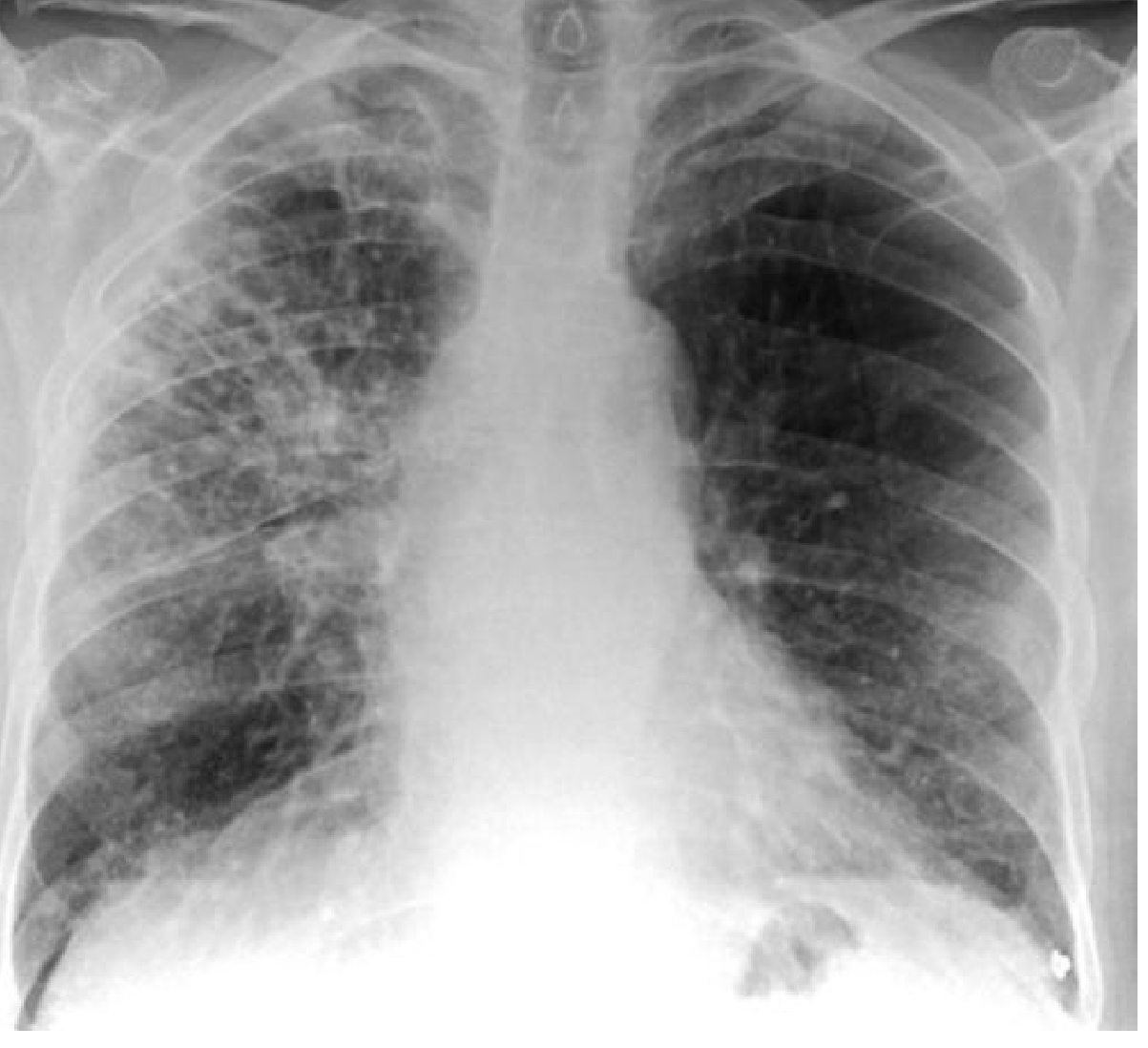
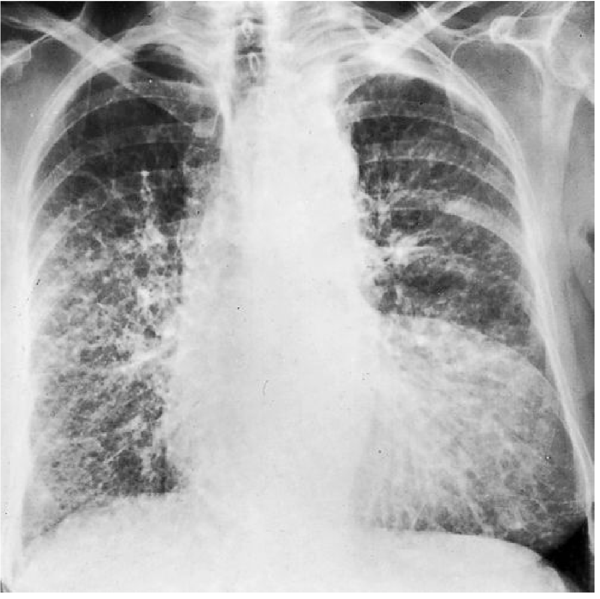
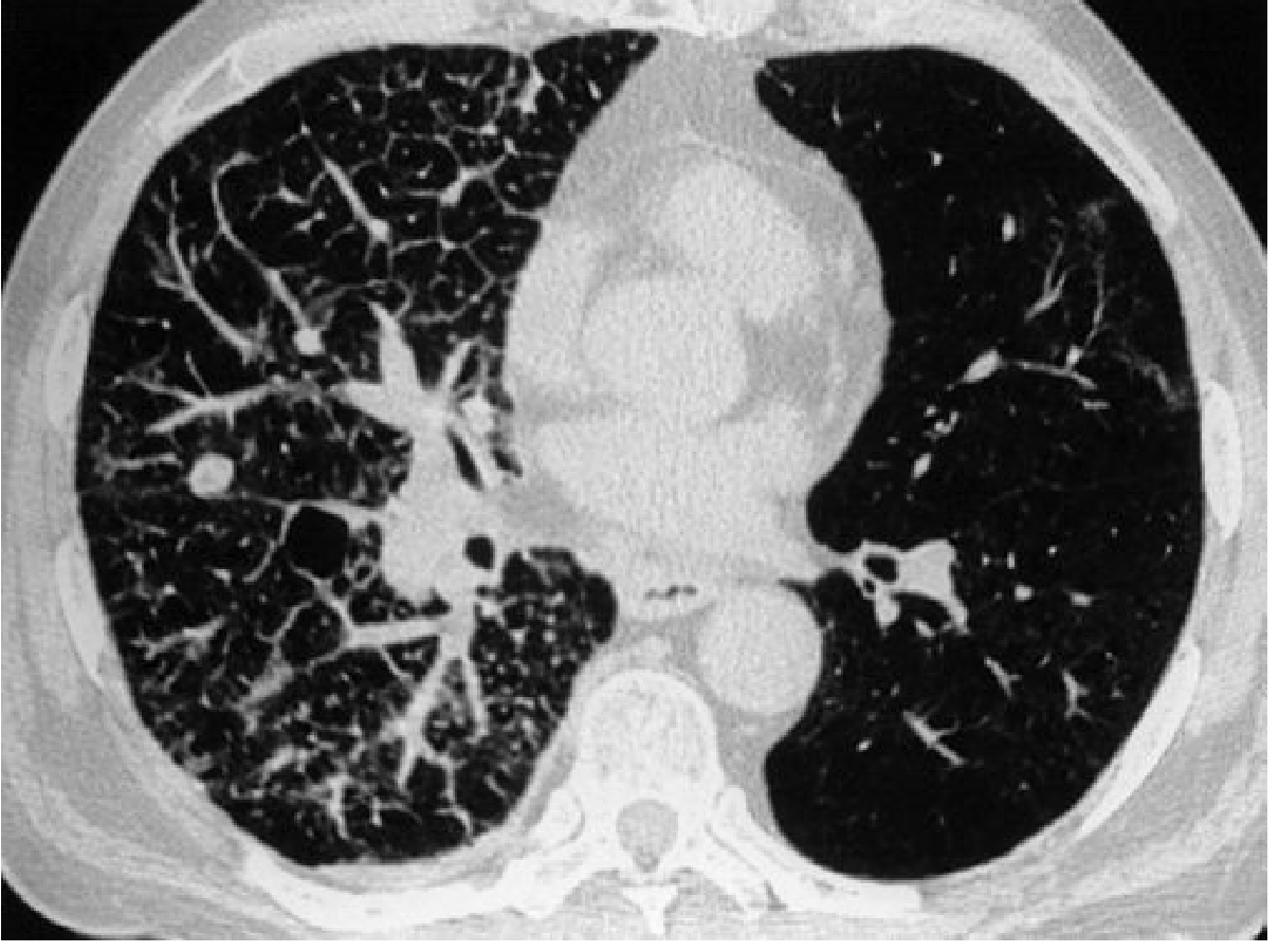
3. Lymphangitic Carcinomatosis
- Bilateral symmetric pattern - from haematogenous emboli spreading via vessel walls into perivascular interstitium
- Unilateral/localised pattern - from direct hilar node extension or pleural invasion
| Modality | Findings |
|---|---|
| CXR | Fine reticulonodular shadowing; thickened septal (Kerley B) lines; fissure thickening (subpleural oedema); pleural effusion (~30%) |
| HRCT | Non-uniform, nodular thickening of interlobular septa; irregular thickening of bronchovascular bundles centrally; scattered parenchymal nodules; patchy airspace shadowing; aerated acini subtended by thickened septa (key difference from pulmonary oedema) |
4. Miliary Metastases
- Innumerable tiny nodules mimicking miliary tuberculosis throughout both lungs
- No large masses, no lymphatic obstruction
- Rarest pattern of metastatic spread
- Primary tumours: thyroid carcinoma, renal carcinoma, bone sarcomas, choriocarcinoma
5. Endobronchial Metastases
- Rare; submucosal deposits cause airway obstruction as dominant radiological feature
- Leads to collapse, consolidation, or obstructive pneumonitis distal to the deposit
- Primary tumours: melanoma, renal, colorectal, breast carcinoma
6. Tumour Emboli
- Radiologically recognisable pulmonary arterial hypertension from tumour emboli blocking small pulmonary arteries
- Tumours: hepatoma, breast, kidney, stomach, prostate, choriocarcinoma
- Rare but high-yield PG MCQ association
7. Pleural Effusion
- Pleural effusion may accompany any pattern
- Especially common with lymphangitic spread (~30%)
- May also result from lymphatic obstruction, direct pleural seeding, or hypoalbuminaemia
Summary Table - High-Yield for PG Exam
| Pattern | Key Primary Tumours | Key Radiological Clue |
|---|---|---|
| Multiple discrete nodules | Breast, kidney, colon, sarcoma | Peripheral, basal, bilateral, well-defined |
| Cannon-ball | RCC, testis, choriocarcinoma | Large rounded bilateral nodules |
| Cavitating nodule | Squamous cell carcinoma | Thick-walled cavity |
| Calcified nodule | Osteosarcoma, chondrosarcoma | Dense calcification in nodule |
| Lymphangitic carcinomatosis | Breast, stomach, prostate, bronchus | Kerley B lines + reticulonodular shadowing; HRCT = septal thickening |
| Miliary | Thyroid, RCC, choriocarcinoma | TB-like tiny bilateral nodules |
| Endobronchial | Melanoma, RCC, colorectal, breast | Collapse/consolidation distal to obstruction |
| Tumour emboli | Hepatoma, choriocarcinoma | Pulmonary hypertension pattern |
Role of Imaging Modalities
- CXR (PA + lateral, high-kV): First-line; detects nodules >1 cm; misses lesions behind cardiac shadow, diaphragm, hilum
- MDCT: Detects lesions >3 mm; gold standard for staging and follow-up; essential for lymphangitic pattern (HRCT)
- PET-CT: FDG uptake; best for detecting distant metastases and monitoring treatment response
- MRI: Not routine for pulmonary metastases; useful for chest wall/pleural invasion assessment
Solitary Pulmonary Nodule in a Known Malignancy
- Smooth, round borders on CT
- Temporal proximity to the primary lesion
- History of RCC, sarcoma, germ cell tumour
- Spiculated edge
- History of head and neck (94%), lung (92%), bladder (89%), or uterine (74%) carcinoma - these carry highest risk of new primary
- Grainger & Allison's Diagnostic Radiology, Chapter 8 (Lung Metastases, Lymphangitic Carcinomatosis, Unusual Patterns)
- Schwartz's Principles of Surgery, 11e, Chapter 19 (Metastatic Lesions to the Lung)
Define hrct describe its techniques and its role in diffuse lung disease describe the diagnostic features of spn
HRCT: Definition, Technique, Role in Diffuse Lung Disease & Diagnostic Features of SPN
PART 1: DEFINITION OF HRCT
PART 2: HRCT TECHNIQUE
1. Thin Collimation (Section Thickness)
- Usually 1 to 2 mm slice thickness
- Reducing below 1 mm gives no significant further improvement in spatial resolution but increases image noise (reduces signal-to-noise ratio)
- Eliminates partial volume averaging that obscures fine lung detail on conventional CT
2. High Spatial Frequency ("Sharp" / "Lung") Reconstruction Algorithm
- A "sharp" or "bone" algorithm is used instead of the standard smooth algorithm
- Reduces image smoothing, making structures visibly sharper
- Image noise becomes more visible but this is acceptable and expected
- Enhances detection of: thickened interlobular septa, ground-glass opacification, abnormal subsegmental airways
3. Single Breath-Hold Imaging (Full Inspiration)
- Images obtained at full inspiration, supine position, apices to lung bases
- Eliminates respiratory motion artefact that degrades fine lung detail
- With MDCT (multi-detector CT): volumetric acquisition in a single breath-hold is now standard, even in tachypnoeic patients
Additional Technical Points
| Parameter | Detail |
|---|---|
| Patient position | Supine (standard); Prone when early interstitial fibrosis suspected (avoids confusion with dependent posterobasal opacification) |
| Expiratory CT | Useful for air trapping and small airways disease; confirms/accentuates mosaic attenuation; essential for diagnosing tracheobronchomalacia |
| IV contrast | Avoided in HRCT - can spuriously increase parenchymal opacification and interfere with comparison studies |
| Window settings | Lung windows (wide window, low centre, e.g. -600 HU centre, 1500 HU width) mandatory for parenchymal assessment |
| Dose reduction | Tube current reduction (40-70 mAs) feasible without significant quality loss; iterative reconstruction reduces noise at low dose |
Post-Processing Techniques
| Technique | Use |
|---|---|
| MIP (Maximum Intensity Projection) | Micronodular disease - better distinction of nodules vs. vessels; slab MIP identifies miliary nodule distribution |
| MinIP (Minimum Intensity Projection) | Emphysema, air trapping - highlights low-density areas |
| MPR/CMPR | Airway evaluation, curved multiplanar for large airways |
| Volume rendering | 3D airway and vascular display |
| CAD systems | Computer-aided detection of pulmonary nodules and emboli |
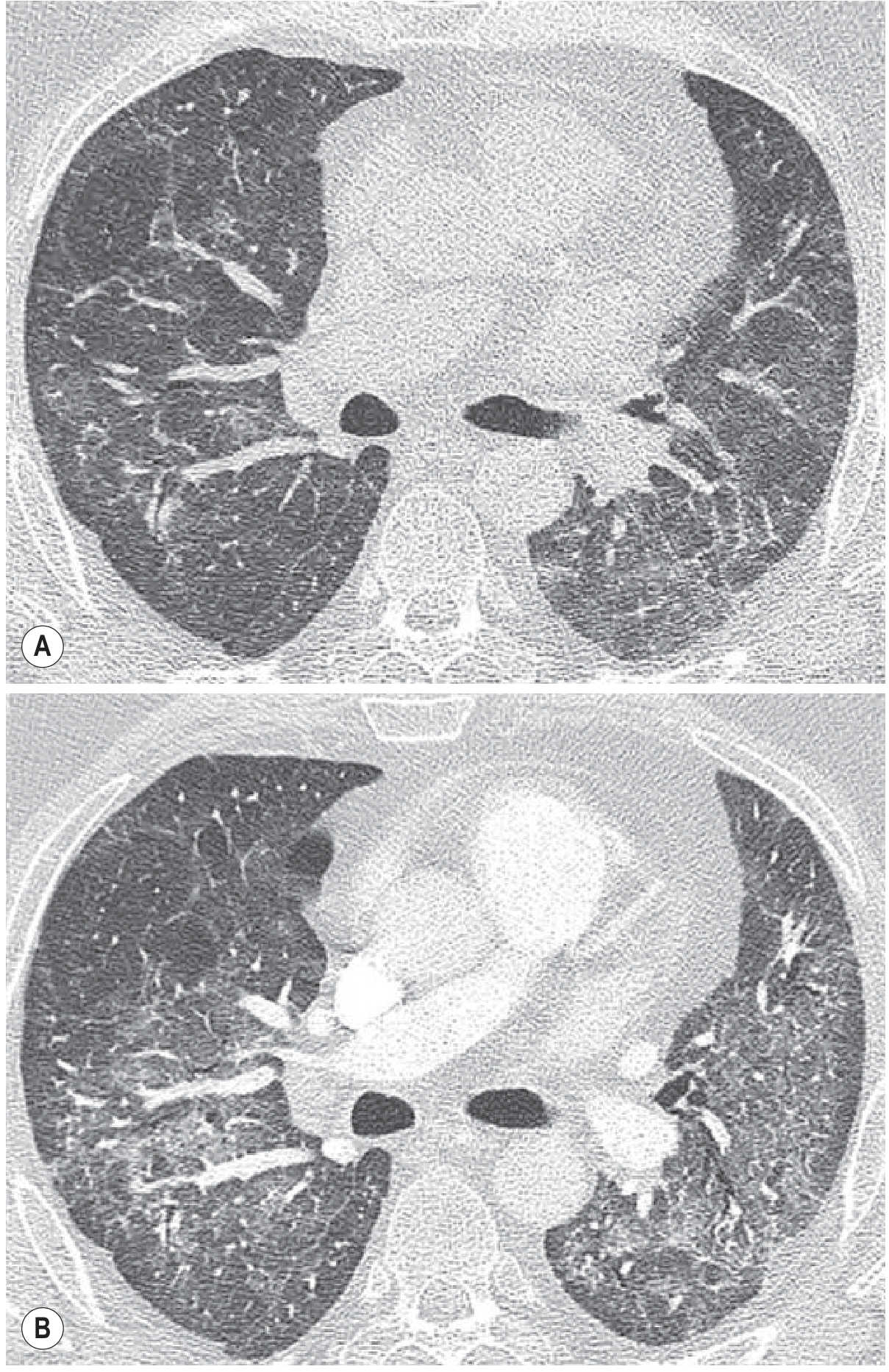
PART 3: ROLE OF HRCT IN DIFFUSE LUNG DISEASE (DLD)
Key HRCT Patterns in DLD
1. Ground-Glass Opacification (GGO)
- Hazy increased attenuation; vessels still visible through it
- Represents alveolar filling or interstitial thickening that does not fill alveoli completely
- Conditions: NSIP, DIP, alveolar haemorrhage, oedema, PCP, acute HP, drug toxicity
2. Reticulation
- Network of fine lines; represents thickened interlobular septa or intralobular interstitium
- In UIP/IPF: bilateral, basal, subpleural reticulation + honeycombing + traction bronchiectasis
3. Honeycombing
- Clustered cystic air spaces (3-10 mm), thick well-defined walls, in subpleural distribution
- Represents end-stage fibrosis; pathognomonic of UIP pattern
- Key finding in IPF
4. Traction Bronchiectasis / Bronchiolectasis
- Irregular airway dilatation due to fibrotic traction
- Indicates established fibrosis; part of UIP and fibrotic NSIP
5. Nodules - Distribution on HRCT is Diagnostic
| Distribution | Pattern | Disease |
|---|---|---|
| Centrilobular (not touching pleura) | Poorly defined, low density | Subacute HP, RB-ILD, endobronchial TB spread |
| Perilymphatic (fissures, septa, subpleural) | Well-defined nodules | Sarcoidosis, lymphangitis carcinomatosa |
| Random | Very small, well-defined, bilateral | Haematogenous TB (miliary), pulmonary metastases, pneumoconiosis |
6. Mosaic Attenuation Pattern
- Regional attenuation differences with well-defined interlobular septal borders
- Three causes:
- Small airways disease (air trapping): "black" areas abnormal; accentuated on expiratory CT; vessels small in black zones
- Chronic occlusive vascular disease (CTEPH): "black" areas abnormal; no air trapping
- Infiltrative lung disease: "grey" (increased attenuation) areas abnormal
7. Consolidation
- Complete alveolar filling; vessels obscured
- Alveolar pattern: pneumonia, cryptogenic organising pneumonia (COP), BAC/adenocarcinoma in situ, alveolar haemorrhage
HRCT Patterns in Specific Diffuse Diseases
| Disease | HRCT Pattern | Distribution |
|---|---|---|
| IPF (UIP) | Honeycombing + traction bronchiectasis + reticulation | Bilateral, basal, subpleural - posterior predominant |
| NSIP | Ground-glass opacity + reticulation; subpleural sparing | Bilateral, basal, subpleural |
| Sarcoidosis | Perilymphatic nodules, upper lobe; perihilar | Upper and mid zones; along fissures and bronchovascular bundles |
| Hypersensitivity Pneumonitis (subacute) | Centrilobular nodules + GGO + mosaic | Diffuse, mid-lower zone predominance |
| DIP | GGO, lower zone, cysts may be present | Bilateral, lower lobe |
| LCH | Upper lobe cysts + nodules; bizarre cyst shapes | Upper lobe predominant |
| LAM | Diffuse thin-walled round cysts | Diffuse, uniform distribution |
| Lymphangitic carcinomatosis | Nodular interlobular septal thickening + bronchovascular bundle thickening + GGO | Unilateral or bilateral |
Clinical Role of HRCT in ILD
- Diagnosis without biopsy: Typical UIP pattern on HRCT eliminates need for surgical lung biopsy in the right clinical context (ATS/ERS guidelines)
- Pattern recognition: Guides multidisciplinary discussion (pulmonologist + radiologist + pathologist)
- Prognostication: UIP pattern = worse prognosis even when not idiopathic
- Disease activity: GGO = active, potentially reversible; honeycombing = irreversible fibrosis
- Biopsy site selection: Identifies areas of active disease (GGO > fibrosis)
- Treatment monitoring: Serial HRCT tracks progression or drug response
PART 4: DIAGNOSTIC FEATURES OF SOLITARY PULMONARY NODULE (SPN)
Definition
Epidemiology
- Found in 0.2% of CXRs incidentally; ~1 million/year in the USA
- 40-50% of SPNs are malignant (Fishman's Pulmonary)
- 80% of benign SPNs are infectious granulomas; hamartoma is the next most common benign cause (~10%)
- Most common malignant type: adenocarcinoma (47%), squamous cell (22%), small cell (4%)
CT Features Suggesting MALIGNANCY
| Feature | Detail |
|---|---|
| Size | Risk <1% for <6 mm; 1-2% for 6-8 mm; significantly higher for ≥8 mm |
| Irregular / spiculated margin | Spiculation = fine linear strands radiating 4-5 mm outward; high specificity for malignancy |
| Corona radiata sign | Fine linear strands extending radially from the nodule surface, like spokes of a wheel; highly suggestive of malignancy |
| Lobulated margin | Indicates uneven growth rates within the tumour |
| Growth over time | Volume-doubling time 20-400 days = malignant; <20 days = infection; >400 days = benign (but can be slow-growing cancer) |
| Part-solid (subsolid) nodule | 40-50% are malignant; higher risk than pure solid |
| Calcification pattern (malignant) | Stippled, amorphous, eccentric calcification = associated with cancer |
| Size ≥3 cm | Mass = malignancy until proved otherwise |
| Invasion of adjacent structures | Pleural tags, chest wall, vascular involvement |
| Associated lymphadenopathy | Hilar / mediastinal nodes = metastatic malignancy |
CT Features Suggesting BENIGNITY
| Feature | Detail |
|---|---|
| Calcification patterns (benign) | Four benign patterns: Diffuse/solid, Central, Laminated (concentric rings), Popcorn |
| Diffuse/solid + Central + Laminated | Granulomatous infection (TB, histoplasmosis) |
| Popcorn calcification | Hamartoma - pathognomonic |
| Small size | <6 mm = <1% malignant risk |
| Stability over 2 years | No change over 2 years = probably benign (most reliable sign of benignity) |
| Smooth, well-defined, round margin | More likely benign; also favours metastasis if known primary exists |
| Fat attenuation | Hamartoma (fat + cartilage) |
Nodule Classification by Composition (CT)
| Type | Definition | Malignancy Risk |
|---|---|---|
| Solid | Completely solid density | Variable by size |
| Ground-glass (non-solid) | No solid component; haziness only | Pure GGO: adenocarcinoma in situ, MIA; FDG-PET unreliable (sensitivity 10%) |
| Part-solid (subsolid) | Both GGO + solid component | Highest risk (40-50%); solid component size guides management |
Role of Imaging Modalities in SPN
| Modality | Role |
|---|---|
| CXR | Initial detection; cannot characterise adequately |
| CT (thin sections 1-2 mm) | Gold standard - characterises number, size, margin, calcification, growth rate |
| PET-CT (FDG) | For solid SPNs >8 mm; sensitivity 97%, specificity 78%; SUVmax >2.5 = malignant; NOT reliable for GGO nodules (sensitivity 10%), nodules <8 mm, carcinoids, AIS/MIA |
| CT contrast enhancement | >15 HU enhancement = malignant (Swensen criteria) |
| Serial CT follow-up | Stability >2 years = benign; growth = suspect malignancy |
| PET-CT biopsy guidance | Fewer inconclusive biopsies vs CT guidance alone |
Summary Table - High-Yield for PG Exam
| SPN Feature | Benign | Malignant |
|---|---|---|
| Margin | Smooth, well-defined | Spiculated, lobulated, corona radiata |
| Calcification | Diffuse, central, laminated, popcorn | Stippled, amorphous, eccentric |
| Size | <6 mm very low risk | >8 mm high risk |
| Growth | Stable >2 years | Doubling time 20-400 days |
| Composition | Solid, stable | Part-solid highest risk |
| FDG-PET | Not helpful <8mm | SUVmax >2.5 |
- Grainger & Allison's Diagnostic Radiology - Chapter 1 (HRCT Technique), Chapter 9 (Diffuse Lung Disease HRCT patterns)
- Schwartz's Principles of Surgery, 11e - Chapter 19 (SPN CT features)
- Fishman's Pulmonary Diseases & Disorders - Chapter 30 (Evaluation of SPN, FDG-PET)
- Goldman-Cecil Medicine - Chapter 177 (SPN evaluation algorithm)
Create a management algorithm flowchart for SPN based on size and imaging features
A detailed clinical management algorithm flowchart for Solitary Pulmonary Nodule (SPN) based on size and imaging features, suitable for radiology PG exam. The flowchart should be clean, professional, medical style with boxes, diamonds for decision points, and arrows. Include the following pathways: Title: "Management Algorithm for Solitary Pulmonary Nodule (SPN)" Start: SPN detected on CXR / CT First branch - SOLID NODULE: - Less than 6mm: Low risk → CT follow-up at 12 months; if stable, no further workup - 6-8mm: Intermediate risk → CT at 3-6 months, then 18-24 months - Greater than 8mm: High risk → CT + PET-CT → if benign features (stable 2yr, benign calcification) → follow-up; if malignant features (spiculated, corona radiata, growing) → Tissue diagnosis (PET-CT guided / CT-guided biopsy) → Malignant → Surgical resection; Benign → Follow-up Second branch - SUBSOLID NODULE: - Pure Ground Glass (non-solid): CT follow-up at 6 months; if stable, annual CT x 5 years - Part-solid (mixed): High malignancy risk (40-50%) → CT at 3-4 months; if solid component persists or grows → PET-CT + Tissue diagnosis → Surgery CT Benign Features box: Diffuse/central/laminated/popcorn calcification, smooth margin, fat attenuation (hamartoma), stable >2 years CT Malignant Features box: Spiculated/lobulated margin, corona radiata sign, growth (doubling time 20-400 days), stippled/eccentric calcification, size >3cm (mass) PET-CT note: Reliable only for solid nodules >8mm; SUVmax >2.5 = malignant; NOT reliable for GGO or nodules <8mm Use color coding: green for benign/follow-up pathways, red/orange for malignant/biopsy/surgery pathways, blue for decision boxes, yellow for imaging steps. Clean white background, bold readable fonts.
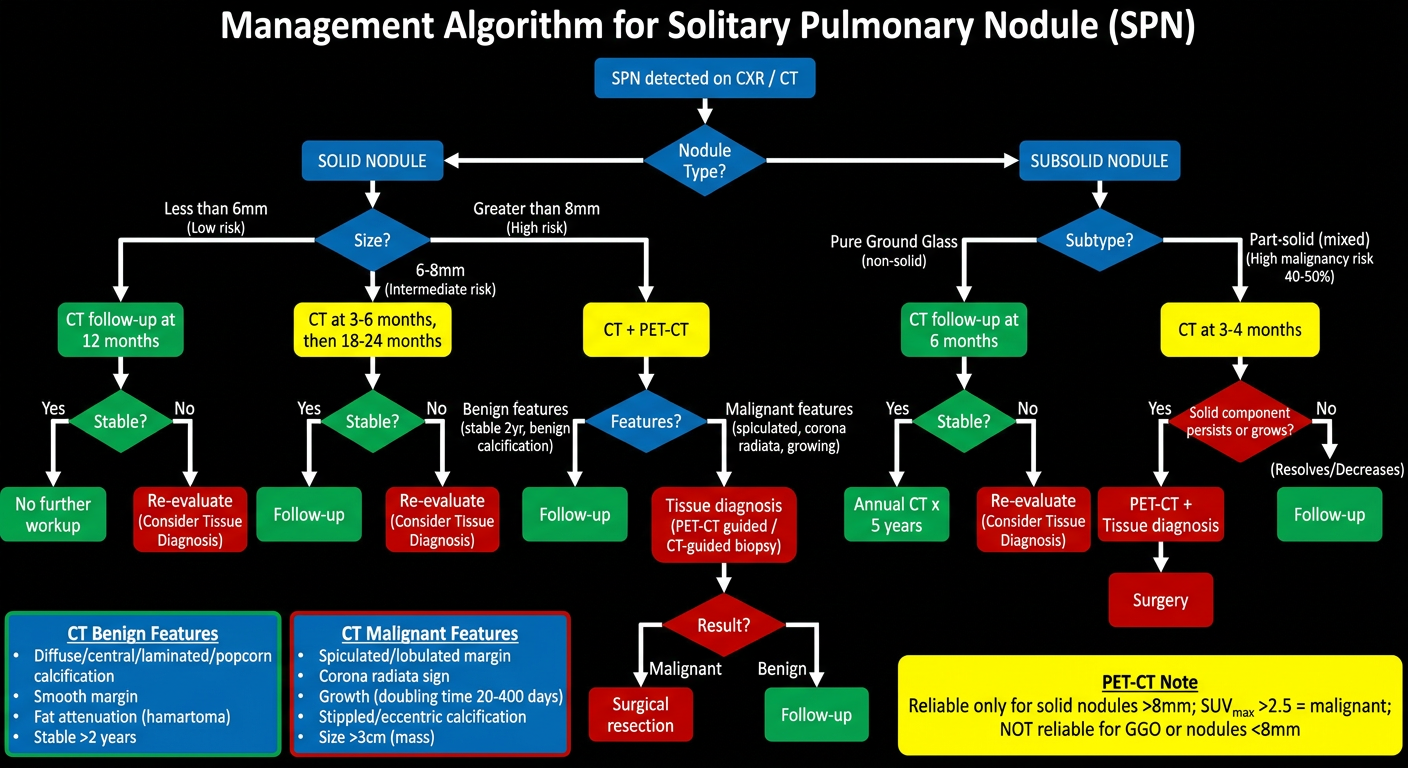
Key Decision Points Summarised
| Size | Risk | Action |
|---|---|---|
| <6 mm | <1% malignancy | CT at 12 months; if stable → discharge |
| 6-8 mm | 1-2% malignancy | CT at 3-6 months, then 18-24 months |
| ≥8 mm | Significant risk | CT + PET-CT → tissue diagnosis if FDG-avid or malignant features |
| >3 cm (mass) | Malignant until proved otherwise | Direct tissue sampling + staging |
| Type | Risk | Action |
|---|---|---|
| Pure GGO | AIS / MIA | CT at 6 months; if stable → annual CT × 5 years |
| Part-solid | 40-50% malignant | CT at 3-4 months; solid component persistence → surgery |
Based on revised fleishner guidelines
Fleischner Society 2017 revised guidelines solitary pulmonary nodule management solid subsolid
Fleischner Society guidelines pulmonary nodule management 2017
https://radiopaedia.org/articles/fleischner-society-pulmonar…
A highly detailed, professional clinical management algorithm flowchart for "Solitary Pulmonary Nodule - Fleischner Society 2017 Guidelines" for radiology PG exam. Clean white background, color-coded boxes, clear arrows, and organized layout. Title at top (bold, dark blue): "FLEISCHNER SOCIETY 2017 GUIDELINES - Incidental Pulmonary Nodule Management" Subtitle (smaller, grey): "Applies to: Age ≥35, immunocompetent, no prior cancer, NOT for lung cancer screening" The flowchart has two main parallel columns: LEFT COLUMN - SOLID NODULES (light blue header) Decision box: "SINGLE solid nodule" → Branch 1: Size < 6 mm (<100 mm³) → Low Risk: NO routine follow-up (green box) → High Risk: Optional CT at 12 months (yellow box) → Branch 2: Size 6-8 mm (100-250 mm³) → Low Risk: CT at 6-12 months → consider CT at 18-24 months (yellow box) → High Risk: CT at 6-12 months → CT at 18-24 months (orange box) → Branch 3: Size > 8 mm (>250 mm³) → Both Low & High Risk: CT at 3 months OR PET-CT OR Tissue sampling (red/orange box) → If malignant → Surgical resection (red box) → If benign → No further follow-up (green box) Decision box: "MULTIPLE solid nodules" → <6 mm: Low Risk: No follow-up. High Risk: Optional CT 12 months → 6-8 mm: CT at 3-6 months → consider CT 18-24 months (both risk groups) → >8 mm: CT at 3-6 months → CT at 18-24 months → manage based on most suspicious nodule RIGHT COLUMN - SUBSOLID NODULES (light purple/lavender header) Decision box: "SINGLE Pure Ground-Glass Nodule (GGO)" → < 6 mm: No routine follow-up (green box) → ≥ 6 mm: CT at 6-12 months → if persistent: CT every 2 years until 5 years (yellow/orange box) → If grows or develops solid component → consider resection (red box) Decision box: "SINGLE Part-Solid Nodule" → < 6 mm: No routine follow-up (green box) → ≥ 6 mm: CT at 3-6 months to confirm persistence → Solid component < 6 mm: Annual CT × 5 years (yellow box) → Solid component ≥ 6 mm: HIGHLY SUSPICIOUS → PET-CT / Tissue sampling / Surgery (red box) Decision box: "MULTIPLE Subsolid Nodules" → < 6 mm: CT at 3-6 months → if stable: CT at 2 and 4 years (high risk) (yellow box) → ≥ 6 mm: CT at 3-6 months → manage based on most suspicious nodule (orange box) BOTTOM BOX - Key Notes (light grey background): • Measurement: Average of long + short axis; thin-slice CT ≤1.5 mm • High-risk features: Smoking history, upper lobe location, spiculated margin, family history • PET-CT: Reliable only for solid nodules >8 mm; NOT for GGO or nodules <8 mm • Part-solid nodules: Malignant in 63% (highest risk); Pure GGO: 18%; Solid: 7% • Transient subsolid nodules (infection/hemorrhage): Confirm persistence at 3-6 months first Color legend box at bottom right: Green = No follow-up / benign Yellow = Short-interval follow-up Orange = Intermediate concern Red = High suspicion / Tissue diagnosis / Surgery
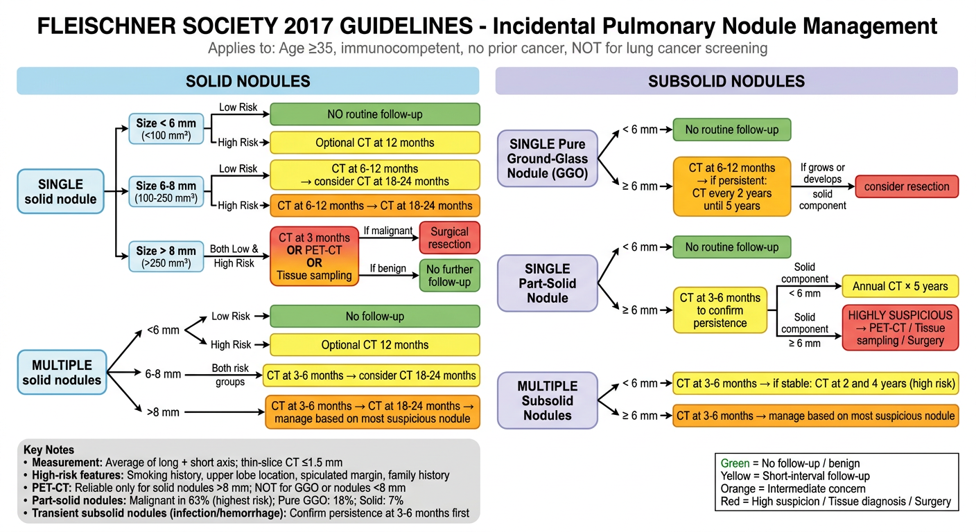
Fleischner Society 2017 - Complete Reference Table
Applies to: Incidental nodules in immunocompetent patients ≥35 years, without prior cancer. NOT for lung cancer screening programmes.
SOLID NODULES
| Size | Single Nodule | Multiple Nodules |
|---|---|---|
| <6 mm (<100 mm³) | Low risk: No follow-up; High risk: Optional CT 12 mo | Low risk: No follow-up; High risk: Optional CT 12 mo |
| 6-8 mm (100-250 mm³) | Low risk: CT 6-12 mo → consider CT 18-24 mo; High risk: CT 6-12 mo → CT 18-24 mo | Both: CT 3-6 mo → consider CT 18-24 mo |
| >8 mm (>250 mm³) | Both: CT 3 mo OR PET-CT OR tissue sampling | CT 3-6 mo → CT 18-24 mo → manage based on most suspicious |
SUBSOLID NODULES
| Type | Size | Management |
|---|---|---|
| Pure GGO (single) | <6 mm | No routine follow-up |
| Pure GGO (single) | ≥6 mm | CT 6-12 months → if persistent: CT every 2 years × 5 years; if grows/solid component develops → resection |
| Part-solid (single) | <6 mm | No routine follow-up |
| Part-solid (single) | ≥6 mm | CT 3-6 months to confirm persistence → if solid component <6 mm: annual CT × 5 years; if solid component ≥6 mm: HIGHLY SUSPICIOUS → PET-CT / biopsy / surgery |
| Multiple subsolid | <6 mm | CT 3-6 months → if stable: CT at 2 and 4 years (high-risk patients) |
| Multiple subsolid | ≥6 mm | CT 3-6 months → manage based on most suspicious nodule |
Key Changes from 2005/2013 Guidelines (High-Yield MCQ Points)
| Change | 2005/2013 | 2017 |
|---|---|---|
| Threshold for follow-up | >4 mm | >6 mm (raised - fewer unnecessary scans) |
| Follow-up intervals | Fixed time points | Ranges (6-12 months, not exact) |
| Solid + subsolid tables | Separate documents | Combined single table |
| Measurement method | Axial plane only | Average of long + short axis, any plane |
| Subsolid follow-up period | 3 years | 5 years (slower growth rate) |
| Initial subsolid follow-up | 3 months | 3-6 months (to confirm persistence) |
Risk Factors (High vs Low Risk)
- Smoking history (current or ex-smoker)
- Upper lobe location
- Spiculated margin
- Family history of lung cancer
- Occupational exposure (asbestos, radon)
- Age >40
Malignancy Risk by Nodule Type (Henschke et al.)
- Part-solid nodules: 63% malignant (highest)
- Pure GGO: 18% malignant
- Solid nodules: 7% malignant
Reference: MacMahon H et al. Guidelines for Management of Incidental Pulmonary Nodules Detected on CT Images: From the Fleischner Society 2017. Radiology 2017;284(1):228-243. [PMID: 28240562]
How to remember
The Master Memory Framework
STEP 1 - Remember the "6-8-MORE" Rule for SOLID nodules
< 6 mm → GREEN = GO (no follow-up needed)
6-8 mm → YELLOW = WAIT (follow-up CT)
> 8 mm → RED = ACT (CT / PET / Biopsy)
- Below Six = Fine (no routine follow-up)
- Above Eight = Late (act now - CT/PET/biopsy at 3 months)
- Between = Wait (CT at 6-12 months)
STEP 2 - Remember the Follow-up Intervals with "3-6-12-18-24"
| Scenario | First scan | Second scan |
|---|---|---|
| Solid >8 mm | 3 months | — (then decide) |
| Part-solid ≥6 mm | 3-6 months | Annual × 5 yr |
| Solid 6-8 mm | 6-12 months | 18-24 months |
| Pure GGO ≥6 mm | 6-12 months | Every 2 yr × 5 yr |
- Bigger = earlier scan
-
8 mm solid = 3 months (most urgent)
- Part-solid ≥6mm = 3-6 months (high malignancy risk 63%)
- 6-8 mm solid = 6-12 months (intermediate)
- Pure GGO ≥6 mm = 6-12 months (slow growing)
STEP 3 - Subsolid: Remember "6 is the Magic Number"
< 6 mm → NO follow-up (same as solid)
≥ 6 mm → Follow-up required
"Confirm it exists at 3-6 months, then watch the solid bit - if the solid component hits 6, it's suspicious"
- Solid component < 6 mm = annual CT × 5 years
- Solid component ≥ 6 mm = HIGHLY SUSPICIOUS → act (PET/biopsy/surgery)
STEP 4 - Multiple Nodules: "Halve the Interval"
| Single solid 6-8 mm | → CT at 6-12 months |
|---|---|
| Multiple solid 6-8 mm | → CT at 3-6 months |
STEP 5 - The "63-18-7" Malignancy Risk Triplet
Part-solid = 63% malignant ← MOST DANGEROUS (looks innocent but isn't)
Pure GGO = 18% malignant ← Moderate risk
Solid = 7% malignant ← Lowest risk per nodule
STEP 6 - What Changed in 2017 (The "RAISE" Mnemonic)
| Letter | Change |
|---|---|
| R | Raised threshold: 4 mm → 6 mm (fewer scans) |
| A | Any plane measurement (not just axial) |
| I | Intervals given as ranges, not fixed dates |
| S | Single table for solid + subsolid (combined) |
| E | Extended subsolid follow-up: 3 yr → 5 yr |
STEP 7 - Visual Story to Anchor It All
Imagine a traffic cop standing at the lung gate:
- Nodule comes up - he measures it.
- Under 6? "You're small, move along" ✅
- 6 to 8? "Wait here, I'll check on you in 6-12 months" 🕐
- Over 8? "Stop right there - scan, PET, or biopsy in 3 months" 🚨
- Part-solid ≥6 with solid bit ≥6? "Surgeon, NOW" 🔴
Quick Recall Card (Write this on your palm before the exam)
SOLID: <6 = none | 6-8 = 6-12mo | >8 = 3mo/PET/biopsy
GGO: <6 = none | ≥6 = 6-12mo → 2yr×5yr
PART-SOLID: <6 = none | ≥6 = 3-6mo → solid<6: annual×5yr | solid≥6: SURGERY
MULTIPLE: halve the first interval, manage worst nodule
RISK: Part-solid 63% > GGO 18% > Solid 7%
2017 CHANGES: threshold ↑ to 6mm, follow-up 5yr, ranges not fixed
Imaging of interstitial lung disease
Imaging of Interstitial Lung Disease (ILD)
Definition
Imaging Modalities
1. Chest Radiograph
- Part of initial assessment but non-specific, considerable observer variation, and low sensitivity for early ILD
- Useful for:
- Detecting gross abnormality
- Establishing disease distribution (zonal predominance)
- Tracking progression by comparison with prior films
- Early: diffuse ground-glass pattern
- Progressive: reticular (linear) infiltrates → reticulonodular infiltrates
- Alveolar filling: ill-defined nodules (acinar rosettes) with air bronchograms
2. HRCT - The Cornerstone of ILD Imaging
- Early detection before CXR becomes abnormal
- Histo-specific diagnosis in select cases (e.g. typical UIP pattern = no biopsy needed)
- Assessment of disease reversibility and prognosis (GGO = potentially reversible; honeycombing = irreversible)
- Biopsy site selection (sample active GGO, not fibrotic areas)
HRCT Patterns in Diffuse Lung Disease
Pattern 1: Reticular Pattern
- Network of fine lines representing thickened intralobular or interlobular septa, or honeycombing
- Intralobular thickening: often appears as faint GGO rather than distinct reticulation (below spatial resolution)
- Numerous thickened interlobular septa = extensive interstitial abnormality (fluid in oedema; cells in lymphangitis; fibrosis in UIP)
- Honeycombing: clustered cystic spaces 3-10 mm with thick well-defined walls; subpleural; represents end-stage fibrosis
Pattern 2: Nodular Pattern
| Distribution | Diseases |
|---|---|
| Perilymphatic (along fissures, septa, bronchovascular bundles, subpleural) | Sarcoidosis, lymphangitis carcinomatosa, silicosis, coal worker's pneumoconiosis |
| Centrilobular (do not touch pleura or fissures) | Hypersensitivity pneumonitis (subacute), RB-ILD, endobronchial TB, diffuse panbronchiolitis |
| Random (uniform bilateral distribution, touch pleura) | Miliary TB, haematogenous metastases, pneumoconiosis |
Pattern 3: Mosaic Attenuation Pattern
| Cause | Abnormal Component | Key Discriminator |
|---|---|---|
| Small airways disease | "Black" (low attenuation) | Air trapping on expiratory CT; small vessels in black zones |
| Chronic occlusive vascular disease (CTEPH) | "Black" | No bronchial abnormality; no air trapping |
| Infiltrative lung disease (ILD) | "Grey" (increased attenuation) | Ground-glass areas are the pathological component |
Pattern 4: Cystic Pattern
- Thin-walled air-containing spaces
- Key: assess morphology (smooth wall = cyst vs. irregular = emphysema) and geometry
- LAM: Diffuse, thin-walled, round, bilateral uniform distribution
- LCH (Langerhans): Upper lobe, bizarre-shaped cysts + nodules
- LIP: Cysts + GGO + centrilobular nodules (often in HIV/Sjögren's)
- DIP: Lower zone cysts + GGO
Idiopathic Interstitial Pneumonias (IIPs) - HRCT Patterns
The 2013 ATS/ERS Classification and HRCT Correlation
| IIP | Histological Pattern | Key HRCT Features | Distribution |
|---|---|---|---|
| IPF | UIP | Honeycombing + traction bronchiectasis + reticulation | Bilateral, basal, subpleural, posterior predominant |
| NSIP | NSIP | Ground-glass ± reticulation; subpleural sparing | Bilateral, basal, symmetrical |
| COP | Organising pneumonia | Patchy subpleural or peribronchial consolidation ± perilobular pattern | Subpleural + peribronchial, lower zone |
| AIP | Diffuse alveolar damage | GGO + consolidation + traction bronchiectasis | Diffuse, dependent + non-dependent |
| DIP | DIP | Extensive lower zone GGO ± reticulation/cysts | Peripheral, lower lobe, smokers |
| RB-ILD | RB-ILD | Poorly defined centrilobular nodules + patchy GGO; bronchial wall thickening | Diffuse, smokers |
| LIP | LIP | GGO + centrilobular nodules + thickened septa + thin-walled cysts | Diffuse; associated with HIV, Sjögren's |
UIP/IPF in Detail - The Most Important Pattern
Typical UIP Pattern (Virtually Pathognomonic = No Biopsy Needed)
- Subpleural bibasal reticulation with honeycombing
- Traction bronchiectasis within areas of fibrosis
- Temporal heterogeneity (areas of fibrosis at different stages of maturity)
- Progressive "propeller blade distribution" - creeps around periphery toward anterior upper lobes
Fleischner Society UIP CT Categories (2018 Guidelines)
| CT Category | CT Distribution | CT Features |
|---|---|---|
| Typical UIP | Basal, subpleural, heterogeneous | Honeycombing + reticular pattern + traction bronchiectasis; no features suggesting alternative diagnosis |
| Probable UIP | Basal, subpleural | Reticular + traction bronchiectasis; no honeycombing |
| Indeterminate for UIP | Variable or diffuse | Some fibrosis features + inconspicuous non-UIP features |
| Non-IPF Diagnosis | Upper/mid lung predominant | Peribronchovascular predominance + subpleural sparing; extensive GGO; mosaic with air trapping; diffuse cysts/nodules |
Key exam point: Typical UIP on HRCT in appropriate clinical context = no surgical biopsy required (ATS/ERS/JRS/ALAT 2018 guidelines)
Other Key ILDs and Their HRCT Features
Sarcoidosis
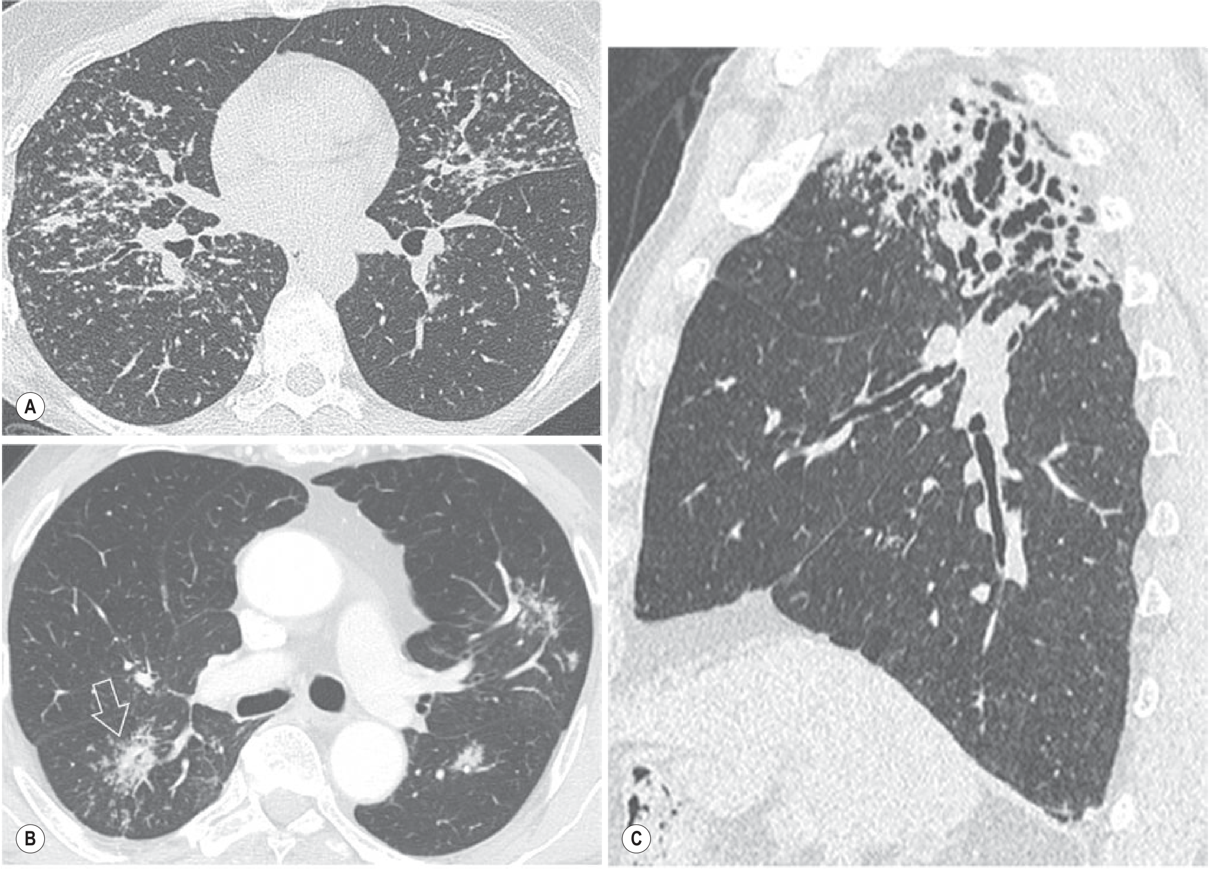
- Well-defined 2-4 mm nodules in perilymphatic distribution (fissures, bronchovascular bundles, interlobular septa)
- Upper lobe predominant
- Bilateral hilar + mediastinal lymphadenopathy (hallmark)
- Galaxy sign: micronodules clustering into larger opacity with peripheral satellite nodules
- Fibrotic stage: upper lobe honeycombing, volume loss, bronchiectasis, fissural displacement
Hypersensitivity Pneumonitis (HP / Extrinsic Allergic Alveolitis)
| Phase | HRCT Features |
|---|---|
| Acute | GGO + consolidation + centrilobular nodules |
| Subacute | Poorly defined centrilobular nodules (~5 mm) + GGO + mosaic attenuation |
| Chronic/Fibrotic | Upper/mid lobe fibrosis + reticulation + traction bronchiectasis + air trapping on expiratory CT |
Langerhans Cell Histiocytosis (LCH)
- Smokers
- Upper lobe nodules (early) → nodules cavitate → bizarre-shaped irregular cysts (late)
- Lower lobe and costophrenic angles spared (distinguishes from LAM)
Lymphangioleiomyomatosis (LAM)
- Young women of childbearing age
- Diffuse, bilateral, uniform thin-walled round cysts throughout all zones
- No nodules, no GGO
- Associated with pneumothorax and chylous effusion
NSIP (Non-Specific Interstitial Pneumonia)
- Favoured over UIP if:
- Central rather than peripheral predominance
- Subpleural sparing (key distinguishing feature from UIP)
- More uniform fibrosis; honeycombing usually absent
- Extensive bilateral symmetrical GGO ± reticulation
- Most commonly associated with CTD (especially SSc, polymyositis/dermatomyositis)
Cryptogenic Organising Pneumonia (COP)
- "Reverse halo" (atoll) sign: central GGO surrounded by rim of consolidation - characteristic but not pathognomonic
- Subpleural or peribronchial patchy consolidation
- Perilobular pattern: arcs of consolidation outlining secondary pulmonary lobule
- Migratory/recurrent infiltrates
- Responds to steroids
Acute Interstitial Pneumonia (AIP) / Diffuse Alveolar Damage
- Idiopathic form of ARDS
- HRCT: Patchy or diffuse GGO + consolidation (dependent predominant)
- Traction bronchiectasis + reticulation after several days
- Temporal homogeneity (unlike UIP)
Zonal Distribution - High-Yield Table
| Distribution | Disease |
|---|---|
| Upper lobe | Sarcoidosis, silicosis, berylliosis, LCH, chronic HP, ankylosing spondylitis, PLCH |
| Lower lobe | IPF (UIP), asbestosis, NSIP, RA-ILD, SSc-ILD |
| Middle + lower | Lymphangitic carcinomatosis, asbestosis, RA-ILD, SSc-ILD |
| Peripheral subpleural | UIP/IPF, COP, eosinophilic pneumonia |
| Perihilar / central | Sarcoidosis, NSIP (subpleural sparing) |
| Diffuse uniform | LAM, miliary TB, metastases |
Role of Other Modalities
| Modality | Role |
|---|---|
| CXR | Initial screen; distribution clues; follow-up |
| HRCT (1-2mm, prone if early fibrosis) | Gold standard; pattern diagnosis; biopsy guidance |
| PET-CT (FDG) | Inflammatory activity in sarcoidosis; predicts 1-year deterioration; cardiac sarcoidosis |
| BAL | Supports diagnosis; excludes infection |
| Surgical lung biopsy | When HRCT pattern indeterminate; NOT needed for typical UIP |
| Pulmonary function tests | Restrictive pattern (reduced TLC, DLCO); complements HRCT |