
This x-ray belongs to digestive system. Analysis the x-ray film and tell What you see in the given x-ray please describe according to given instructions and you can you given documents also Sequence :- narrow or diffuse Barium :- Border :- well defined or I'llI define and even and uneven Size :- Constrast:- Shaped :- Diagnosis :
Reading File
Finding Sources
Finding Sources
Reading File
Reading File
Searching Images
barium enema apple core lesion carcinoma colon
Loading Image
Loading Image
Reading File
Now I have everything I need to give a thorough structured analysis. Let me synthesize the findings.
Barium Enema X-Ray Analysis — Double Contrast Study (Large Bowel)
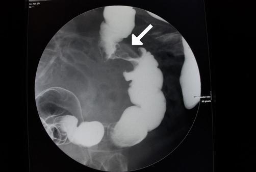
These are two views (AP projections) of a double-contrast barium enema (DCBE) study showing the large intestine. The right panel (post-evacuation or delayed view) reveals the abnormality most clearly.
🔬 Structured Analysis
Sequence: Narrow (Focal/Segmental)
A short-segment, focal narrowing is visible in the region of the sigmoid/descending colon on the right-side film. The remainder of the colon appears of normal calibre and haustration in the left film, making this a focal narrowing rather than diffuse colitis or stricture.
Barium:
Double-contrast technique — barium coats the mucosal wall while air distends the lumen. In the narrowed segment, barium column is interrupted / narrowed. There is mucosal destruction in the involved segment with loss of normal haustral folds. The proximal colon shows normal barium coating with intact haustration.
Border: Ill-defined, Uneven
The margins of the narrowed segment are ill-defined and irregular, with:
- Overhanging ("shouldering") edges at both ends of the lesion — the classic "hooks" or "shelf sign"
- Abrupt transition from normal to narrowed bowel (not a smooth, gradual taper as in benign stricture)
- Mucosal pattern is destroyed/absent within the lesion
Size:
The narrowed segment appears short in length (approximately 3–6 cm), which is characteristic of an annular constricting carcinoma (as opposed to the long-segment narrowing seen in Crohn's disease or radiation stricture).
Contrast (Filling Defect):
There is a filling defect — the barium fails to fill the narrowed lumen normally. The lesion appears as a central constriction with loss of luminal width, surrounded by irregular mucosal destruction. There is no significant pre-stenotic dilatation visible on the left film, but the right film shows altered bowel calibre at the lesion site.
Shape: Apple-Core / Napkin-Ring
The lesion has the classic "apple-core" (or napkin-ring) appearance:
- Circumferential narrowing of the bowel lumen
- Irregular mucosal borders
- Abrupt shouldering at both edges
- Short segment involved
- Resembles an apple with a bite taken out — the most pathognomonic radiological sign of annular carcinoma of the colon
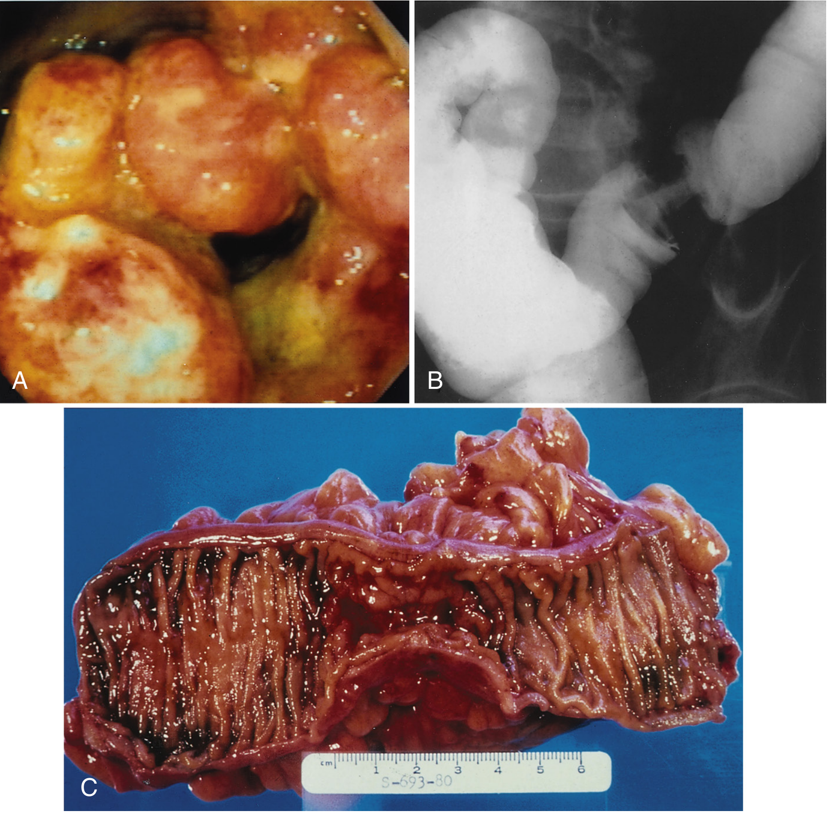
"A barium enema of a constricting carcinoma of the descending colon presents what is best described as an 'apple core' lesion. On the barium enema, you will note the loss of mucosal patterns, the 'hooks' at the margins of the lesion, the relatively short length of the lesion, and the abrupt ending of the lesion." — Swanson's Family Medicine Review, p. 205
✅ Diagnosis
Carcinoma of the Colon (Annular/Constricting Adenocarcinoma) — Most likely sigmoid or descending colon location — Radiological type: Apple-core / Napkin-ring lesion
Reference Images for Comparison
The textbook image (Sleisenger & Fordtran, Fig. 127.16) shows the same triad:
Summary Table
| Parameter | Finding |
|---|---|
| Sequence | Narrow (focal, short-segment) |
| Barium | Interrupted; mucosal coating absent in lesion; haustral folds destroyed |
| Border | Ill-defined, uneven; abrupt overhanging "shelf" margins (shouldering) |
| Size | Short segment (~3–6 cm); circumferential |
| Contrast | Filling defect; barium fails to traverse normally |
| Shape | Apple-core / Napkin-ring |
| Diagnosis | Carcinoma of the Colon (Annular Adenocarcinoma — sigmoid/descending colon) |
Next steps clinically: Colonoscopy with biopsy for histological confirmation, CEA level, CT chest/abdomen/pelvis for staging, and surgical/oncological referral.
- Swanson's Family Medicine Review, p. 205
- Sleisenger and Fordtran's Gastrointestinal and Liver Disease, Fig. 127.16, p. 2620
Is there anything else on rectum part
Loading Image
Finding Sources
Searching Images
barium enema rectum carcinoma filling defect narrowing
Yes — looking carefully at the rectal region in both films, particularly the pelvic/lower portion, there are additional findings worth noting:
Rectal Region — Focused Analysis
What is Visible in the Rectum
Left film (barium-filled view):
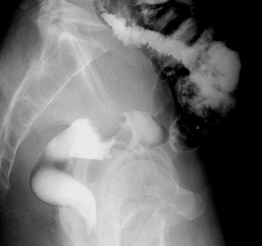
The rectum appears distended and well-filled with barium, forming a smooth, rounded barium pool in the pelvis — this appears relatively normal in calibre on this view.
Right film (post-evacuation / double-contrast view):
This is where the rectum becomes more informative. In the lower pelvic area, the rectal outline shows:
- Irregular mucosal pattern — the normally smooth rectal wall mucosa appears disrupted and uneven
- Possible additional narrowing/deformity at the rectosigmoid junction or upper rectum — there is an area of irregular barium coating with loss of smooth contour
- Asymmetric filling — the barium does not coat the rectal wall uniformly, suggesting a mural lesion or mucosal irregularity
Structured Analysis of the Rectal Finding
| Parameter | Finding |
|---|---|
| Sequence | Focal narrowing at rectosigmoid / upper rectum |
| Barium | Irregular, asymmetric coating; mucosal destruction |
| Border | Ill-defined, uneven; irregular margins |
| Size | Short segment; localized |
| Contrast | Partial filling defect / mucosal irregularity |
| Shape | Irregular intraluminal defect — possible second apple-core or ulcerating mass |
Clinical Significance
This raises the important concern of a synchronous colorectal carcinoma — two separate tumours in the same patient at the same time.
Synchronous colorectal cancers occur in 3–5% of colorectal cancer cases. The double-contrast barium enema is specifically valued for its ability to detect synchronous lesions in the rest of the colon when one primary tumour is identified. — Yamada's Textbook of Gastroenterology (colorectal cancer section)
Two possible explanations for the rectal finding:
- Second primary carcinoma (synchronous) — given the irregular mucosal pattern and asymmetric filling
- Tumour extension — if the primary sigmoid lesion has spread inferiorly to involve the rectosigmoid junction
Reference Image — Rectal Carcinoma on Barium Enema
Bottom Line
Yes, the rectum is NOT normal on this study. The rectal region — most visible on the right (post-evacuation) film — shows irregular mucosal coating and asymmetric filling, strongly suggesting either:
- A second synchronous carcinoma of the rectum/rectosigmoid, OR
- Direct extension of the sigmoid tumour downward
This should be further evaluated with:
- Rigid/flexible sigmoidoscopy + colonoscopy (to biopsy both lesions)
- MRI rectum (gold standard for rectal cancer staging — T and N staging)
- CT chest/abdomen/pelvis for distant metastasis staging
This is a shared conversation. Sign in to Orris to start your own chat.