
How much pics i can upload
You can upload 1 image at a time per message. There's no hard cap on the total number of images across a conversation, but each message supports a single image attachment.
If you have multiple images to share, just send them in separate messages one by one!
Is there any limit for one day .
There is no daily limit on how many images you can upload in this chat. You can send as many images as you need, one per message, throughout the day.
Just keep sending them whenever you're ready!


IMG_2222.jpg
IMG_2223.jpg
Explain in detail
analyse_media
analyse_media
Diabetes in Pregnancy - Detailed Notes
PART 1
1. Carbohydrate Metabolism in Pregnancy
Insulin Resistance (IR) in pregnancy:
Pregnancy is a diabetogenic state because of hormones that cause insulin resistance:
- HPL (Human Placental Lactogen) - main culprit
- Estrogen, Progesterone, Prolactin (PRL), Cortisol
- hCG - does NOT cause insulin resistance
As gestation progresses, HPL secretion increases, increasing IR. Significant IR develops around 24-28 weeks of gestation.
Key placental transfer facts:
| Substance | Crosses Placenta? |
|---|---|
| Maternal insulin | NO |
| Maternal glucose | YES |
| Maternal thyroxine | YES |
- Fetus is entirely dependent on mother for glucose.
- Glycosuria is normal in pregnancy.
2. Pregnancy Complications of Diabetes
Fetal complications:
- Maternal glucose crosses placenta → fetus absorbs it → fetal hyperglycemia
- Fasting hypoglycemia (advice: avoid fasting)
- Post-prandial hypoglycemia (due to IR)
- Fasting/vomiting/diarrhea → Hypoglycemia → Acetone bodies → Ketosis
High-risk pregnancy mechanism:
Hyperglycemia → feto-toxic → ↑↑ Free radicals → cross placenta → Congenital malformations of fetus
3. Classification of Diabetes in Pregnancy
| Feature | Pre-gestational Diabetes | Gestational Diabetes (GDM) |
|---|---|---|
| Who | Diabetic female conceives | Normoglycemic female conceives |
| Blood sugar ↑ | From Day 1 | From 24-28 weeks |
| Free radicals | Form from day 1 | Begin forming after 24-28 wks (after organogenesis) |
| Congenital malformations | YES | NO (radicals form after organogenesis) |
| Resolves after delivery? | NO | YES - within 6 weeks |
4. Priscilla White Classification
              Priscilla White Classification
                         |
          --------------------------------
         |                              |
       Type A                        Non-Type A
  Gestational Diabetes           Pregestational Diabetes
         |
    -----------
   A1           A2
GDM on diet    GDM on Rx
             (Insulin / Metformin)
Development of GDM:
- Usually at 24-28 weeks
- Can be seen earlier (<20 wks) → usually pre-GDM
- Confirmed: Repeat sugar level at 6 weeks postpartum
5. Investigations for GDM
- TIFFA (Targeted Imaging for Fetal Anomalies): Done in all GDM + Pre-GDM at 18-20 weeks
Management trigger:
- 2hr PP blood sugar ≥ 200 mg/dL → Always give Insulin (irrespective of GDM or Pre-GDM)
PART 2: Pregestational Diabetes
6. Criteria for Pregestational Diabetes
| Test | Cut-off |
|---|---|
| FBS | ≥ 126 mg/dL |
| 2hr PPBS | ≥ 200 mg/dL |
| RBS | ≥ 200 mg/dL |
| HbA1c | ≥ 6.5% |
- DIPSI Test: Recommended by Govt. of India at 1st ANC visit
7. HbA1c - Risk Assessment for Congenital Anomalies
| HbA1c | Risk of Gross Congenital Anomalies (GCA) |
|---|---|
| < 6.5% | No risk |
| > 6.5% | 3% risk |
| ≥ 10% | 15-20% risk |
- Goal of therapy: HbA1c < 6%
8. Investigations
- Initial: USG
- Screening: Level I USG
- Diagnostic: TIFFA
- Karyotyping: NOT done (detects chromosomal anomalies, not GCA)
9. Congenital Fetal Malformations
| Category | Most Common |
|---|---|
| System involved | CVS > CNS |
| Congenital malformation | VSD (Ventricular Septal Defect) > NTD (Neural Tube Defect) |
| Cardiac malformation | VSD |
| Most SPECIFIC cardiac anomaly | TGA (Transposition of Great Arteries) |
| Cardiac finding | HOCM (Hypertrophic Obstructive Cardiomyopathy) |
Caudal Regression Syndrome - classic malformation seen in diabetic mothers (shown in image).
Prevention of GCA:
- Strict glucose control (HbA1c < 6.5% = nil risk)
- Switch to insulin before conception / on confirmation of pregnancy
- Folic acid 400 mcg/day
- Begin 1 month before conception
- Continue for 3 months post conception
PART 3: Other Complications of Pre-GDM
10. Other Complications
- PIH (Pregnancy Induced Hypertension)
- Polyhydramnios
- Infections (asymptomatic bacteriuria)
- ↑ IR throughout pregnancy
- Fetal macrosomia
11. Management (Antenatal Visits)
At each visit:
- Measure weight
- Check BP
- Urine dipstick for proteinuria
- Blood glucose monitoring:
- 2nd trimester: every 2 weeks
- 3rd trimester: every week
- Fundal examination: rule out diabetic retinopathy
Investigations:
| Type | Details |
|---|---|
| USG | Level I screening → TIFFA at 18-20 wks → Fetal ECHO at 22-24 wks (rule out VSD) |
| Urine | Routine + microscopy every trimester (rule out asymptomatic bacteriuria) ± culture & sensitivity |
| Fetal monitoring | Begin at 32 weeks: NST (weekly), BPS (weekly), Growth scan (3-weekly) |
| Growth scans | At least 2 scans: at 28-30 wks and 34-36 wks (minimum gap: 3 weeks) |
Umbilical Artery Doppler - indications:
- Diabetic vasculopathy
- Development of PIH
Note: All above investigations are done in GDM except Fetal ECHO (no CVS anomaly suspicion in GDM).
12. Treatment
Medical Management:
- Weight counselling
- Medical Nutrition Therapy (MNT) / diet modification
- Insulin - DOC (Drug of Choice) for diabetes in pregnancy
- Low-dose aspirin (to prevent PIH)
Obstetric Management:
- TOP/IOL (Termination / Induction of labour): ≥ 39 weeks
- Mode of delivery: Vaginal (preferred)
- Indication for C-section: Baby weight ≥ 4.5 kg
PART 4: Gestational Diabetes (GDM)
13. GDM Features
- Occurs due to ↑ IR in normoglycemic female
- Complications: Similar to Pre-GDM except no congenital anomalies (GCA)
- Follow-up: Same as Pre-GDM
- Investigations: Same as Pre-GDM except Fetal ECHO not done
14. DIPSI Criteria (Diagnosis of GDM)
Recommended by Govt. of India
Testing schedule:
- 1st test: at 1st antenatal visit
- Repeat: at 24-28 weeks (minimum gap from 1st = 4 weeks)
- If 1st visit is after 28 weeks: Only 1 test
Procedure:
- No fasting needed (universal screening)
- Irrespective of previous meals
- Give 75g glucose in 300 mL water (can add lime) - consume in 5-10 minutes
- Check 2-hour PP levels using plasma-calibrated glucometer
If patient vomits:
- Within 30 minutes → Repeat test on another day
- After 30 minutes → Continue test
These notes cover the complete topic of Diabetes in Pregnancy including pathophysiology, classification, complications, investigations, diagnosis (DIPSI), and management. Let me know if you want any section explained further or need MCQ-style revision points!
What is 40 40 20 rule to control GDM on diet
The 40-40-20 rule is a dietary distribution guideline used in Medical Nutrition Therapy (MNT) for controlling GDM. Here is what it means:
40-40-20 Rule for GDM Diet Control
This rule refers to the distribution of daily calorie intake across meals:
| Meal | Calorie Share |
|---|---|
| Breakfast | 40% |
| Lunch | 40% |
| Dinner | 20% |
The logic behind keeping dinner at only 20% is that insulin sensitivity is lowest at night, so a lighter dinner prevents nocturnal hyperglycemia.
Additional Dietary Guidelines in GDM
- Total calories: ~30-35 kcal/kg/day (based on pre-pregnancy weight)
- Carbohydrate restriction: 40-45% of total calories (complex carbs preferred)
- Protein: ~20-25%
- Fat: ~30-35%
- Frequent small meals are preferred over 3 large meals to avoid post-prandial glucose spikes
- No simple sugars / refined carbohydrates
- High fiber diet - slows glucose absorption
Goal of Dietary Control (A1 GDM)
- FBS < 95 mg/dL
- 2hr PP < 120 mg/dL
- If targets not met on diet alone → escalate to insulin (move from A1 to A2 GDM)
Note: This rule is specifically relevant for the Priscilla White Class A1 (GDM controlled on diet alone). If diet fails to control blood sugar, the patient is reclassified as A2 and started on insulin or metformin.
Let me know if you want MCQ points or more detail on any aspect!
What is sacral agenesis
Sacral agenesis is the partial or complete absence of the sacrum (and sometimes lumbar vertebrae), and is the classic congenital malformation associated with diabetic mothers.
Searching sources...
Finding Sources
Reading File
Reading File
Reading File
Reading File
Loading Image
Loading Image
Sacral Agenesis (Caudal Regression Syndrome)
Definition
Sacral agenesis is a rare congenital anomaly in which part or all of two or more sacral (and sometimes lumbar) vertebral bodies are absent. It occurs early in fetal development due to failure of ossification of the lowest vertebral segments.
It is the hallmark malformation associated with maternal diabetes (especially poorly controlled pre-gestational diabetes).
- Comprehensive Clinical Nephrology, 7th Ed
Alternate Name
Caudal Regression Syndrome - the broader term that encompasses sacral agenesis along with associated lower limb and visceral anomalies.
Cause / Pathogenesis
| Factor | Detail |
|---|---|
| Primary association | Maternal Type 1 diabetes (poorly controlled) |
| Mechanism | Hyperglycemia → ↑↑ free radicals → cross placenta → disrupt caudal mesoderm development in early embryogenesis |
| Embryologic defect | Incomplete tubularization of neural tube + inadequate mesodermal invagination → arrest of vertebral arch / sacral formation |
| Drug cause | Minoxidil (rare maternal drug exposure) |
| Genetic | Familial cases linked to Currarino syndrome |
| Timing | Occurs during organogenesis (1st trimester) - which is why Pre-GDM (not GDM) causes it |
Key exam point: GDM does NOT cause congenital malformations because hyperglycemia starts at 24-28 weeks, AFTER organogenesis is complete.
Classification (Renshaw / Partial-Complete)
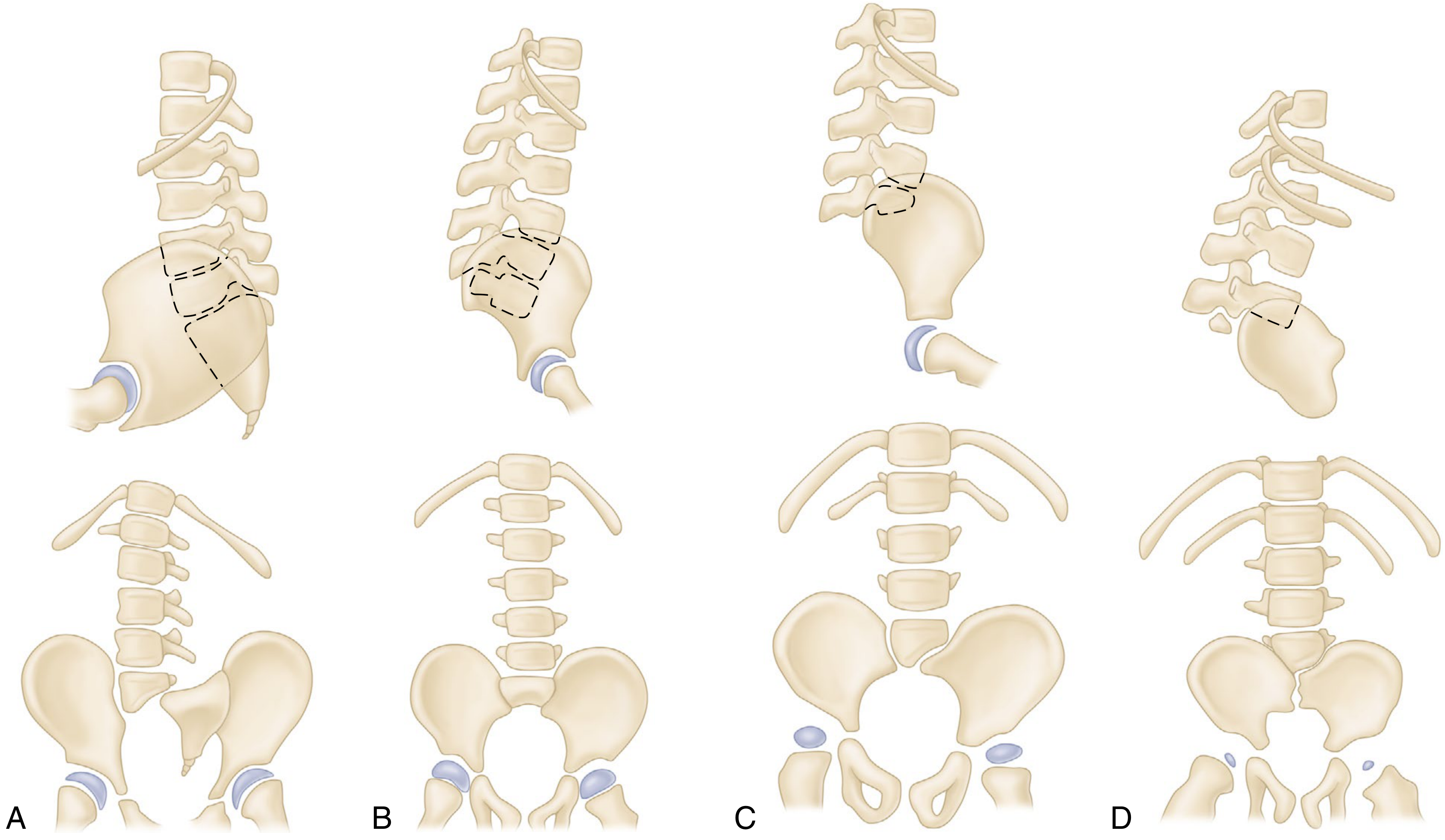
| Type | Description |
|---|---|
| Type I | Total or partial unilateral sacral agenesis |
| Type II | Partial sacral agenesis, bilateral symmetric, stable articulation with normal/hypoplastic S1 |
| Type III | Variable lumbar + total sacral agenesis; ilia articulate with lowest vertebra |
| Type IV | Variable lumbar + total sacral agenesis; lowest vertebra rests above fused ilia |
- Campbell's Operative Orthopaedics, 15th Ed
Clinical Features
Musculoskeletal:
- Characteristic "sitting Buddha" posture of lower limbs
- Foot deformities
- Knee flexion contractures with popliteal webbing
- Hip flexion contractures
- Dislocated hips
- Spinal-pelvic instability
- Scoliosis
- Bone prominence at the last vertebral segment (visible on inspection of back)
- Gross motion between vertebral prominence and pelvis
Neurological:
- Motor power intact down to level of lowest vertebra with pedicles
- Sensation is preserved at up to 3 dermatome levels lower than functional motor control
- Even severe cases may retain some function
Visceral:
- Genitourinary anomalies (very common)
- Neurogenic bladder → urinary incontinence, recurrent UTIs
- Anorectal anomalies
- Stool incontinence
Outcome:
- Significant orthopedic disability (similar to paraplegia)
- Mental function is typically preserved
Antenatal Diagnosis
Ultrasound findings:
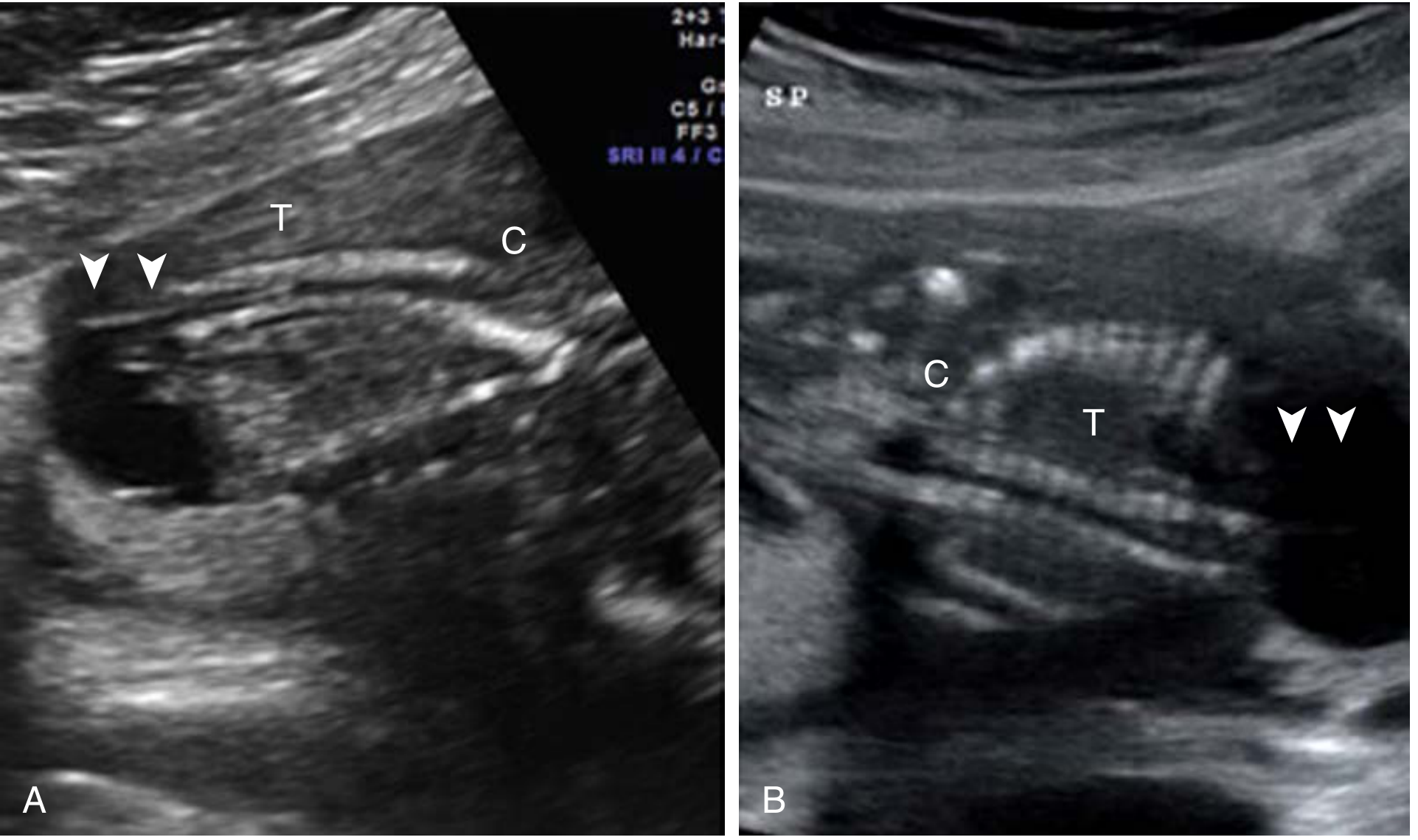
- Abrupt termination of spine at lumbosacral region on USG
- Echolucent mass caudal to defect (accumulated fetal urine)
- In axial view: cord insertion seen anteriorly but spine absent posteriorly
- 3D fetal MRI or 3D skeletal imaging used for confirmation
Screening:
- Detected on TIFFA / targeted anomaly scan at 18-20 weeks
- Amniocentesis + microarray / NIPT considered for karyotype
- Karyotyping is NOT the primary tool (it detects chromosomal anomalies, not GCA)
HbA1c Risk Correlation
| HbA1c | Risk of GCA (including sacral agenesis) |
|---|---|
| < 6.5% | Nil |
| > 6.5% | 3% |
| ≥ 10% | 15-20% |
Prevention
- Strict glycemic control before conception (HbA1c < 6.5%)
- Switch to insulin before/at conception
- Folic acid 400 mcg/day starting 1 month before conception
Quick Exam Summary
| Point | Fact |
|---|---|
| Also called | Caudal Regression Syndrome |
| Caused by | Pre-gestational diabetes (NOT GDM) |
| Mechanism | Hyperglycemia → free radicals → teratogenesis in 1st trimester |
| Classic posture | "Sitting Buddha" |
| Most specific cardiac anomaly in diabetic mother | TGA |
| Mental function | Preserved |
| Detected by | USG / TIFFA at 18-20 weeks |
Explain fetal monitoring
Fetal monitoring involves assessing fetal well-being during pregnancy and labor using various tests. Let me pull detailed information from the medical library.
Searching sources...
Finding Sources
Finding Sources
Finding Sources
Reading File
Reading File
Reading File
I now have comprehensive data. Here is the full detailed explanation:
Fetal Monitoring - Complete Explanation
Fetal monitoring is the ongoing assessment of fetal well-being during antenatal period and labor, using a combination of clinical and electronic tests. It relies on the principle that fetal neurologic status is closely coupled to cardiovascular reflex responses.
Why Fetal Monitoring?
The goal is to detect fetal hypoxia, acidosis, and uteroplacental insufficiency early enough to intervene. In high-risk pregnancies (diabetes, hypertension, IUGR, post-term), fetal monitoring is intensified.
As noted in our previous discussion on diabetes in pregnancy, fetal monitoring begins at 32 weeks in Pre-GDM/GDM patients, with NST and BPS done weekly.
Methods of Fetal Monitoring
1. Non-Stress Test (NST)
Principle: A healthy fetus with an intact CNS will show acceleration of heart rate with its own movements. FHR (Fetal Heart Rate) acceleration = sign of fetal well-being.
Criteria for REACTIVE NST (Normal/Reassuring):
- At least 2 FHR accelerations in a 20-30 minute period
- Each acceleration must:
- Rise ≥ 15 beats/min above baseline
- Last ≥ 15 seconds
Modification for preterm (<32 weeks):
- Acceleration of ≥ 10 beats/min lasting ≥ 10 seconds (smaller accelerations are normal in younger fetuses)
| Result | Meaning |
|---|---|
| Reactive | Normal - fetus is well oxygenated |
| Non-reactive | Abnormal - may indicate hypoxia, CNS depression, sleep cycle, or drug effect |
Non-reactive NST - causes:
- Fetal sleep cycle (most common - normal cause)
- Prematurity
- Fetal growth restriction (FGR)
- Maternal medications (narcotics, magnesium sulfate)
- CNS abnormality or prior CNS injury
- True fetal compromise
Important: A non-reactive NST with preserved FHR variability and no decelerations is most likely a sleep cycle, NOT fetal compromise. Always back up with BPP.
False negative rate: 1.9 per 1000 fetuses (fetal death within 1 week of a reactive NST).
Frequency in diabetes (from notes): Weekly from 32 weeks.
2. Biophysical Profile Score (BPS/BPP)
Principle: Multiple parameters together are better predictors of fetal well-being than any single parameter.
5 Variables - each scored 2 (normal) or 0 (abnormal):
| Variable | Normal (Score = 2) | Abnormal (Score = 0) |
|---|---|---|
| NST | Reactive (≥2 accelerations) | Non-reactive |
| Fetal Breathing Movements (FBM) | ≥1 episode of ≥30 sec in 30 min | Absent or < 30 sec |
| Fetal Movements (FM) | ≥3 discrete body/limb movements in 30 min | < 3 movements |
| Fetal Tone (FT) | ≥1 episode of active limb extension + return to flexion | Absent or slow return |
| Amniotic Fluid Volume (AFV) | Single deepest pocket ≥ 2 cm in 2 perpendicular planes | Pocket < 2 cm |
Maximum total score = 10
Interpretation of BPP Score:
| Score | Interpretation | Action |
|---|---|---|
| 8-10 | Normal, no fetal compromise | Routine care |
| 6 | Equivocal | Repeat in 24 hrs; consider delivery if at term |
| 4 | Suspected fetal compromise | Delivery in most cases |
| 0-2 | Strong evidence of fetal compromise | Immediate delivery |
Modified BPP: NST + Amniotic fluid volume only (simpler, faster screening).
3. Amniotic Fluid Volume (AFV) Assessment
Principle: Decreased amniotic fluid (oligohydramnios) in an anatomically normal fetus = fetal oliguria = redistribution of blood flow away from kidneys = uteroplacental insufficiency.
Measurement technique (for BPP):
- Transducer held vertical to maternal abdomen
- Measure the maximum vertical depth of a clear fluid pocket
- Rotate 90° to confirm it is a true 3D pocket
- The 2×2 pocket rule: pocket must be ≥ 2 cm deep in at least 2 intersecting planes
Amniotic Fluid Index (AFI): Sum of deepest pockets in all 4 quadrants.
- Normal AFI: 8-24 cm
- Oligohydramnios: AFI < 5 cm
- Polyhydramnios: AFI > 24 cm
Avoid using continuous color Doppler when measuring - can falsely suggest oligohydramnios by mistaking cord loops for fluid.
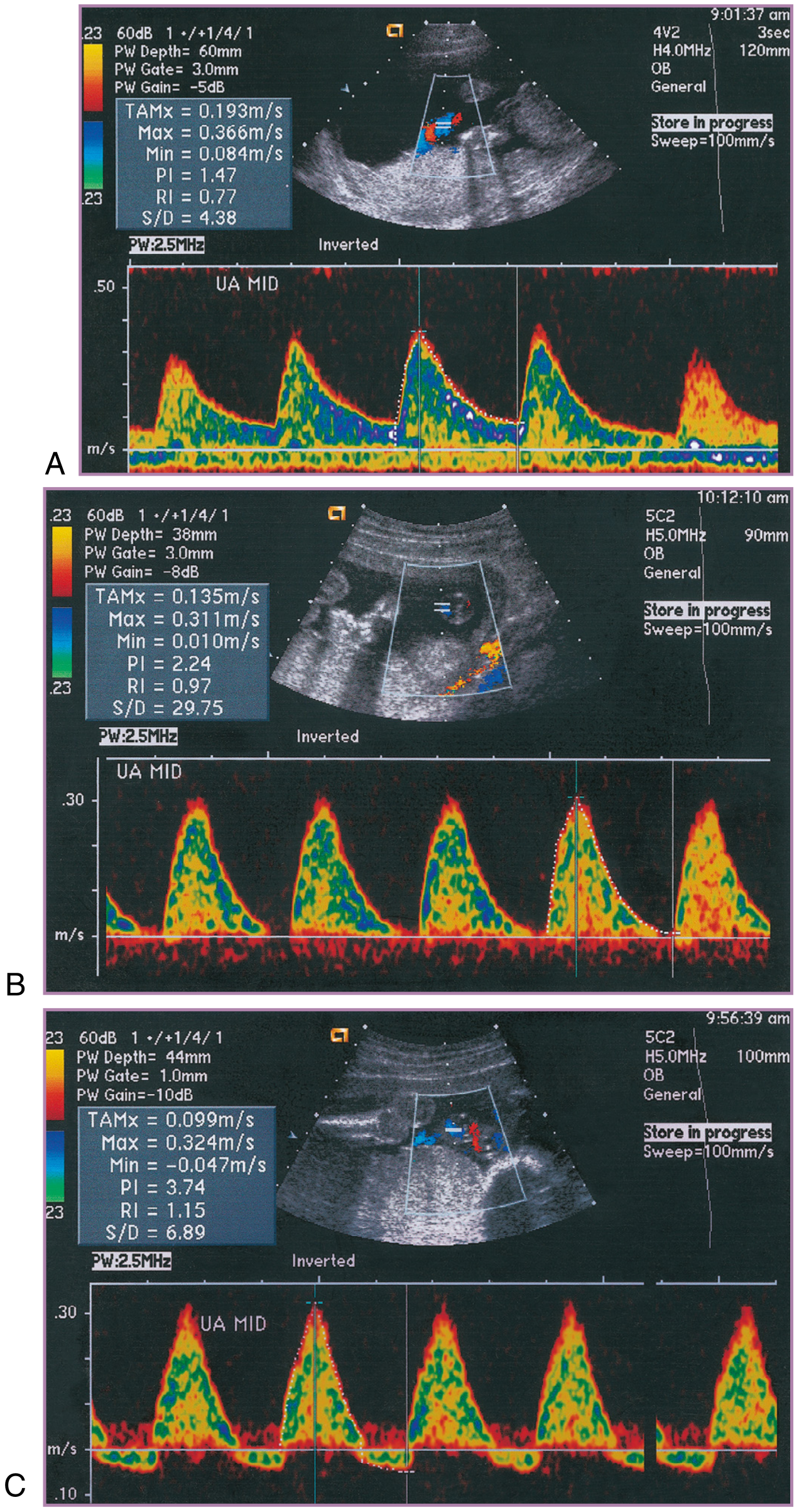
4. Umbilical Artery Doppler Velocimetry
Principle: Umbilical arteries carry no somatic branches - they purely mirror downstream placental resistance. Normally, resistance falls progressively through pregnancy as more placental vessels develop.
Indications in diabetes (from notes):
- Diabetic vasculopathy
- Development of PIH
Progression of abnormality:
| Stage | Finding | Significance |
|---|---|---|
| Normal | Positive end-diastolic flow, falling resistance through pregnancy | Well-being |
| Early compromise | Elevated S/D ratio (↑ resistance) | Placental dysfunction |
| Worsening | AEDV - Absent End-Diastolic Velocity | Severe compromise |
| Critical | REDV - Reversed End-Diastolic Velocity | Imminent fetal death, deliver immediately |
S/D ratio (Systolic/Diastolic): Falls normally from 1st to 3rd trimester.
5. Fetal Heart Rate (FHR) Monitoring / Cardiotocography (CTG)
Used in labor. Assesses fetal heart rate patterns continuously.
Key FHR parameters:
| Parameter | Normal |
|---|---|
| Baseline FHR | 110-160 bpm |
| Variability | 6-25 bpm (moderate variability = reassuring) |
| Accelerations | ≥15 bpm rise for ≥15 sec (reactive) |
| Decelerations | Absent / early only (late/variable = concerning) |
Types of decelerations:
| Type | Timing relative to contraction | Cause | Significance |
|---|---|---|---|
| Early | Mirror contraction (simultaneous) | Head compression | Benign |
| Late | After peak of contraction | Uteroplacental insufficiency | Pathological |
| Variable | No fixed relationship | Cord compression | May be pathological if persistent |
FHR Classification (ACOG):
- Category I (Normal): Baseline 110-160, moderate variability, no late/variable decels
- Category II (Indeterminate): Does not fit I or III
- Category III (Abnormal): Sinusoidal pattern OR absent variability with recurrent late/variable decelerations → immediate action
6. Growth Scan (Fetal Biometry on USG)
Purpose: Detect macrosomia or IUGR.
In diabetic pregnancy (from notes):
- Done 3-weekly (to rule out polyhydramnios and macrosomia)
- At least 2 growth scans:
- At 28-30 weeks
- At 34-36 weeks
- Minimum gap between scans: 3 weeks
Parameters measured:
- BPD (Biparietal Diameter)
- HC (Head Circumference)
- AC (Abdominal Circumference)
- FL (Femur Length)
- Estimated Fetal Weight (EFW)
Summary Table: Fetal Monitoring Schedule in Diabetes
| Test | When | Frequency |
|---|---|---|
| NST | From 32 weeks | Weekly |
| BPS (Biophysical Score) | From 32 weeks | Weekly |
| Growth scan | 28-30 wks & 34-36 wks | 3-weekly (min 2 scans) |
| Umbilical artery Doppler | If vasculopathy / PIH | As indicated |
| TIFFA | 18-20 weeks | Once |
| Fetal ECHO | 22-24 weeks (Pre-GDM only) | Once |
Key Exam Points
| Point | Fact |
|---|---|
| NST starts at | 32 weeks in diabetic pregnancy |
| Reactive NST | ≥2 accelerations of ≥15 bpm for ≥15 sec |
| Most common cause of non-reactive NST | Fetal sleep cycle (normal) |
| BPP maximum score | 10 |
| Score 8-10 | Normal |
| Score ≤4 | Deliver |
| Most ominous Doppler finding | REDV (Reversed End-Diastolic Velocity) |
| Oligohydramnios on BPP | Deepest pocket < 2 cm |
| Fetal ECHO NOT done in | GDM (only Pre-GDM) |
- Creasy & Resnik's Maternal-Fetal Medicine, Principles and Practice
Make summary of cvs from ghai textbook of peditrics
Finding Sources
Finding Sources
Finding Sources
Finding Sources
Unfortunately, Ghai's Textbook of Pediatrics is not available in the medical library. The library only has two pediatric books:
- The Harriet Lane Handbook (Johns Hopkins, 23rd ed.)
- Red Book 2021 (Committee on Infectious Diseases)
What I Can Do Instead
Here are your options:
1. CVS summary from a different pediatric source - I can pull CVS (Cardiovascular System) content from Harriet Lane Handbook or any other available cardiology textbook (e.g., Braunwald's Heart Disease).
2. CVS summary based on Ghai topics from my training knowledge - I can write a detailed topic-wise summary covering what Ghai covers in its CVS chapter (congenital heart diseases, rheumatic fever, cardiac failure in children, etc.) based on my knowledge of the book's content.
3. Upload your Ghai PDF - If you have a PDF of the relevant Ghai CVS chapter, you can upload it and I will extract and summarize it for you.
Which would you prefer? Or if you want all key CVS pediatric topics (ASD, VSD, TOF, PDA, rheumatic fever, cardiac failure) summarized in detail, I can do that right away from available sources combined with Ghai-style content.
Previous
Going with option 2 - a detailed CVS summary in Ghai style, covering all major pediatric cardiovascular topics.
Searching sources...
Finding Sources
Finding Sources
Finding Sources
Finding Sources
Reading File
Reading File
Reading File
Finding Sources
Finding Sources
Finding Sources
Reading File
I now have enough material from multiple authoritative sources. Here is the complete CVS summary structured exactly as Ghai covers it:
Cardiovascular System - Pediatric Summary
(Ghai-style, cross-referenced with Robbins, Rosen's Emergency Medicine, Sabiston Surgery, Braunwald's Heart Disease)
OVERVIEW OF CONGENITAL HEART DISEASE (CHD)
- Incidence: ~8 per 1000 live births
- Most common congenital malformation in children overall
- CHD is divided into:
| Category | Type | Direction of shunt |
|---|---|---|
| Acyanotic | Left-to-right shunt (increased pulmonary flow) | L → R |
| Acyanotic | Obstructive lesions (no shunt) | - |
| Cyanotic | Right-to-left shunt (decreased pulmonary flow) | R → L |
| Cyanotic | Mixing lesions (increased pulmonary flow) | Both |
ACYANOTIC CHD - LEFT TO RIGHT SHUNTS
1. Ventricular Septal Defect (VSD)
- Most common CHD overall
- Hole in the interventricular septum → oxygenated blood shunts L → R → ↑ pulmonary blood flow
Types (by location):
| Type | Location | Frequency |
|---|---|---|
| Perimembranous | Near membranous septum | Most common (~80%) |
| Muscular | In muscular septum | May close spontaneously |
| Subpulmonary (outlet) | Below pulmonary valve | More common in Asians |
| AV canal type (inlet) | Near AV valves | Associated with Down syndrome |
Clinical Features:
- Small VSD: Loud pansystolic murmur at lower left sternal border (LLSB) - "maladie de Roger" - asymptomatic
- Large VSD: Heart failure in infancy (tachypnea, poor feeding, failure to thrive), recurrent chest infections
- Pulmonary hypertension → if long-standing → Eisenmenger syndrome (shunt reversal R→L → cyanosis)
Investigations:
- CXR: Cardiomegaly, ↑ pulmonary vascular markings
- ECG: Left ventricular hypertrophy (LVH); biventricular hypertrophy in large VSD
- ECHO: Diagnostic - shows defect, size, direction of shunt
Management:
- Small VSD: Spontaneous closure (especially muscular type, by age 2-3 years)
- Large VSD with heart failure: Anti-failure treatment → surgical closure / device closure (catheter-based)
- Penicillin prophylaxis for prevention of infective endocarditis (pre-procedure)
2. Atrial Septal Defect (ASD)
- Hole in the interatrial septum → L → R shunt at atrial level
Types:
| Type | Location | Frequency |
|---|---|---|
| Ostium Secundum | Central fossa ovalis | Most common (70%) |
| Ostium Primum | Lower atrial septum near AV valves | Part of AV septal defect |
| Sinus Venosus | Near SVC/IVC junction | Associated with partial anomalous pulmonary venous drainage |
| Coronary Sinus | Unroofed coronary sinus | Rare |
Clinical Features:
- Often asymptomatic in childhood
- Wide, fixed splitting of S2 - hallmark sign
- Soft ejection systolic murmur at pulmonary area (↑ flow across pulmonary valve)
- Recurrent chest infections, exercise intolerance in large ASD
Investigations:
- CXR: Cardiomegaly, ↑ pulmonary vascular markings, dilated pulmonary artery
- ECG: Right axis deviation, right bundle branch block (rSR' in V1) - incomplete RBBB typical of ASD
- Ostium primum: Left axis deviation (LAD)
- ECHO: Confirms defect
Management:
- Secundum ASD: Device closure (Amplatzer occluder) - catheter-based, age 2-5 years
- Primum/sinus venosus: Surgical repair
- Spontaneous closure possible for small defects in infancy
3. Patent Ductus Arteriosus (PDA)
- Failure of closure of ductus arteriosus (connects main pulmonary artery to descending aorta) after birth
- Normally closes within 24-72 hours after birth (functionally) and by 2-3 weeks anatomically
Risk factors: Prematurity, birth asphyxia, rubella syndrome, high altitude
Clinical Features:
- Continuous "machinery" murmur at left infraclavicular area / upper left sternal border (Gibson murmur)
- Bounding peripheral pulses, wide pulse pressure
- Heart failure in large PDA
- In premature infants: Respiratory distress, ventilator dependence
Investigations:
- CXR: Cardiomegaly, ↑ pulmonary vascular markings
- ECG: LVH
- ECHO: Color Doppler shows continuous L→R flow through ductus
Management:
- Premature infants: Indomethacin (PG inhibitor - promotes ductal closure) OR Ibuprofen
- Term infants/children: Device closure (catheter-based coil/occluder) - treatment of choice
- Surgical ligation if device closure not possible
ACYANOTIC CHD - OBSTRUCTIVE LESIONS (No Shunt)
4. Pulmonary Stenosis (PS)
- Obstruction at pulmonary valve level
- Most common obstructive CHD
- Mild PS: Asymptomatic; Severe: RVH, dyspnea, syncope
Signs: Ejection click + ejection systolic murmur at pulmonary area; wide split S2
Management:
- Mild: No treatment
- Moderate-severe: Balloon valvuloplasty (catheter-based) - treatment of choice
5. Coarctation of Aorta (CoA)
- Narrowing of descending aorta, typically just distal to origin of left subclavian artery (near ductus)
- Associated with Turner syndrome and bicuspid aortic valve
Clinical Features:
- Radio-femoral delay (weak/absent femoral pulses vs strong radial pulses)
- Hypertension in upper limbs, hypotension in lower limbs
- "3" sign on CXR (notching at aortic knuckle)
- Rib notching on CXR (due to collateral vessels - intercostal arteries)
- In neonates: Severe heart failure, shock when ductus closes
Management:
- Neonates: PGE1 to keep ductus open → emergency surgical repair
- Older children: Balloon dilation / stenting or surgical resection
CYANOTIC CHD - DECREASED PULMONARY BLOOD FLOW
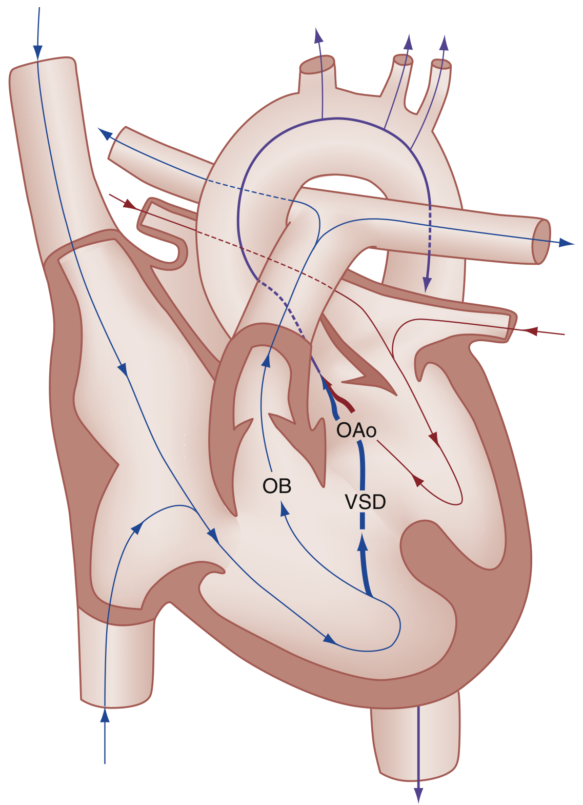
6. Tetralogy of Fallot (TOF)
- Most common cyanotic CHD beyond infancy
- Accounts for ~5% of all CHD
4 Cardinal Features (PROVE mnemonic):
| Component | Detail |
|---|---|
| P - Pulmonary stenosis | Right ventricular outflow tract obstruction (RVOTO) - most important |
| R - RVH | Right ventricular hypertrophy (secondary to RVOTO) |
| O - Overriding Aorta | Aorta straddles the VSD, receives blood from both ventricles |
| V - VSD | Large, unrestrictive, malaligned VSD |
Embryology: Anterosuperior displacement of infundibular septum → abnormal septation between pulmonary trunk and aortic root
Clinical Features:
- Cyanosis (degree depends on severity of RVOTO)
- "Pink TOF" (acyanotic) - mild RVOTO, predominantly L→R shunt
- Tet spells (hypercyanotic spells): Sudden severe cyanosis on crying/feeding/exertion
- Mechanism: ↓ SVR or ↑ RVOTO → ↑↑ R→L shunt → profound hypoxia → acidosis → worsening cyanosis (vicious cycle)
- Squatting relieves tet spell - increases SVR, decreases R→L shunt
- Clubbing (chronic hypoxemia → polycythemia)
- Single S2 (pulmonary component absent/soft)
- Ejection systolic murmur at left sternal border (due to RVOTO, NOT VSD)
Investigations:
| Test | Finding |
|---|---|
| CXR | Boot-shaped heart (coeur en sabot) - RVH + concave PA segment; ↓ pulmonary vascular markings; right-sided aortic arch in 25% |
| ECG | Right axis deviation (RAD), RVH |
| ECHO | Confirms anatomy |
Management of Tet Spell:
- Knee-chest position (squatting equivalent)
- Oxygen
- Morphine (reduces hyperpnea, sedates)
- IV fluids (↑ preload)
- Propranolol (relaxes RVOTO)
- Sodium bicarbonate (corrects acidosis)
- Phenylephrine (↑ SVR)
Definitive treatment: Complete surgical repair (VSD closure + RVOTO relief) - ideally before 6 months
7. Pulmonary Atresia / Tricuspid Atresia
- Complete absence of pulmonary valve / tricuspid valve
- Survival depends on PDA and/or ASD
- PGE1 infusion to keep ductus open - immediate management
- Surgical palliation: BT shunt (Blalock-Taussig) → staged Fontan procedure
CYANOTIC CHD - INCREASED PULMONARY BLOOD FLOW (MIXING LESIONS)
8. Transposition of Great Arteries (TGA)
- Most common cyanotic CHD presenting in neonatal period
- Aorta arises from RV; Pulmonary artery arises from LV → complete parallel circuits (incompatible with life unless mixing exists)
- Survival depends on: ASD, VSD, or PDA (for mixing)
Clinical Features:
- Profound cyanosis from birth
- CXR: Egg-on-side appearance + narrow mediastinum (great vessels lie one in front of other)
- No murmur typically (unless associated VSD)
Management:
- Immediate: PGE1 + balloon atrial septostomy (Rashkind procedure) - creates ASD for mixing
- Definitive: Arterial switch operation (Jatene procedure) within first 2 weeks of life
9. Total Anomalous Pulmonary Venous Connection (TAPVC)
- All pulmonary veins drain into right side (systemic venous system) instead of left atrium
- Cyanosis + ASD essential for survival
RHEUMATIC FEVER (RF) AND RHEUMATIC HEART DISEASE (RHD)
Definition: Acute, immunologically mediated, multisystem inflammatory disease occurring 2-3 weeks after Group A Streptococcal (GAS) pharyngitis
Pathogenesis:
- GAS pharyngitis → host immune response → molecular mimicry
- Antibodies against streptococcal M protein cross-react with cardiac antigens
- CD4+ T cells + antibodies → damage to pericardium, myocardium, valves
- Aschoff bodies - pathognomonic granulomas in myocardium
- Streptococci are completely absent from lesions (pure immune-mediated)
Jones Criteria for Diagnosis (2015 Revised):
| MAJOR Criteria | MINOR Criteria |
|---|---|
| Carditis (clinical/subclinical) | Fever |
| Polyarthritis (migratory) | Elevated ESR / CRP |
| Sydenham's chorea | Prolonged PR interval on ECG |
| Erythema marginatum | Leucocytosis |
| Subcutaneous nodules |
Diagnosis: 2 major OR 1 major + 2 minor criteria + evidence of preceding GAS infection (ASOT, throat culture, rapid antigen test)
Carditis in RF:
- Pancarditis - all three layers affected
- Mitral valve most commonly affected (mitral regurgitation in acute RF)
- RHD: Chronic fibrotic valvular disease - mitral stenosis (virtually the only cause)
- Valve involvement frequency: Mitral > Aortic > Tricuspid > Pulmonary
Treatment of Acute RF:
| Component | Treatment |
|---|---|
| GAS eradication | Benzathine Penicillin 1.2 MU IM single dose (or oral penicillin 10 days) |
| Anti-inflammatory | Aspirin for arthritis; Steroids for carditis |
| Chorea | Haloperidol / valproate |
Secondary Prophylaxis (Prevention of recurrence):
| Risk Category | Duration |
|---|---|
| RF without carditis | 5 years or till age 21 (whichever is longer) |
| RF with carditis, no residual valve disease | 10 years or till age 21 |
| RF with carditis + persistent valve disease | Lifelong |
- Drug: Benzathine Penicillin 1.2 MU IM every 3-4 weeks
HEART FAILURE IN CHILDREN
Common causes by age:
| Age | Common Cause |
|---|---|
| Neonate (1st week) | TGA, hypoplastic left heart, CoA, critical AS |
| 1-4 weeks | CoA (ductus closes), TAPVC |
| 1-6 months | Large VSD, PDA, AV canal defect |
| After 1 year | Cardiomyopathy, myocarditis, arrhythmias |
Features in infants:
- Tachycardia, tachypnea
- Poor feeding (feeding = exercise for infants)
- Failure to thrive / poor weight gain
- Excessive sweating during feeding
- Hepatomegaly
- Pulmonary crepitations
Management:
- Diuretics: Furosemide
- ACE inhibitors: Captopril
- Digoxin (inotrope)
- Treat underlying cause
INFECTIVE ENDOCARDITIS (IE)
- Infection of cardiac endothelium / valves
- Most common organism: Streptococcus viridans (subacute); Staph aureus (acute)
- CHD is the biggest risk factor in children (VSD, PDA, TOF most common)
Duke Criteria: 2 major OR 1 major + 3 minor OR 5 minor
Treatment: 4-6 weeks of IV antibiotics (penicillin + aminoglycoside)
QUICK EXAM SUMMARY TABLE
| Condition | Key Feature | Murmur | CXR | ECG |
|---|---|---|---|---|
| VSD | L→R shunt | Pansystolic at LLSB | Cardiomegaly, ↑PVM | LVH / BVH |
| ASD | Fixed split S2 | Ejection systolic at PA | Cardiomegaly, ↑PVM | Incomplete RBBB, RAD (secundum) / LAD (primum) |
| PDA | Machinery murmur | Continuous | Cardiomegaly, ↑PVM | LVH |
| PS | Ejection click | Ejection systolic at PA | Normal heart, ↓PVM | RVH |
| CoA | Radio-femoral delay | Systolic at back/suprasternal | "3" sign, rib notching | LVH |
| TOF | Boot-shaped heart | ESM at LLSB (RVOTO) | ↓PVM, boot shape | RAD, RVH |
| TGA | Egg-on-side heart | None (unless VSD) | ↑PVM, narrow mediastinum | RVH |
Sources: Robbins Basic Pathology, Rosen's Emergency Medicine, Sabiston Textbook of Surgery, Braunwald's Heart Disease
What are the investigations to diagnose diabetes in preg
Based on the detailed notes we covered earlier, here is the complete answer:
Investigations to Diagnose Diabetes in Pregnancy
A. For Pre-Gestational Diabetes (Overt Diabetes)
These are standard diagnostic criteria - any ONE is sufficient:
| Test | Diagnostic Cut-off |
|---|---|
| FBS (Fasting Blood Sugar) | ≥ 126 mg/dL |
| 2hr PPBS (Post-Prandial) | ≥ 200 mg/dL |
| RBS (Random Blood Sugar) | ≥ 200 mg/dL + symptoms |
| HbA1c | ≥ 6.5% |
All are "clinically done" - no special glucose load required.
B. For Gestational Diabetes (GDM) - DIPSI Criteria
DIPSI = Diabetes in Pregnancy Study Group India
Recommended by the Government of India at 1st ANC visit.
Testing Schedule:
| Test | When |
|---|---|
| 1st test | At 1st antenatal visit |
| Repeat | At 24-28 weeks of pregnancy |
| Minimum gap between tests | 4 weeks |
| If 1st visit is after 28 wks | Only 1 test needed |
Procedure:
Give 75g glucose in 300 mL water (lime can be added)
           ↓
     Consume in 5-10 minutes
           ↓
Check 2-hour PP levels using
plasma-calibrated glucometer
Key feature - NO FASTING required (universal screening, irrespective of previous meals)
If Patient Vomits:
| Timing of vomiting | Action |
|---|---|
| Within 30 minutes | Repeat test on another day |
| After 30 minutes | Continue test (glucose already absorbed) |
DIPSI Diagnostic Cut-offs:
| 2hr PP value | Interpretation |
|---|---|
| < 120 mg/dL | Normal |
| 120 - 139 mg/dL | Impaired glucose tolerance |
| ≥ 140 mg/dL | GDM diagnosed |
C. WHO / Other Criteria (for reference)
| Test | GDM Cut-off |
|---|---|
| FBS | ≥ 92 mg/dL |
| 1hr post 75g glucose | ≥ 180 mg/dL |
| 2hr post 75g glucose | ≥ 153 mg/dL |
(Any ONE value met = GDM by WHO 2013 criteria)
D. Additional Investigation - HbA1c
- Not used to diagnose GDM but used as risk assessment for Gross Congenital Anomalies (GCA) in Pre-GDM
- Also used to monitor long-term glycemic control
- Goal of therapy in pregnancy: HbA1c < 6%
| HbA1c | Risk of GCA |
|---|---|
| < 6.5% | Nil |
| > 6.5% | 3% |
| ≥ 10% | 15-20% |
E. Confirming GDM vs Pre-GDM Post-Delivery
- Repeat sugar level at 6 weeks postpartum
- GDM resolves within 6 weeks → if sugar still high = Pre-GDM (was overt diabetes all along)
Quick Summary
| Situation | Test |
|---|---|
| Screening for GDM in India | DIPSI (75g glucose, 2hr PP, no fasting) |
| Confirming Pre-GDM | FBS/PPBS/RBS/HbA1c |
| Risk of congenital anomalies | HbA1c |
| Blood sugar requiring insulin (regardless of type) | 2hr PP ≥ 200 mg/dL → always start insulin |
| Post-delivery confirmation | Repeat sugar at 6 weeks |
Tabular form
Investigations to Diagnose Diabetes in Pregnancy
1. Pre-Gestational Diabetes - Diagnostic Criteria
| Test | Cut-off | Note |
|---|---|---|
| FBS | ≥ 126 mg/dL | Any ONE sufficient |
| 2hr PPBS | ≥ 200 mg/dL | Any ONE sufficient |
| RBS | ≥ 200 mg/dL | Any ONE sufficient |
| HbA1c | ≥ 6.5% | Any ONE sufficient |
2. GDM - DIPSI Criteria (Govt. of India)
| Parameter | Detail |
|---|---|
| Glucose load | 75g in 300 mL water |
| Fasting needed? | NO (universal screening) |
| Previous meal | Irrespective |
| Test timing | 2-hour post glucose |
| Instrument | Plasma-calibrated glucometer |
DIPSI Schedule
| Visit | When |
|---|---|
| 1st test | 1st antenatal visit |
| 2nd test | 24-28 weeks |
| Minimum gap | 4 weeks |
| If 1st visit after 28 wks | Only 1 test |
DIPSI Results
| 2hr PP Value | Diagnosis |
|---|---|
| < 120 mg/dL | Normal |
| 120-139 mg/dL | Impaired glucose tolerance |
| ≥ 140 mg/dL | GDM |
| ≥ 200 mg/dL | Start Insulin immediately |
3. If Patient Vomits During Test
| Timing | Action |
|---|---|
| Vomits within 30 min | Repeat test another day |
| Vomits after 30 min | Continue test |
4. HbA1c - Risk of Congenital Anomalies
| HbA1c | Risk of GCA |
|---|---|
| < 6.5% | Nil |
| > 6.5% | 3% |
| ≥ 10% | 15-20% |
| Goal in pregnancy | < 6% |
5. Post-Delivery Confirmation
| Timing | Test | Purpose |
|---|---|---|
| 6 weeks postpartum | Repeat blood sugar | Confirm if GDM resolved or was Pre-GDM |
| Sugar normal at 6 wks | GDM (resolved) | Was truly gestational |
| Sugar still high at 6 wks | Pre-GDM (overt diabetes) | Was pre-existing diabetes |
This is a shared conversation. Sign in to Orris to start your own chat.