
I am a dermatology resident. Make me a note on vascular and nerve supply of dermis including blood supply, venous drainage , lymphatic drainage and nerve supply of dermis. Use file attached along with information from rooks,Fitzpatrick,Andrew’s and bolognia. Give diagram as well . Just write
ls "/home/daytona/workspace/attachments/ed8531f3-07bc-4c63-ad76-f8c43bc751bb/"
python3 << 'EOF' import pdfplumber moschella_path = "/home/daytona/workspace/attachments/ed8531f3-07bc-4c63-ad76-f8c43bc751bb/Moschella & Hurley_s Dermatology 4th Ed.pdf" with pdfplumber.open(moschella_path) as pdf: print(f"Total pages: {len(pdf.pages)}") # Search for relevant content in first 60 pages (anatomy chapters) for i, page in enumerate(pdf.pages[:60]): text = page.extract_text() if text and any(kw in text.lower() for kw in ['blood supply', 'vascular', 'plexus', 'lymphat', 'nerve supply', 'innervat', 'venous', 'arteriole', 'dermis']): print(f"\n=== PAGE {i+1} ===") print(text[:3000]) EOF
python3 << 'EOF' import pdfplumber iadvl_path = "/home/daytona/workspace/attachments/ed8531f3-07bc-4c63-ad76-f8c43bc751bb/IADVL new edition .pdf" with pdfplumber.open(iadvl_path) as pdf: print(f"Total pages: {len(pdf.pages)}") # Search first 80 pages for anatomy content for i, page in enumerate(pdf.pages[:80]): text = page.extract_text() if text and any(kw in text.lower() for kw in ['blood supply', 'vascular', 'plexus', 'lymphat', 'nerve supply', 'innervat', 'venous', 'arteriole', 'dermis']): print(f"\n=== PAGE {i+1} ===") print(text[:3000]) EOF
python3 << 'EOF' import pdfplumber # Re-read the Moschella pages with vascular content more fully (pages 3 and 4 were truncated) moschella_path = "/home/daytona/workspace/attachments/ed8531f3-07bc-4c63-ad76-f8c43bc751bb/Moschella & Hurley_s Dermatology 4th Ed.pdf" with pdfplumber.open(moschella_path) as pdf: # Page 3 (index 2) has vascular architecture detail for i in [2, 3]: text = pdf.pages[i].extract_text() print(f"\n=== FULL PAGE {i+1} ===") print(text) EOF
Detailed anatomical cross-section diagram of the dermis showing vascular and nerve supply. The diagram should show from top to bottom: epidermis at top, then papillary dermis, then reticular dermis, then subcutaneous fat at bottom. Include labeled structures: 1) Dermal papilla with capillary loop (ascending arterial limb and descending venous limb), 2) Superficial subpapillary vascular plexus (arterioles and post-capillary venules) at junction of papillary and reticular dermis, 3) Deep vascular plexus at reticular dermis-subcutaneous fat junction, 4) Communicating/perforating vessels connecting deep to superficial plexus perpendicularly, 5) Small subcutaneous artery and vein at bottom, 6) Superficial lymphatic plexus just below superficial venous plexus, 7) Deep lymphatic plexus at reticular dermis level, 8) Nerve plexus: deep dermal nerve plexus and superficial subpapillary nerve plexus, 9) Free nerve endings extending to epidermis, 10) Glomus body (Sucquet-Hoyer canal) in acral dermis. Color code: red for arteries/arterioles, blue for veins/venules, green for lymphatics, yellow for nerves. Clean medical illustration style with clear labels and arrows.
Vascular and Nerve Supply of the Dermis
DIAGRAM
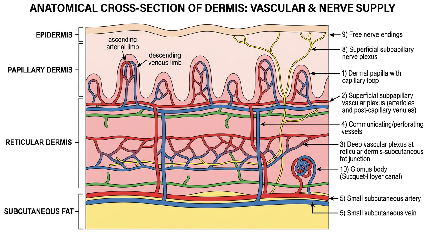
I. BLOOD SUPPLY (Arterial Supply)
Origin and Entry
- Cutaneous blood is supplied by musculocutaneous arteries and direct cutaneous arteries that penetrate the subcutaneous tissue (hypodermis) from underlying muscle and fascia.
- These vessels travel within the fibrous septa separating subcutaneous fat lobules before entering the deep reticular dermis.
The Two-Plexus System
1. Deep Horizontal Vascular Plexus (Deep Plexus)
- Located at the junction of the reticular dermis and subcutaneous fat
- Consists of larger-caliber arterioles and venules
- Communicates with branches extending into fibrous septa of the subcutaneous fat
- Originates from musculocutaneous arteries that penetrate upward from below
- Vessels here have two layers of smooth muscle, a discontinuous internal elastic lamina, pericytes, and adventitial Veil cells
2. Superficial Horizontal Vascular Plexus (Sub-papillary Plexus)
- Located in the upper part of the reticular dermis, just beneath the papillary dermis - defines the boundary between papillary and reticular dermis (IADVL)
- Forms a layer of anastomosing arterioles and venules
- Extends within an adventitial mantle to envelop adnexal structures (folliculosebaceous-apocrine units, eccrine glands)
- Anastomoses are most developed in the upper dermis and around adnexal structures (Moschella & Hurley)
3. Communicating / Connecting Vessels
- Connect the deep and superficial plexuses
- Oriented perpendicular to the skin surface and to one another
- Are ascending arterioles and descending venules
4. Papillary Capillary Loops
- From the superficial plexus, arcade capillary loops project upward into each dermal papilla
- Each loop has:
- An ascending arterial limb - continuous endothelium, basal lamina, few pericytes
- A hairpin turn at the apex (thinnest portion, attenuated endothelium and basal lamina - where transcapillary exchange occurs)
- A descending venous (post-capillary) limb - multiple layers of basal lamina separated by collagen fibrils, loose sheath of pericytes and Veil cells (IADVL)
Microstructure of Dermal Vessels
- Ascending arterioles: two layers of smooth muscle, discontinuous internal elastic lamina, pericytes, Veil cells (thin adventitious cells resembling fibroblasts, no basement membrane)
- Anastomosing arterioles: single layer of smooth muscle, lack elastic fibers
- Capillaries in superficial dermis: continuous endothelium; transport via pinocytosis
- Venous capillaries in adventitial dermis: fenestrated (allow transport of solutes)
- Endothelial cells are rich sources of angiotensin-converting enzyme and cytokines (IADVL)
Vascular Control
- Adrenergic (sympathetic): alpha-receptor activation on vascular smooth muscle causes vasoconstriction; predominant control in acral vessels
- Non-adrenergic control (non-acral): includes nitric oxide, local temperature, humoral agents
- Vasoconstrictors: angiotensin II, vasopressin, epinephrine
- Vasodilators: ethanol, histamine, prostaglandin-E, local heating
- Total skin blood flow ~250 mL/min at thermoneutral; rises to 6-8 L/min during hyperthermia (Moschella & Hurley)
II. GLOMUS BODY (Arteriovenous Anastomosis)
- A specialized arteriovenous (AV) shunt bypassing the capillary bed entirely (Moschella & Hurley; IADVL)
- Location: Deep reticular dermis; most abundant in acral skin - nail beds, finger pads, palms, soles, toes, ears, nose
- Function: Temperature regulation (shunting blood away from capillary bed in cold); also contributes to blood pressure regulation
- Structure:
- Arterial segment (Sucquet-Hoyer canal): narrow lumen, thick wall of rows of contractile glomus cells (modified smooth muscle cells)
- Venous segment: thin-walled, wide lumen draining into a subpapillary venule
- Innervated by both myelinated and unmyelinated adrenergic sympathetic nerves
- Clinical associations: Glomus tumors (painful subungual tumors), Klippel-Trenaunay syndrome, Sturge-Weber syndrome (defects in vascular development) (IADVL)
III. VENOUS DRAINAGE
Sequence of Drainage (Deep to Superficial, then back Deep)
- Capillary loop apex (venous limb) → post-capillary venules of the superficial plexus
- Superficial post-capillary venules → dermal communicating venules (vertically oriented)
- Communicating venules → large venules of the deep plexus
- Deep plexus venules → small veins of subcutaneous fat
- → Regional veins → systemic venous circulation (Moschella & Hurley)
Post-capillary Venules - Key Points
- The predominant vessel type in the upper dermis
- Most permeable of all cutaneous vessels
- Surrounded by other cellular components of the dermal microvascular unit
- Sites of immune complex deposition in vasculitis (allergic vasculitis)
- Vasoactive substances (prostaglandins, histamine) from leukocytes, mast cells, platelets increase venular permeability by inducing endothelial cell and pericyte contraction, widening intercellular spaces
- This favors extravasation of fluid and inflammatory cells into surrounding connective tissue (Moschella & Hurley)
Functional Significance of Venous Side
- The red color of inflammatory eruptions is due to blood in dilated vessels of the upper dermis
- Post-capillary venule dilation underlies urticaria, erythema, and the wheal-flare reaction
IV. LYMPHATIC DRAINAGE
General Overview
- Clear interstitial fluid and waste products
- Circulate Langerhans cells, lipids, proteins, immune complexes
- Failure leads to recurrent infections, impaired immunity, edema, and fibrosis (Moschella & Hurley)
Architecture - Two-Plexus System (Parallels Vascular Plexuses)
1. Superficial Lymphatic Plexus
- Situated in the upper part of the reticular dermis, just beneath the superficial venous plexus
- Depth: approximately 200 µm from the epidermis (Moschella & Hurley)
- Corresponds to the zone where elastic fiber orientation changes from vertical (papillary dermis) to horizontal (reticular dermis)
- Begins as blind-ending lymphatic capillaries (initial lymphatics / prelymphatics) starting in the papillary dermis
- The lymphatic capillaries then drain into collecting vessels forming this subpapillary plexus
2. Deep Lymphatic Plexus
- Located in the reticular dermis
- Drains the superficial plexus vessels downward
- These vessels then drain into subcutaneous lymphatic vessels, which drain into regional lymph nodes
Drainage Sequence
Structural Features of Lymphatic Vessels
- Wall: single layer of attenuated endothelial cells
- No pericytes in smallest lymphatics (unlike blood capillaries)
- Discontinuous basal lamina (vs. continuous in blood capillaries)
- No fenestrae; very few pinocytotic vesicles or Weibel-Palade bodies
- Wide gaps between overlapping endothelial cell ends act as one-way flap valves - allow filtration and removal of fluids, macromolecules, inflammatory cells, and foreign material from dermal extracellular compartment (Moschella & Hurley)
- Bicuspid intraluminal valves prevent backflow and stasis (IADVL)
- Smooth muscle in vessel wall present only at subcutaneous tissue level
- Electron microscopy: lymphatic endothelial cells contain vimentin filaments
Anchoring Filaments
- Fine collagen fibrils attach at right angles to lymphatic capillary walls and extend into surrounding connective tissue
- These act as anchoring filaments preventing lymphatic vessel collapse under interstitial edema pressure (Moschella & Hurley)
- An outer network of reticulin and elastic fibers also envelops the lymphatics
- Elastic fibers around dermal lymphatics are attached to the subepidermal basement membrane zone by fine oxytalan fibers
- In the uppermost dermis: elastic fibers oriented perpendicular to skin surface; in bulk of reticular dermis: oriented horizontally - this provides preferential pathways of low resistance through extracellular matrix for lymph flow (Moschella & Hurley)
Important Points
- No lymphatic vessels exist in the epidermis (Moschella & Hurley)
- Lymphogenesis (adult): hair cycle, wound healing, menstrual cycle
- Pathological lymphogenesis: tumor growth, metastatic spread, atopic dermatitis, psoriasis
- Regulated by VEGF (vascular endothelial growth factor) acting on lymphatic endothelium during embryogenesis (Moschella & Hurley)
V. NERVE SUPPLY OF THE DERMIS
Origin
- Nerves of the skin are part of the peripheral nervous system
- Derived mostly from the neural crest - giving rise to dorsal root and sympathetic ganglia, their nerve processes, Schwann cells, endoneurial sheath cells, and laminar cells of specialized sensory end organs (Moschella & Hurley)
- Spinal nerves innervate the skin via branches entering the dermis with an epineural, perineural, and endoneural sheath and Schwann cell envelope (IADVL)
Two Functional Components
A. Somatic Sensory System
B. Autonomic Motor System (Unmyelinated Sympathetic Postganglionic Nerves)
- Cutaneous vascular tone
- Piloerection (arrector pili muscles) - adrenergic
- Apocrine gland secretion - adrenergic
- Eccrine sweating - cholinergic (thermoregulatory), adrenergic (emotional/acral)
Plexus Architecture - Mirrors Vascular Architecture
- Deep Dermal Nerve Plexus - in deep dermis
- Superficial Subpapillary Nerve Plexus - in upper dermis
- Branches from both plexuses supply different areas and structures
- Sympathetic motor fibers travel with sensory fibers until they branch to innervate hair follicles, sweat glands, arrector pili, and blood vessel smooth muscle
Nerve Fiber Types and Their Functions
| Fiber Type | Diameter | Myelination | Function |
|---|---|---|---|
| A-Beta (Aβ) | 10-14 µm | Heavily myelinated | Touch discrimination, vibration, proprioception |
| A-Gamma (Aγ) | Narrower than Aβ | Myelinated | Light touch, pressure |
| A-Delta (Aδ) | Even narrower | Thinly myelinated | Pain, temperature, physiologic itch |
| C fibers | <5 µm (narrowest) | Unmyelinated | Pain, temperature, pathologic itch |
Sensory Receptors of the Dermis
A. Free Nerve Endings
- Most widespread and important sensory receptors (IADVL)
- Found predominantly in the papillary dermis
- Unmyelinated axons, many terminate near the DEJ making contact with basal lamina of epidermis
- Usually have a Schwann cell sheath and basal lamina
- Particularly abundant in glans penis (almost every dermal papilla) and scattered in deeper dermis
- Detect: pain, temperature, itch
B. Encapsulated (Corpuscular) Receptors
| Receptor | Location | Structure | Modality |
|---|---|---|---|
| Meissner's Corpuscle | Tips of dermal papillae, volar skin (fingertips, soles, eyelids, lips, genitalia) | 50 x 150 µm; layers of flattened modified Schwann cells; non-myelinated branches from large myelinated nerves | Light touch (rapidly adapting) |
| Pacinian Corpuscle | Deep dermis and subcutaneous tissue (fingertips, toes) | Up to 1 mm ovoid; 30+ concentric lamellae (perineurium); single axon; onion-like in cross section | Deep pressure, vibration (250-350 Hz); rapidly adapting |
| Ruffini's End Organ | Deep dermis, parallel to skin tension lines | Spindle-shaped capsulated receptor | Cutaneous tension, skin stretch; slowly adapting |
| Merkel's Disc / Touch Dome | Hairy + glabrous skin (digits, lips, oral cavity, outer root sheath of hair follicle) | Merkel cells + specialized basal keratinocytes forming "touch dome" or haarscheibe | Fine touch, tactile discrimination, stereognosis; slowly adapting |
| Mucocutaneous End Organs | Papillary dermis at mucocutaneous junctions (glans, prepuce, clitoris, labia minora, perianal, eyelids, lips) | Similar to Meissner's; 2-6 myelinated nerves forming loops | Touch at skin-mucosa junctions |
| Krause End Bulbs | Superficial dermis | Encapsulated swelling of myelinated fibers | Cold sensation (historical; now reclassified) |
| Golgi-Mazzoni Corpuscle | Subcutaneous tissue (finger) | Like Pacinian but simpler organization | Pressure |
C. Penicillate Nerve Endings
- Found in hair-bearing skin in subepidermal location
- Separate unmyelinated branches from one or more myelinated axons, collectively ensheathed by Schwann cell processes
- Collagen fibrils surround each branch to its end
- Very few organelles → rapidly adapting receptors (IADVL)
Receptor Classification by Adapting Property
- Slowly adapting (SA): Merkel's disc, Ruffini's end organs - encode sustained stimuli
- Rapidly adapting (RA): Meissner's corpuscles, Pacinian corpuscles, penicillate endings - encode onset/offset of stimuli
Mechanoreceptors: Threshold
- Meissner's, Pacinian, Merkel's, and Ruffini's are low-threshold mechanoreceptors
- Even weak stimuli can elicit sensations
- Innervated by large myelinated Aβ fibers (IADVL)
VI. APPLIED/CLINICAL RELEVANCE
| Condition | Relevance |
|---|---|
| Post-capillary venulitis (leukocytoclastic vasculitis) | Immune complex deposition in walls of post-capillary venules; vasoconstrictors/dilators alter permeability |
| Raynaud's phenomenon | Exaggerated adrenergic vasoconstriction of arterioles |
| Glomus tumor | Painful subungual tumor from glomus body |
| Klippel-Trenaunay syndrome | Unilateral vascular malformations, venous varicosities; defect in vascular development |
| Sturge-Weber syndrome | Capillary malformations; mutation in endoglin / ACVRL1 (TGF-β receptor) genes |
| Hereditary hemorrhagic telangiectasia | Mutations in endoglin and ACVRL1 |
| Lymphedema / elephantiasis nostra | Recurrent cellulitis → impaired lymphatic drainage, dermal fibrosis |
| TEN / SJS | Weakening of DEJ connections; removal of protective barrier; disrupts vascular and nerve supply |
| Psoriasis / atopic dermatitis | Pathological lymphangiogenesis and angiogenesis |
| Syringomyelia, leprosy | Selective loss of temperature and pain (A-delta and C fiber damage) |
SUMMARY TABLE: Plexus Architecture at a Glance
| Layer | Vascular | Lymphatic | Nerve |
|---|---|---|---|
| Papillary dermis | Capillary loops ascending from superficial plexus | Blind-ending initial lymphatics (prelymphatics) | Free nerve endings, Meissner's corpuscles (papilla tips) |
| Upper reticular dermis | Superficial (sub-papillary) arteriolo-venular plexus | Superficial lymphatic plexus (~200 µm depth) | Superficial subpapillary nerve plexus |
| Mid reticular dermis | Communicating perforating vessels | Lymphatic collecting vessels | Communicating nerve branches |
| Deep reticular dermis | Deep arteriolo-venular plexus | Deep lymphatic plexus | Deep dermal nerve plexus |
| Subcutaneous tissue | Musculocutaneous arteries/small subcutaneous veins | Subcutaneous lymphatics → regional nodes | Main nerve trunks + autonomic fibers |