
Can I upload an pdf file
- Look for the attachment/paperclip icon in the chat input area
- Click it and select your PDF file
- Then type your question or instruction alongside it
- Extract and read text from the document
- Summarize the content
- Answer questions based on what's in the PDF
- Analyze data, tables, or figures
- Extract specific sections or information
- OCR scanned/image-based PDFs to make text searchable
- Merge, split, or manipulate PDF pages if needed
Prostate Cancer 1. The Core Pathology & Anatomy Prostate cancer is the most common noncutaneous malignancy in men and the second most common cause of cancer death in men.over 60% of patients are completely asymptomatic at the time of diagnosis Histology: 95% of prostate cancers are adenocarcinomas developing in the acini of prostatic ducts. The "Danger Zone": 70% of these cancers arise in the Peripheral Zone (PZ) mcqs . Cancers here are generally more aggressive and tend to invade periprostatic tissues. Anatomical Boundaries: The prostate is separated from the rectum by a layer of fascia called the Denonvilliers aponeurosis. The neurovascular bundle (crucial for erectile function) lies on either side of the prostate on the rectum. Protective Factors: Diets high in soy protein, lycopene, vitamin E, and selenium, along with reduced fat intake, have a protective effect. 2. Clinical Manifestations (Vignette Clues) You can usually stage the disease in an exam question just by reading the patient's symptoms. Early Stage (Organ Confined): Completely asymptomatic. Locally Advanced: Presents with obstructive voiding symptoms (hesitancy, decreased force of stream), hematuria, or hematospermia. Advanced Stage: Look for lower extremity edema, pelvic/perineal discomfort, and severe bone pain. Metastasis to the bone mcqs is the most common spread and can result in pathologic fractures or spinal cord compression. 3. Detection and Diagnosis (Highly Tested) Over 60% of patients are asymptomatic and diagnosed solely due to an elevated screening PSA. PSA Thresholds: The standard threshold is 4 ng/mL. PSA Velocity: A rate increase of > 0.75 ng/mL per year warrants a biopsy. Free PSA: This helps distinguish cancer from benign processes like BPH. A free PSA cutoff of less than 25% can detect 95% of cancers while avoiding unnecessary biopsies. The Biopsy: Transrectal ultrasound (TRUS) with sextant biopsies (taking samples from the base, midgland, and apex on each side) is the standard. Imaging Rules: Do not order a bone scan unless the PSA is > 10 ng/mL. CT or MRI of the abdomen/pelvis is usually unrevealing if the PSA is < 20 ng/mL. Palliative Bone Care: For widespread, painful bone metastases that don't respond to standard painkillers, you can use Strontium-89 and Samarium-153 . These radioactive elements selectively concentrate in the bone tumors and alleviate pain in >70% of patients 4. Grading the Cancer Prognosis heavily relies on the Gleason Score, which is the sum of the two most common histologic patterns seen on the biopsy. Score 2 to 4: Well-differentiated. Score 5 to 7: Intermediate differentiation. Score 8 to 10: Poorly differentiated. 5. Management Strategies Treatment must be individualized based on life expectancy, comorbidity, and tumor grade. Low/Intermediate Risk Disease: Watchful Waiting: Can be used judiciously for men with Gleason scores of 2 to 4 and a life expectancy of 10 years or less. Definitive Therapy: Radical prostatectomy, External Beam Radiation Therapy (EBRT), or Brachytherapy (radioactive seed implants). Advanced/Metastatic Disease: Androgen Deprivation: This is the mainstay of treatment for newly diagnosed metastatic disease mcqs, achieving symptom improvement in 80 to 90% of patients. The "Tumor Flare" Trap: Medical castration uses LHRH agonists (like leuprolide or goserelin). Exam Point: LHRH agonists cause an initial surge of LH and testosterone within the first 72 hours, transiently worsening symptoms. You MUST give an antiandrogen (like flutamide or bicalutamide) with the first LHRH injection to prevent this tumor flare. Hormone-Resistant Disease: This is defined by a climbing PSA despite castrate levels of testosterone. First Step: Discontinue the antiandrogen (flutamide) but continue the LHRH agonist. This simple step often results in a PSA decline that lasts 4 to 24 months. 🧠 Tricks to Remember the Material 1. The "P.P.P." Anatomy Rule: Prostate cancer arises in the Peripheral zone, and you can easily Palpate it on a digital rectal exam. (Recall that BPH arises in the Transitional zone and causes early urinary symptoms, whereas peripheral cancers grow silently before causing obstruction). 2. The Flare Prevention Pairing: Think of the LHRH agonist as lighting a match (the testosterone surge). You always need an extinguisher ready. LHRH Agonist + Antiandrogen (Flutamide) = Halts the Flare. 3. Bone Scan Cutoffs (The Rule of 10 and 20): PSA > 10: Order a bone scan. PSA > 20: Order a CT/MRI (unrevealing below this).
prostate cancer anatomy peripheral zone diagram
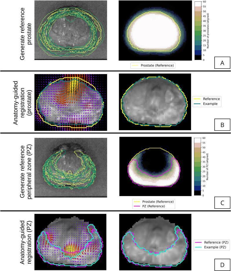
Educational diagram illustrating the medical imaging registration and model geometry framework for the prostate. The image is divided into four panels (A-D). Panels A and C demonstrate the generation of reference geometry for the whole prostate and the peripheral zone (PZ), respectively. These panels show multiple overlapping contours from different patients overlaid on axial T2-weighted MRI (left) and a corresponding heatmap (right) where color intensity (blue to white) represents the number of aligned structures, with the final reference template outlined in yellow. Panels B and D illustrate anatomy-guided deformable registration for the whole prostate and PZ. The left side of these panels displays a vector field map (color-coded arrows indicating deformation magnitude and direction) overlaid on the MRI, while the right side shows the resulting deformed MRI with aligned patient and reference boundaries. This methodology highlights advanced medical image processing techniques used to create a probabilistic anatomical atlas for prostate cancer research and clinical diagnostic mapping.
![A multi-panel medical graphic illustrating the diagnostic workflow for prostate cancer staging and image fusion. On the left, axial views of the pelvis demonstrate two imaging modalities: a [68Ga]Ga-PSMA-11 PET scan showing metabolic activity (hotspot) in the prostate gland with a green contour, and an MRI-derived Apparent Diffusion Coefficient (ADC) map showing tissue density with a red contour. The central element is an anatomical schematic diagram of the prostate base, divided into labeled zones: anterior fibromuscular stroma (AFS), anterior/posterior transition zones (TZa/p), anterior/posterior peripheral zones (PZa/p), and the central zone (CZ). A red circle on the schematic identifies a lesion in the right posterior peripheral zone (PZp), corresponding to histological findings of acinar adenocarcinoma (ISUP 3). The right panel shows the fusion of PET and ADC images using MIM maestro software, overlaying metabolic and structural data to refine tumor localization. This image serves as an educational resource for radiomics, multi-modal image registration, and prostate zonal anatomy in the context of oncological pathology.](https://cdn.orris.care/cdss_images/pmc_clinical_VQA_a2397d96820e4e51076095061aee44f42d68a092e79dc64a3a722a4202bfa832.jpg)
A multi-panel medical graphic illustrating the diagnostic workflow for prostate cancer staging and image fusion. On the left, axial views of the pelvis demonstrate two imaging modalities: a [68Ga]Ga-PSMA-11 PET scan showing metabolic activity (hotspot) in the prostate gland with a green contour, and an MRI-derived Apparent Diffusion Coefficient (ADC) map showing tissue density with a red contour. The central element is an anatomical schematic diagram of the prostate base, divided into labeled zones: anterior fibromuscular stroma (AFS), anterior/posterior transition zones (TZa/p), anterior/posterior peripheral zones (PZa/p), and the central zone (CZ). A red circle on the schematic identifies a lesion in the right posterior peripheral zone (PZp), corresponding to histological findings of acinar adenocarcinoma (ISUP 3). The right panel shows the fusion of PET and ADC images using MIM maestro software, overlaying metabolic and structural data to refine tumor localization. This image serves as an educational resource for radiomics, multi-modal image registration, and prostate zonal anatomy in the context of oncological pathology.
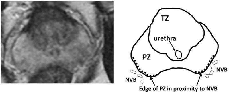
An educational figure combining an axial MRI image (left) and a corresponding anatomical schematic (right) of the prostate gland. The content illustrates the zonal anatomy of the prostate and its spatial relationship to adjacent structures. The diagram identifies the transition zone (TZ) as the central region, containing the urethra. Surrounding the posterior and lateral aspects of the TZ is the peripheral zone (PZ). Notably, the schematic highlights the 'edge of PZ in proximity to NVB' using a dotted line, where the peripheral zone borders the bilateral neurovascular bundles (NVB). This clinical illustration is designed to teach the anatomical landmarks relevant to focal therapies, such as High-Intensity Focused Ultrasound (HIFU), where the distance between the treatment area in the peripheral zone and the neurovascular bundle is a critical factor for preserving post-operative erectile function. The modality demonstrated is T2-weighted magnetic resonance imaging, which provides the soft-tissue contrast necessary to distinguish between prostatic zones.
Gleason score histology prostate adenocarcinoma grading
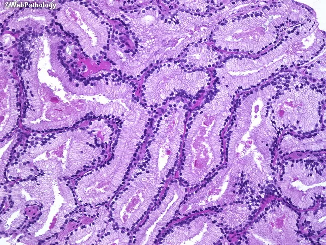
High-power histology image of prostatic acinar adenocarcinoma (Gleason score 3+3=6) viewed under bright-field light microscopy. The specimen demonstrates well-formed, back-to-back glands infiltrating the periglandular stroma, consistent with low-grade malignant acinar architecture. Gland lumina are generally round to slightly elongated, lined by a single layer of cuboidal to columnar epithelial cells with mild cytologic atypia, small central nuclei, and occasional stratification. The basal cell layer characteristic of benign prostatic acini is not evident within the malignant glands, and there is minimal desmoplastic reaction. The glands vary in size but maintain a predominantly tubular, well-differentiated appearance without prominent cribriform, fused, or solid growth patterns, which aligns with Gleason pattern 3. Nuclei show modest enlargement; nucleoli are rarely conspicuous in this field. Overall morphology supports a low-grade prostatic adenocarcinoma, with infiltration into surrounding stroma but without high-grade features locally evident. This image captures a classic example applicable to educational discussions of prostate cancer grading, prognosis, and management implications. Clinically relevant keywords include prostate cancer, acinar adenocarcinoma, Gleason 3+3, tumor architecture, well-formed glands, low grade, and active surveillance considerations. It complements histology teaching of tumor grading systems and is suitable for radiologic-pathologic correlation and pathology review.
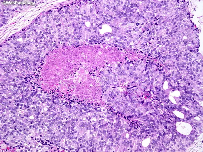
Histopathology: Prostate tissue examined by light microscopy after Hematoxylin and Eosin staining reveals high-grade prostatic adenocarcinoma with solid nests exhibiting comedonecrosis. The center shows necrotic ghost remnants surrounded by a layer of karyorrhectic debris; at the periphery, layers of viable malignant epithelial cells display enlarged, hyperchromatic nuclei and prominent nucleoli. The architectural pattern includes solid and cribriform nests with occasional back-to-back glands, consistent with Gleason pattern 5. Overall Gleason score is 10 (5+5). Notable features include extensive tumor necrosis, conspicuous cytologic atypia, and a high nuclear-to-cytoplasmic ratio. Tumor invasion of the adjacent stroma is evident in places, with limited mitotic activity but aggressive phenotype suggested by necrotic cores and dense cellularity. Clinically, this histology indicates a highly aggressive, poorly differentiated lesion with elevated metastatic potential and poorer prognosis compared with lower Gleason scores. This finding supports definitive management with multimodal therapy and close staging. For educational and diagnostic use, this image demonstrates comedonecrosis in prostatic adenocarcinoma, cribriform/high-grade patterns, and the cytologic features that contribute to Gleason grading and prognostic assessment.
Prostate Cancer - Complete Study Notes
1. Core Pathology & Anatomy
- Most common noncutaneous malignancy in men; 2nd most common cause of cancer death in men (after lung cancer)
- Over 60% are asymptomatic at diagnosis, found incidentally via PSA screening
- Major risk factors: age (>65 years), African American race (70% higher incidence than whites), family history
- 95% adenocarcinomas arising in the acini of prostatic ducts
- 70% arise in the Peripheral Zone (PZ) - palpable on DRE, generally more aggressive
- BPH arises in the Transitional Zone - causes early obstructive urinary symptoms
- The Central Zone is less commonly involved
- Prostate separated from the rectum by Denonvilliers' aponeurosis (fascia)
- Neurovascular bundles (NVB) lie bilaterally at the posterolateral aspect - critical for preserving erectile function in surgery
2. Clinical Manifestations (Vignette Clues)
| Stage | Symptoms |
|---|---|
| Early (organ-confined) | Completely asymptomatic |
| Locally advanced | Obstructive voiding (hesitancy, weak stream), hematuria, hematospermia |
| Advanced/Metastatic | Lower limb edema, pelvic/perineal pain, bone pain (most common met site), pathologic fractures, spinal cord compression |
3. Detection & Diagnosis
- Standard cutoff: 4.0 ng/mL
- Note: 15% of men with PSA <4 ng/mL still have prostate cancer (15% of those are high-grade) - PSA has both high false-positive and false-negative rates - [Family Medicine 9e, p. 715]
- PSA velocity: >0.75 ng/mL/year warrants biopsy
- Free PSA <25% favors cancer over BPH; helps avoid unnecessary biopsies
- Abnormal DRE predicts cancer in only 18-28% of cases
- Up to 25% of biopsy-detected cancers after abnormal DRE occur on the opposite side from the palpated nodule
- TRUS-guided sextant biopsy (base, midgland, apex - both sides = 6 cores minimum)
| PSA Level | Action |
|---|---|
| PSA > 10 ng/mL | Order bone scan |
| PSA > 20 ng/mL | Order CT/MRI abdomen-pelvis (usually unrevealing below this) |
- For widespread painful bone mets not responding to standard analgesics:
- Strontium-89 and Samarium-153 - radioactive elements concentrating selectively in bone tumors, alleviating pain in >70% of patients
4. Gleason Scoring
| Gleason Score | Differentiation | Prognosis |
|---|---|---|
| 2-4 | Well-differentiated | Low risk of cancer death |
| 5-7 | Intermediate | Intermediate |
| 8-10 | Poorly differentiated | High probability of death within 10 years |
5. Management Strategies
Risk-Stratified Approach
- Watchful waiting: For Gleason 2-4 with life expectancy ≤10 years
- Active surveillance: PSA monitoring + repeat biopsy (for low-risk, eligible patients)
- Definitive therapy options:
- Radical prostatectomy
- External Beam Radiation Therapy (EBRT)
- Brachytherapy (radioactive seed implants)
- Androgen Deprivation Therapy (ADT) is the mainstay - achieves symptom improvement in 80-90%
- Methods:
- Surgical castration (bilateral orchiectomy) - immediate, irreversible
- Medical castration - LHRH agonists (leuprolide, goserelin)
⚠️ The LHRH Tumor Flare Trap (High-Yield Exam Point)
Think: LHRH agonist = lighting a match. Antiandrogen = the extinguisher. Always have both.
- Defined as: rising PSA despite castrate testosterone levels
- First step: Discontinue the antiandrogen (antiandrogen withdrawal syndrome) - PSA decline occurs in a significant proportion and can last 4-24 months
- Continue the LHRH agonist
- Further options: enzalutamide, abiraterone, docetaxel chemotherapy, sipuleucel-T
6. Memory Anchors
| Rule | Details |
|---|---|
| P.P.P. Rule | Prostate cancer → Peripheral zone → Palpable on DRE |
| BPH vs PCa | BPH = Transitional zone (early urinary sx); PCa = Peripheral zone (silent until advanced) |
| Rule of 10/20 | PSA >10 → bone scan; PSA >20 → CT/MRI |
| Flare pair | LHRH agonist + antiandrogen always together at initiation |
| Gleason shortcut | 2-4 = good; 8-10 = bad; 5-7 = middle |
| Bone mets type | Osteoblastic (sclerotic) - unique to prostate cancer |
| Vitamin E | Does NOT prevent PCa - actually increases risk (SELECT trial) |
The text which I sent, make it simple and easy for me and also make some scenario based mcqs
🫀 Prostate Cancer - Made Simple
Part 1: What Is It?
- The #1 cancer in men (excluding skin)
- The #2 killer cancer in men (lung is #1)
- Most men don't even know they have it - over 60% have zero symptoms when first found
Part 2: WHERE Does It Start? (The Zones)
         ┌─────────────────────┐
         │   PERIPHERAL ZONE   │  ← Cancer starts here (70%)
         │  ┌───────────────┐  │     Palpable on DRE
         │  │  TRANSITION   │  │  ← BPH starts here
         │  │     ZONE      │  │     Causes early urinary symptoms
         │  │  ┌─────────┐  │  │
         │  │  │ CENTRAL │  │  │  ← Rarely affected
         │  │  │  ZONE   │  │  │
         │  │  └─────────┘  │  │
         │  └───────────────┘  │
         └─────────────────────┘
Part 3: Symptoms by Stage
| Level | What the Patient Says |
|---|---|
| 🟢 Early | "I feel completely fine" (nothing) |
| 🟡 Local spread | "I have trouble peeing, weak stream, blood in urine" |
| 🔴 Spread to bones | "My back/hips are killing me" + leg swelling + could fracture bones |
Part 4: How Is It Found?
The PSA Blood Test
| PSA Level | Meaning |
|---|---|
| < 4 ng/mL | Normal (but 15% of men still have cancer!) |
| > 4 ng/mL | Abnormal - investigate |
| Rising >0.75/year | Get a biopsy (PSA velocity) |
The Biopsy
- Use TRUS (ultrasound probe in rectum) to guide needle
- Take 6 samples (sextant) from base, middle, and tip - both sides
Imaging Rules (Super Easy to Remember)
"The Rule of 10 and 20"
- PSA > 10 → order a Bone Scan
- PSA > 20 → order CT/MRI of abdomen & pelvis
For Bone Pain That Won't Go Away
- Use Strontium-89 or Samarium-153 - radioactive elements that home in on bone tumors like a GPS and kill pain in >70% of patients
Part 5: Grading - The Gleason Score
| Score | What It Means | Think of it as... |
|---|---|---|
| 2-4 | Well-organized cells | "Still looks like normal prostate" |
| 5-7 | Getting messy | "Something is wrong" |
| 8-10 | Totally chaotic | "Very aggressive, act fast" |
Part 6: Treatment
Low-Risk (Gleason 2-4, no spread, life expectancy ≤10 years)
- Just watch and wait (watchful waiting) - don't over-treat
Medium to High-Risk (but still in the prostate)
- 🔪 Surgery - Radical Prostatectomy (remove the whole prostate)
- ☢️ Radiation - EBRT (beam from outside) or Brachytherapy (seeds planted inside)
Metastatic Disease (Spread outside prostate)
- Cut off the testosterone - cancer FEEDS on it
- Options:
- Surgery to remove testicles (orchiectomy)
- Drugs called LHRH agonists (leuprolide, goserelin)
⚠️ THE MOST IMPORTANT EXAM TRAP: The Testosterone Flare
First 72 hours → Testosterone SURGES before it drops
                       ↓
            Cancer gets TEMPORARILY WORSE
                       ↓
         Bone pain spikes, symptoms worsen
🔑 Memory trick: LHRH = "lighting a match." Antiandrogen = "fire extinguisher." Never light without it.
When Cancer Stops Responding (Hormone-Resistant)
- PSA keeps rising even with castrate testosterone levels
- First move: STOP the antiandrogen (flutamide) but KEEP the LHRH agonist
- This simple step often drops PSA for 4-24 months (called "antiandrogen withdrawal")
🧠 Cheat Sheet Summary
| Topic | Key Fact |
|---|---|
| Most common zone | Peripheral Zone (70%) |
| Bone mets type | Osteoblastic (sclerotic/white) |
| PSA cutoff | 4 ng/mL |
| Bone scan trigger | PSA > 10 |
| CT/MRI trigger | PSA > 20 |
| Free PSA for cancer | < 25% |
| PSA velocity | > 0.75 ng/mL/year = biopsy |
| Flare prevention | LHRH + Antiandrogen together |
| Hormone-resistant step 1 | Stop antiandrogen, keep LHRH |
| Vitamin E | Does NOT protect - increases risk! |
🧪 Scenario-Based MCQs
- A) Reassure him and repeat PSA in 1 year
- B) Order a bone scan immediately
- C) Refer for TRUS-guided prostate biopsy
- D) Start androgen deprivation therapy
- E) Order CT scan of abdomen and pelvis
✅ Answer: C PSA > 4 ng/mL with no symptoms is the classic asymptomatic screening catch. Next step is biopsy via TRUS. A bone scan is only needed if PSA > 10. CT/MRI is only useful if PSA > 20.
- A) Given a higher dose of leuprolide
- B) Added flutamide (antiandrogen) at the time of the first injection
- C) Started chemotherapy instead
- D) Performed surgical castration first
- E) Ordered an MRI before starting treatment
✅ Answer: B This is the classic tumor flare from LHRH agonist use. The initial testosterone surge worsens symptoms. Giving an antiandrogen (flutamide/bicalutamide) alongside the first injection blocks this surge and prevents the flare.
- A) Add chemotherapy (docetaxel)
- B) Switch to a different LHRH agonist
- C) Stop the LHRH agonist and monitor
- D) Discontinue flutamide, continue LHRH agonist
- E) Start radiation therapy to the prostate
✅ Answer: D This is castration-resistant prostate cancer (CRPC). The first and simplest step is antiandrogen withdrawal - stop flutamide while keeping the LHRH agonist. This paradoxically causes PSA to drop and can last 4-24 months before further escalation is needed.
- A) High-dose corticosteroids
- B) Strontium-89 or Samarium-153
- C) Surgical decompression of the spine
- D) Brachytherapy
- E) Orchiectomy
✅ Answer: B When pain from widespread bone metastases fails standard analgesics, Strontium-89 or Samarium-153 are indicated. These radioactive elements selectively concentrate in bone tumor sites and relieve pain in >70% of patients.
- A) The DRE was performed incorrectly
- B) Prostate cancer always spreads from left to right
- C) Up to 25% of prostate cancers detected after abnormal DRE are found on the opposite side
- D) The biopsy result is a false positive
- E) Peripheral zone cancers always involve both lobes
✅ Answer: C DRE is imprecise. Studies show up to 25% of biopsy-detected cancers after an abnormal DRE are found on the opposite side from the palpated nodule. This is why sextant biopsies sample both lobes regardless of which side feels abnormal.
- A) Radical prostatectomy
- B) External beam radiation therapy
- C) Androgen deprivation therapy
- D) Watchful waiting
- E) Brachytherapy
✅ Answer: D For low-grade (Gleason 2-4), organ-confined prostate cancer in a man with life expectancy ≤10 years, watchful waiting is the preferred approach. Aggressive treatment carries risks that outweigh benefit in this setting.
- A) CT scan of the chest only
- B) MRI of the prostate
- C) Radionuclide bone scan
- D) PET-FDG scan
- E) No imaging needed yet
✅ Answer: C PSA > 10 ng/mL = order a bone scan. A CT/MRI of the abdomen/pelvis is usually unrevealing unless PSA > 20. FDG-PET has little role in prostate cancer imaging. A bone scan with Tc-99m is the standard first-line imaging for suspected bone metastases.