
Approach to angular deformity in age less than 8 years
Reading File
Now let me search the medical library for content on angular deformity in children.
Finding Sources
Finding Sources
Finding Sources
Reading File
This is about femoral fractures. Let me search more broadly for the approach to angular deformity in children - genu valgum/varum, and remodeling potential.
Finding Sources
Finding Sources
Reading File
Finding Sources
Reading File
Finding Sources
Finding Sources
Finding Sources
Reading File
Reading File
Now let me look at more content on Blount disease and genu varum, and read the genu valgum section more completely. Also need content on physiologic patterns.
Reading File
Loading Image
Excellent! Now I have comprehensive content. Let me also fetch the Langenskiold classification image and get the table image referenced for tibial bowing types.
Loading Image
Loading Image
I now have comprehensive information to provide a detailed answer. Let me compile the full response.
Approach to Angular Deformity in Children Under 8 Years
In this age group, the most important principle is understanding physiologic vs. pathologic angular deformity. Children under 8 years have enormous remodeling potential, and the majority of angular deformities seen in this age group are physiologic and self-resolving. Intervention is reserved for those outside the physiologic range or with an identifiable pathologic cause.
1. Normal Physiologic Sequence of Lower Limb Alignment
Understanding what is "normal" at each age is the foundation of the approach:
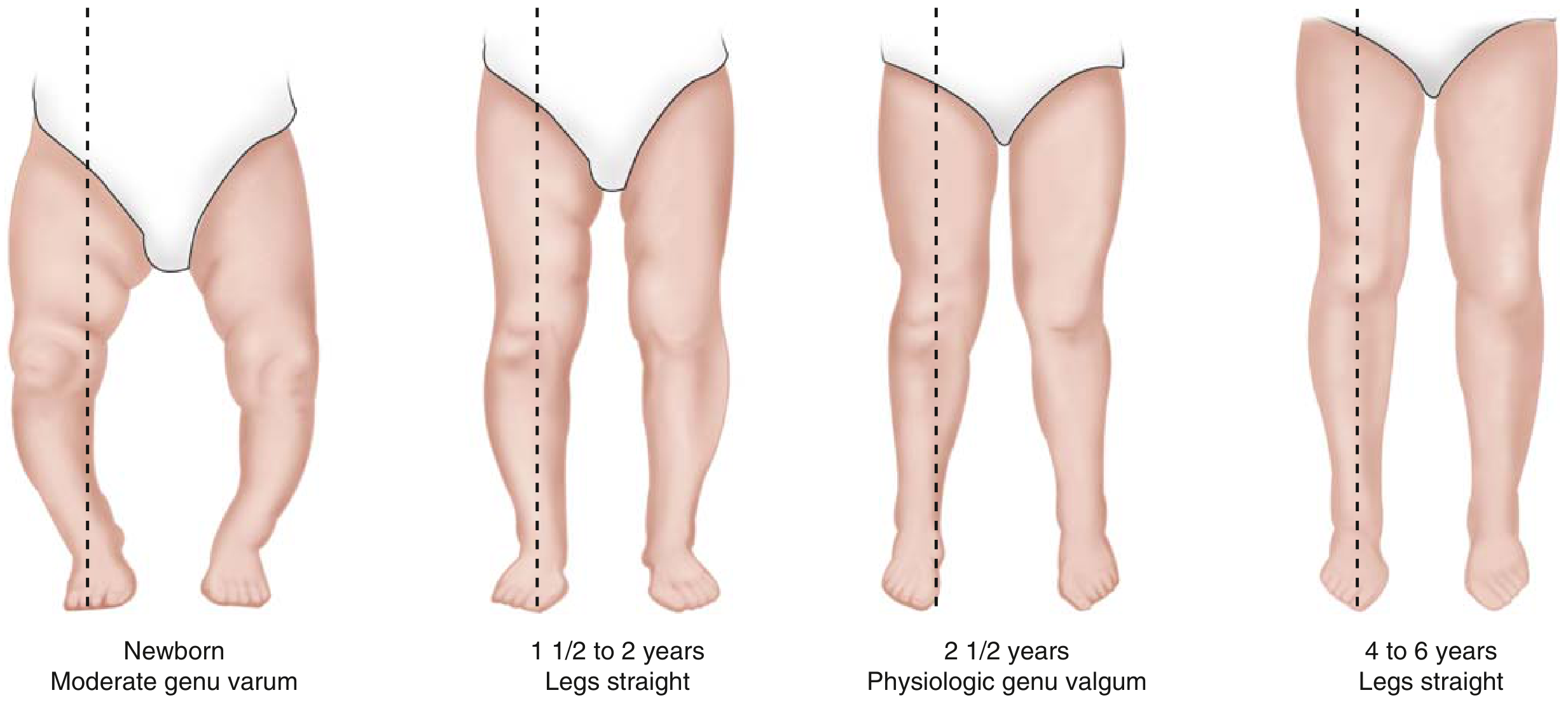
FIG. 3.15: In children with physiologic genu varum, bowing begins to slowly improve at ~18 months and continues as the child grows. By age 3-4 years, bowing has corrected and legs typically have a normal appearance. - Miller's Review of Orthopaedics 9th Ed.
| Age | Normal Alignment |
|---|---|
| Newborn | Moderate genu varum (bowed legs) |
| 1.5 - 2 years | Legs relatively straight |
| 2.5 years | Physiologic genu valgum (knock-knees) - peak ~15° |
| 3 - 4 years | Maximum valgus |
| 4 - 6 years | Legs straight; adult alignment approached |
Key facts:
- Genu varum normally evolves to genu valgum by age 2.5 years with gradual transition to physiologic valgus by age 4 years
- Up to 15 degrees of valgus at the knee is common in children 2-6 years of age
- Cases within this physiologic range do not require treatment
2. Causes of Angular Deformity in Children <8 Years
Genu Varum (Bowed Legs)
- Physiologic - normal in children <2 years (most common cause)
- Blount disease (infantile type) - age 0-4 years (most common pathologic cause)
- Osteogenesis imperfecta
- Osteochondromas
- Trauma / physeal injury
- Various dysplasias (rickets, skeletal dysplasias)
Genu Valgum (Knock-Knees)
- Physiologic - common between 2-6 years
- Renal osteodystrophy (most common pathologic bilateral cause)
- Tumors (e.g., osteochondromas)
- Infections - may stimulate proximal asymmetric tibial growth
- Trauma
3. Key Diagnostic Tool: Differentiating Physiologic from Pathologic
Red flags suggesting pathologic deformity:
- Deformity outside the expected age range
- Asymmetric bowing (unilateral)
- Progressive rather than improving
- Obesity + early walking + internal tibial torsion → suspect Blount disease
- Associated metabolic disease (rickets: cupping/fraying of metaphyses on X-ray)
- Severe deformity (>15° genu varum in a toddler, metaphyseal-diaphyseal angle >16°)
Radiographic evaluation (when indicated):
- AP standing X-ray of both lower extremities
- Drennan metaphyseal-diaphyseal angle: >16° is abnormal - formed between the metaphyseal beaks; differentiates Blount from physiologic bowing
- Physiologic bowing shows symmetric flaring of tibia and femur
4. Infantile Blount Disease (Age 0-4 Years)
The most important pathologic cause to recognize in this age group.
Classic presentation: Overweight child who started walking before 1 year of age, with genu varum + internal tibial torsion.
Langenskiold Classification (based on degree of metaphyseal-epiphyseal changes):
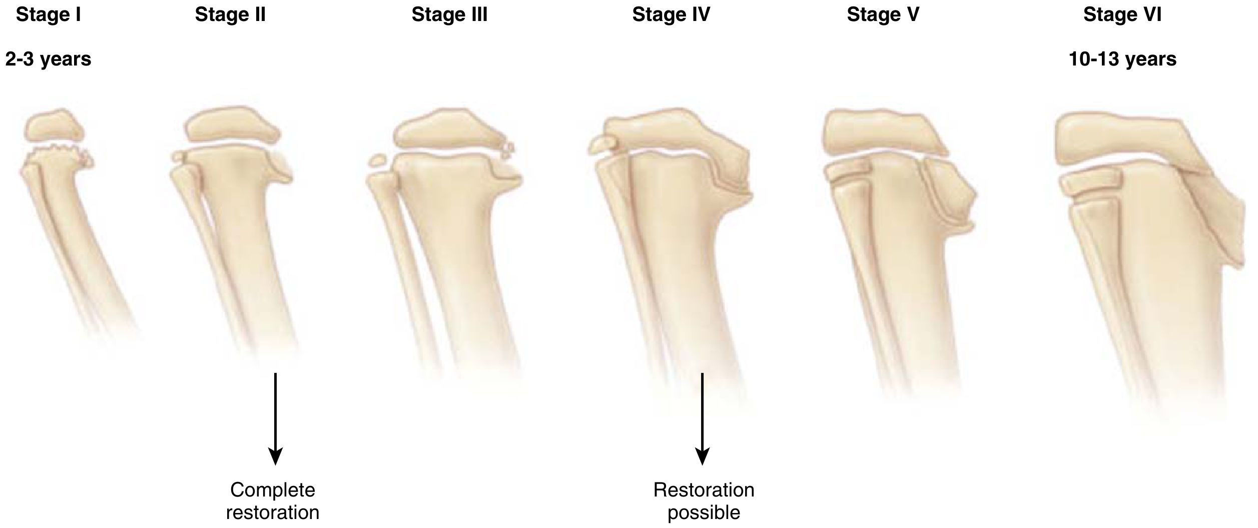
Langenskiold classification of infantile tibia vara - Stages I-VI with increasing age. Stages I-II: complete restoration possible. Stage IV: restoration possible. Stages V-VI: complex, requiring multiple procedures.
Treatment by Langenskiold stage:
- Stage I or II + patient <3 years: Bracing (KAFO - knee-ankle-foot orthosis)
- Stage II (age >3 years) and Stage III: Proximal tibial/fibular valgus osteotomy to overcorrect (because medial physeal growth abnormalities persist)
- Stages IV-VI: Complex, multiple procedures required; epiphysiolysis also needed for stages V and VI
5. Tibial Bowing - Classification and Approach
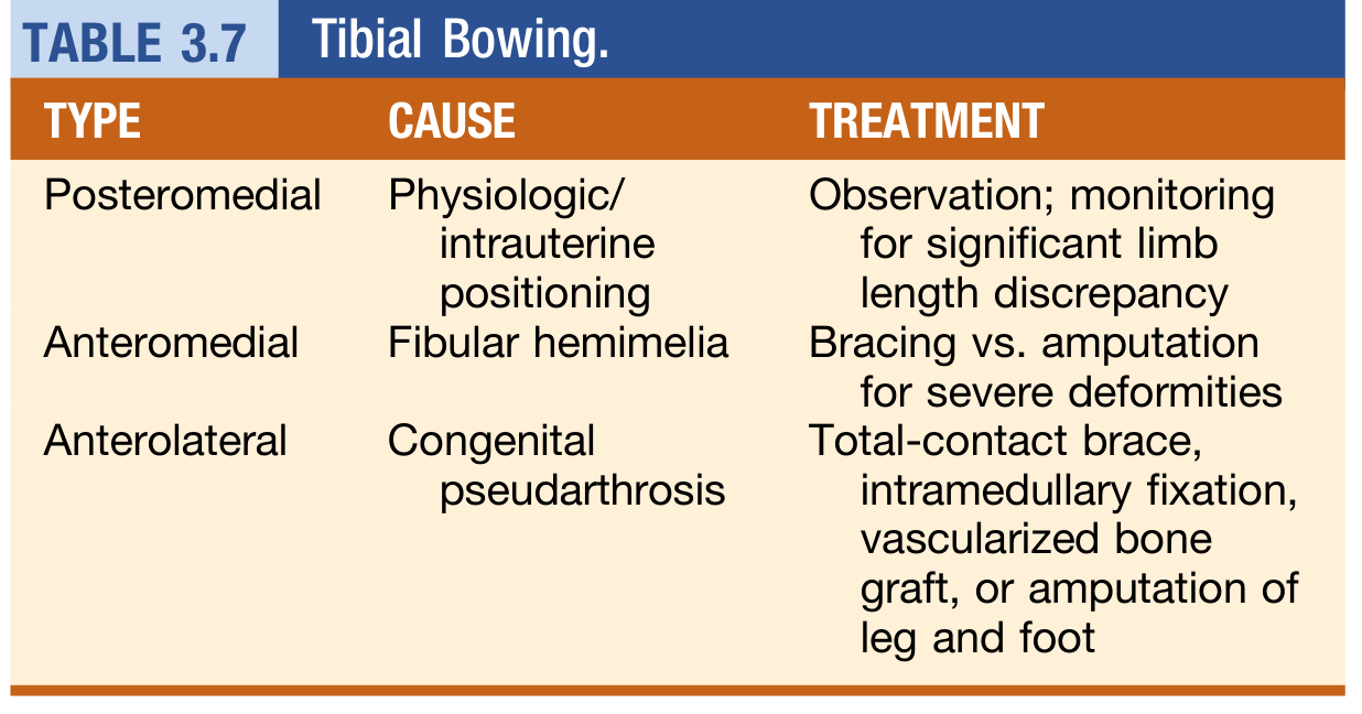
Table 3.7: Tibial Bowing - Type, Cause, and Treatment. - Miller's Review of Orthopaedics 9th Ed.
| Type | Cause | Treatment |
|---|---|---|
| Posteromedial | Physiologic/intrauterine positioning | Observation; monitor for LLD (average 3-4 cm) |
| Anteromedial | Fibular hemimelia | Bracing vs. amputation for severe cases |
| Anterolateral | Congenital pseudarthrosis of tibia (often NF1) | Total-contact brace, IM fixation, vascularized graft, or amputation |
6. Genu Valgum (Knock-Knees) - When to Intervene
- Conservative treatment is ineffective in pathologic genu valgum
- Surgery is considered in children older than 10 years with:
-
10 cm between the medial malleoli, OR
-
15 degrees of valgus angulation
-
- In children under 8 years, hemiepiphysiodesis (temporary/timed) of the medial distal femoral physis is effective for severe deformities if performed before end of growth
- Mechanism: Guided growth relies on the Hueter-Volkmann law (growth is decreased by increased compression); physeal growth returns to normal when compression is removed
7. Remodeling Potential: Fracture-Related Angular Deformity
For fractures in children <8 years, angular deformity within the plane of an adjacent joint is often completely remodeled with growth. Key principles:
- Young children have extensive remodeling potential - many angular deformities from fractures correct spontaneously
- Older children do NOT remodel as well (e.g., 10° angulation in forearm bones at age >10 years may cause significant rotation limitation)
- Corrective osteotomy for post-fracture angular deformity should be delayed at least 1 year unless function is impaired
- Torsional deformities after femoral fractures: most are mild (<10°) and asymptomatic, rarely requiring treatment
Summary: Decision Framework for Angular Deformity <8 Years
Child with angular deformity
        ↓
Is it within physiologic age range?
    YES → Observe, reassure, follow up
    NO  ↓
Is it progressive, asymmetric, or severe?
    ↓
Investigate for pathologic cause:
  - Blount disease (Drennan angle, Langenskiold stage)
  - Metabolic bone disease (rickets, renal osteodystrophy)
  - Congenital: fibular hemimelia, pseudarthrosis
  - Trauma / physeal injury
        ↓
Pathologic confirmed?
  - Age <3 yrs + Stage I/II Blount → KAFO Bracing
  - Age >3 yrs / Stage II-III Blount → Proximal tibial osteotomy
  - Genu valgum: hemiepiphysiodesis if severe (before growth ends)
  - Posteromedial tibial bowing → Observe, monitor LLD
  - Anterolateral bowing (pseudarthrosis) → Total-contact brace, then surgery
Sources:
- [Miller's Review of Orthopaedics, 9th Ed.](Miller's Review of Orthopaedics, pp. 276-279) - Physiologic patterns, Blount disease, tibial bowing
- [Schwartz's Principles of Surgery, 11th Ed.](Schwartz's, p. 1944) - Remodeling potential in pediatric fractures
- [Campbell's Operative Orthopaedics, 15th Ed. 2026](Campbell's, p. 1856) - Acceptable angulation and corrective osteotomy timing
Plate induced osteoporosis structured answer
Plate-induced osteoporosis (also called stress protection osteoporosis) refers to bone resorption and cortical porosity beneath a rigid fixation plate, caused by the plate shielding the bone from normal physiological mechanical loads. The key mechanism is that rigid plates carry most of the stress, depriving the underlying cortex of the mechanical stimulation needed to maintain bone density (Wolff's law). This leads to cortical thinning, porosity, and increased refracture risk after plate removal.
Searching sources...
Finding Sources
Finding Sources
Finding Sources
Finding Sources
Finding Sources
Finding Sources
Finding Sources
Finding Sources
Reading File
Reading File
Reading File
Finding Sources
Reading File
Finding Sources
Loading Image
I now have all the material needed for a comprehensive structured answer. Let me compile it.
Plate-Induced Osteoporosis (Stress Protection Osteoporosis)
1. Definition
Plate-induced osteoporosis (also called stress protection osteoporosis or stress shielding) is the progressive loss of bone density, cortical porosity, and structural weakening of bone that occurs beneath a rigid fixation plate, as a result of the plate shielding the underlying bone from its normal physiological mechanical loads.
2. Basis: Wolff's Law
The entire phenomenon is rooted in Wolff's Law: bone remodels in response to the mechanical stresses placed on it.
- Bone subjected to normal physiological loading - compressive and tensile forces - maintains or increases its density
- Bone deprived of mechanical stimulation undergoes resorption and becomes osteoporotic
- A rigid plate carries most of the applied load, so the underlying cortex receives far less strain than normal → net bone resorption dominates over formation
3. Mechanism / Pathophysiology
Rigid plate applied to bone
        ↓
Plate has high stiffness (especially steel > titanium)
        ↓
Plate carries majority of applied mechanical load
(stress bypasses the underlying bone cortex)
        ↓
Underlying cortex experiences reduced strain
(below the threshold required to stimulate osteoblastic activity)
        ↓
Osteoclastic resorption > Osteoblastic formation
        ↓
Cortical thinning + increased intracortical porosity
        ↓
Structurally weakened bone beneath and around the plate
Additional contributing factors:
- Periosteal stripping during plate application disrupts the periosteal blood supply, further impairing bone nutrition and remodeling
- Rigidity of fixation suppresses secondary (callus) bone healing and forces primary/direct bone healing, which is a slower process with no radiographic callus evidence of strength
- Atrophy of bone under screw holes from local pressure necrosis and vascular disruption
4. Factors That Determine the Severity
| Factor | Higher Risk | Lower Risk |
|---|---|---|
| Plate material | Stainless steel (stiffer) | Titanium / PEEK (more flexible) |
| Plate design | Conventional DCP (direct bone contact) | LCP / locking plate (elevated off bone surface) |
| Duration in situ | Long periods | Short periods |
| Fracture site | Diaphyseal (cortical bone) | Metaphyseal |
| Soft tissue handling | Extensive periosteal stripping | Minimally invasive (MIPO) |
| Number of screws | More screws, stiffer construct | Fewer screws, working length preserved |
5. Radiographic / Clinical Features
- Radiographs: Cortical thinning and osteopenia visible beneath and adjacent to plate; screw holes appear lucent and widened; absence of periosteal callus (primary bone healing leaves no radiographic trace of consolidation)
- Clinically: Bone appears healed radiographically but is mechanically weak
- Key danger: Early or premature plate removal → refracture through the original fracture site (through osteoporotic, screw-holed bone)
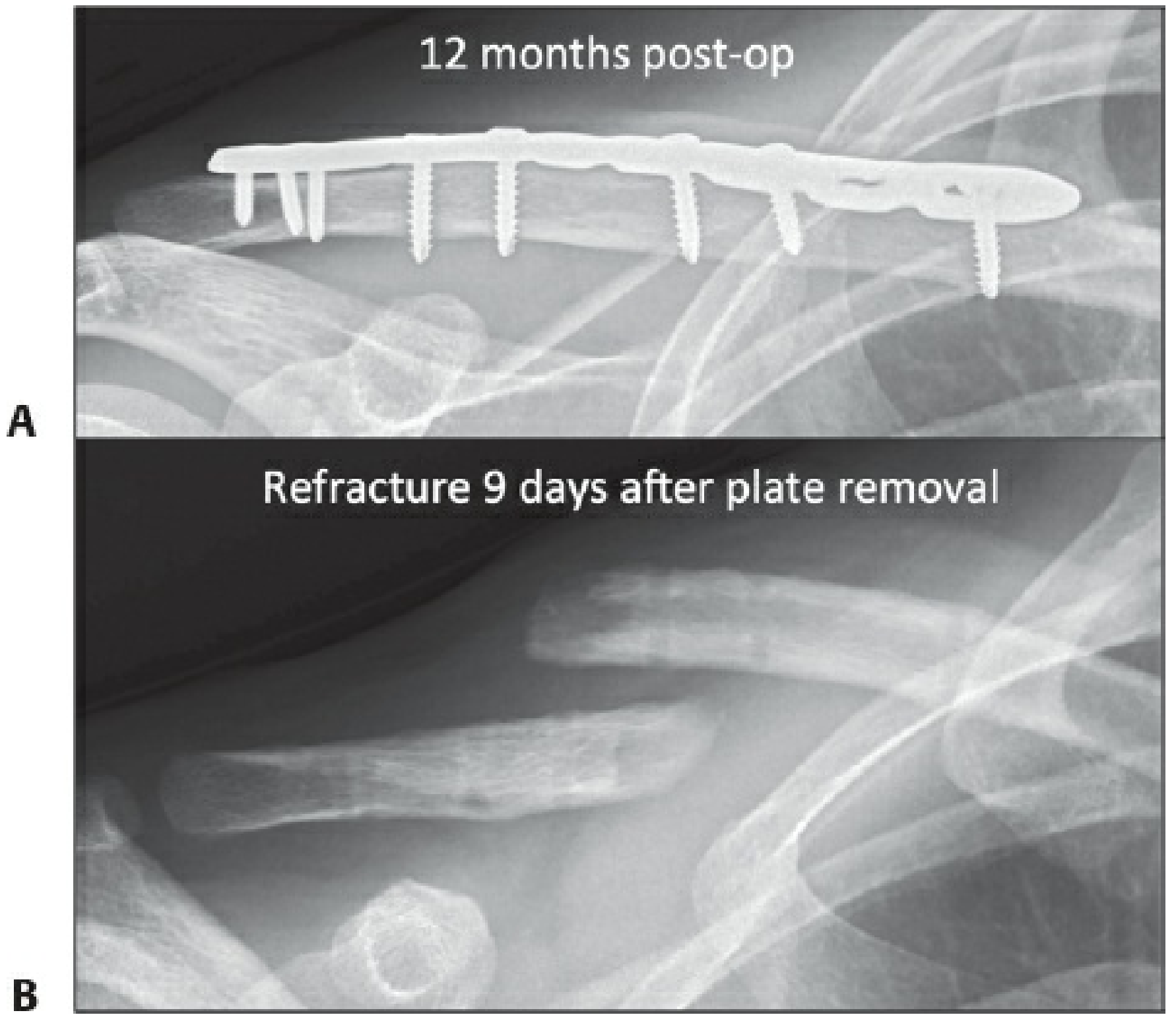
Fig. 1-17: Plating of a clavicle fracture. A: At 12 months, primary bone healing is largely invisible on radiographs (no periosteal callus). B: Refracture occurred just 9 days after plate removal through the original fracture site. - Rockwood & Green's Fractures in Adults, 10th Ed. 2025
6. Why Primary (Direct) Bone Healing Makes This Worse
- Rigid plates that achieve absolute stability promote primary/contact bone healing, which proceeds without intermediate callus
- This healing process is slower and does not produce the structural buttress that callus provides
- When loaded to failure, radiographically "healed" bones stabilized with compression plates failed through the original fracture site at 48 weeks in one-third of specimens (animal studies)
- Because the bone looks "healed" on X-ray but is structurally weak, the surgeon may be falsely reassured - plate removal therefore carries high refracture risk
7. Prevention Strategies
A. Implant Material Choice
- Titanium plates are less stiff than stainless steel → less stress shielding
- PEEK (polyetheretherketone) plates - even more flexible, approaching bone stiffness; decrease bending stiffness significantly
- Note: changing material mainly affects bending stiffness; axial motion adjacent to plate remains deficient unless implant design specifically addresses it
B. Plate Design
- Locking Compression Plates (LCP): Locked screws do not compress plate to bone → preserves periosteal blood supply; acts as internal-external fixator
- MIPO (Minimally Invasive Plate Osteosynthesis): Avoids periosteal stripping → better biologic environment
- Bridge plating: Longer working length + fewer screws per fragment → allows relative motion → stimulates callus formation
C. Dynamic / Active Implants
- Dynamic Locking Screws (DLS) / Far Cortical Locking (FCL) screws: Thinner screw shaft elastically flexes relative to near cortex → enables controlled axial dynamization at fracture site → stimulates callus without shear motion
- Active locking plates: Elastomeric elements within locking holes allow controlled axial motion
- Both screw dynamization and plate dynamization have shown faster, stronger, and more circumferential healing compared to rigid locking constructs in animal models
D. Timing of Plate Removal
- Plate removal should be delayed for up to 2 years after surgery to allow adequate time for the bone to regain structural integrity
- Early removal (before 2 years, especially in the first year) carries the highest risk of refracture through the original fracture site
- After removal, patient should be counselled to protect the limb (avoid contact sports, heavy loading) for 6-12 weeks while screw holes fill in with remodeled bone
8. Consequences of Plate-Induced Osteoporosis
| Consequence | Details |
|---|---|
| Refracture | Through fracture site or screw holes after plate removal |
| Stress riser | Empty screw holes act as stress concentrators |
| Atrophic nonunion | Overly stiff construct + residual gap → insufficient motion → no callus → fibrous union |
| Implant failure | Cyclically loaded osteoporotic bone cannot maintain screw purchase → toggle, loosening |
| Delayed union | Reduced biologic stimulus from poor periosteal vascularity |
9. Summary Table: Plate Stiffness vs. Fracture Healing
| Fixation Type | Stability | Healing Mode | Callus? | Stress Shielding |
|---|---|---|---|---|
| Rigid DCP (compression) | Absolute | Primary / Direct | None | High |
| Locking plate (stiff) + gap | Absolute | Atrophic nonunion risk | None | High |
| Bridge plate / MIPO | Relative | Secondary (callus) | Yes | Low |
| Dynamic screw/plate | Controlled axial | Secondary (callus) | Yes | Low |
| Intramedullary nail | Relative | Secondary (callus) | Yes | Minimal |
Sources:
- Rockwood & Green's Fractures in Adults, 10th Ed. 2025 - Primary bone healing, stress shielding, strategies for improvement
- Bailey & Love's Short Practice of Surgery, 28th Ed. - Plates and screws: advantages/disadvantages
This is a shared conversation. Sign in to Orris to start your own chat.