
Rheumatoid arthritis
I'll start by reading the medical library skill, then search for comprehensive information on rheumatoid arthritis.
Searching sources...
Reading File
Now I'll run parallel searches across the library and PubMed simultaneously.
Finding Sources
Searching PubMed
"rheumatoid arthritis"[MeSH Terms] AND treatment
Searching the Web
rheumatoid arthritis 2025 2026 guidelines treatment update
Good - I found relevant headings. Now let me search for a dedicated rheumatology textbook and read the core RA content in parallel.
Finding Sources
Finding Sources
Searching PubMed
"rheumatoid arthritis"[MeSH Terms] AND "treatment outcome"[MeSH Terms]
Excellent - I found Firestein & Kelley's Textbook of Rheumatology and Harrison's. Let me search these for RA content now.
Finding Sources
Searching PubMed
"rheumatoid arthritis"[MeSH Terms] AND "biologics"
Now let me read the core RA content from Firestein & Kelley's in parallel - the most authoritative rheumatology textbook.
Reading File
Reading File
Reading File
Let me read the most important clinical RA sections - block6 for clinical features/management and block3 for pathogenesis/diagnosis.
Reading File
Reading File
Now let me read the clinical features, diagnosis, and management sections from block6 (the most content-rich section at lines 532-654).
Reading File
Reading File
Now let me search specifically for clinical features, diagnosis and management content from the Family Medicine textbook which will have a clinical summary, and also search for RA treatment/management.
Finding Sources
Finding Sources
I found the KEY POINTS section for management (block18) and the 2010 ACR/EULAR classification criteria (block10). Let me read these critical sections.
Reading File
Reading File
Now let me read the detailed management section from block18 and the ACR/EULAR classification criteria table.
Reading File
Finding Sources
I found the KEY POINTS from block17 which has the clinical overview. Let me read it and also continue reading the management section.
Reading File
Reading File
I now have excellent content. Let me do one final read to get the biologic/treatment ladder content.
Reading File
Reading File
I now have comprehensive data to write a thorough response. Let me compile it.
Rheumatoid Arthritis
A complete clinical overview based primarily on Firestein & Kelley's Textbook of Rheumatology (2-Volume Set) and supplemented by current guidelines.
Definition and Overview
Rheumatoid arthritis (RA) is a chronic, systemic autoimmune condition characterized by a polyarticular small joint inflammatory arthritis, as well as diverse extra-articular manifestations. The primary pathology occurs in the synovial tissues, producing symmetric polyarticular inflammation that can progress to joint destruction, functional disability, and premature mortality.
Epidemiology
- Prevalence: 0.5-1% in most developed countries; global age-standardized prevalence ~0.25% (Global Burden of Disease 2017).
- Regional variation: Highest in North America, Western Europe, and the Caribbean (0.3-0.4%); lowest in Southeast Asia and Sub-Saharan Africa (~0.1%).
- Special population: Native Americans (Pima and Chippewa tribes) have rates approaching 5-7%.
- Sex ratio: ~3:1 female predominance.
- Peak onset: 4th-5th decade, though it can occur at any age.
Pathogenesis
Genetic Risk Factors
The HLA-DRB1 "shared epitope" is the strongest genetic risk factor, and its frequency explains much of the population variation in RA prevalence. Multiple non-HLA loci are also implicated (PTPN22, STAT4, CTLA4, etc.).
Pre-Clinical Phase
RA has a recognizable pre-clinical phase. Autoantibodies (RF and ACPA) can be detected in serum years before clinical disease onset. Evidence suggests early inflammatory events begin at extra-articular mucosal sites - lung, oral cavity, and gut:
- In seropositive unaffected first-degree relatives of RA patients, subclinical mucosal inflammation is demonstrable
- RA-related autoantibodies occur in diseases characterized by mucosal inflammation (e.g., bronchiectasis)
- ACPA specificity expands in a time-dependent manner pre-clinically, peaking at symptom onset
Citrullination and ACPAs
- Citrullination is a post-translational modification converting positively charged arginine to neutral citrulline, catalyzed by peptidylarginine deiminase (PAD) enzymes (PAD2, PAD4).
- In genetically predisposed individuals, citrullinated proteins trigger autoimmunity.
- Anti-citrullinated protein antibodies (ACPAs) target: collagen type II, fibrinogen, vimentin, and α-enolase.
- The interaction of HLA-DRB1 shared epitope + smoking + periodontitis drives ACPA generation.
Role of Oral Microbiome
Porphyromonas gingivalis is a key periodontal pathogen that expresses its own PAD enzyme, directly citrullinating bacterial and host proteins. Antibodies to this bacterium are found in 11-23% of RA patients. Gut microbiome dysbiosis (e.g., Prevotella copri enrichment) is also observed in early, untreated RA.
Synovial Pathology
- Fibroblast-like synoviocytes (FLS) are activated, producing pro-inflammatory cytokines (TNF, IL-1, IL-6) and matrix metalloproteinases leading to cartilage/bone destruction.
- FLS undergo epigenetic reprogramming (global DNA hypomethylation), acquiring an aggressive, invasive "inflammatory memory" phenotype.
- NETs (Neutrophil Extracellular Traps): Neutrophils are abundant in RA synovium and overexpress PAD2/PAD4. NETs activate FLS, releasing citrullinated autoantigens and perpetuating autoimmunity.
Autoantibodies and Complement
- Rheumatoid factor (RF): IgM (usually) against the Fc portion of IgG.
- ACPAs: More specific than RF; predict aggressive, erosive disease.
- Anti-carbamylated protein (anti-CarP) antibodies: Additional highly specific marker.
- Complement is activated primarily within the joint (via alternative and lectin pathways), with C3a, C5a, and MAC all contributing to synovial inflammation.
- Platelet activation: Thrombocytosis and platelet microparticles are markers of active RA; platelet-leukocyte aggregates, soluble P-selectin, and soluble CD40L are elevated.
Clinical Features
Articular Manifestations
- Symmetric polyarticular inflammatory arthritis involving the small joints of the hands and feet (MCPs, PIPs, wrists, MTPs)
- Prolonged morning stiffness (>1 hour) - cardinal feature
- Swelling, warmth, and tenderness of joints; "boggy" synovitis on exam
- Characteristic deformities (late disease): ulnar deviation at MCPs, swan-neck (PIP hyperextension, DIP flexion), Boutonniere deformity, Z-deformity of thumb
- Large joint involvement (knees, elbows, shoulders, ankles, hips) also occurs
- Cervical spine involvement: atlantoaxial subluxation - important perioperative concern
- Spares DIP joints (distinguishing from OA and psoriatic arthritis)
Extra-Articular Manifestations
| System | Manifestation |
|---|---|
| Pulmonary | Interstitial lung disease (ILD), pleuritis, pulmonary nodules, bronchiectasis |
| Cardiovascular | Accelerated atherosclerosis, pericarditis, myocarditis - RA is an independent CV risk factor |
| Skin | Rheumatoid nodules (subcutaneous, extensor surfaces, seropositive patients) |
| Eyes | Keratoconjunctivitis sicca (secondary Sjogren's), episcleritis, scleritis |
| Hematologic | Anemia of chronic disease, thrombocytosis (with active disease); Felty syndrome (RA + splenomegaly + neutropenia) |
| Neurologic | Peripheral neuropathy, carpal tunnel syndrome, mononeuritis multiplex, cervical myelopathy |
| Vasculitis | Rheumatoid vasculitis (severe seropositive disease) |
Key extra-articular features that shorten lifespan: ILD, cardiovascular disease, and malignancy (especially lymphoma).
Diagnosis and Classification
2010 ACR/EULAR Classification Criteria
Used for classifying RA in patients with at least one joint with definite synovitis not explained by another disease. A score ≥6 is needed for classification as definite RA.
| Domain | Finding | Score |
|---|---|---|
| Joint involvement | 1 medium-large joint | 0 |
| 2-10 medium-large joints | 1 | |
| 1-3 small joints | 2 | |
| 4-10 small joints | 3 | |
| >10 joints (including ≥1 small) | 5 | |
| Serology | Negative RF and ACPA | 0 |
| Low positive RF or ACPA | 2 | |
| High positive RF or ACPA | 3 | |
| Acute phase reactants | Normal CRP and ESR | 0 |
| Abnormal CRP or ESR | 1 | |
| Duration of symptoms | <6 weeks | 0 |
| ≥6 weeks | 1 |
Note: If incontrovertible radiographic evidence of RA exists, the diagnosis can be made even without meeting criteria numerically.
Key Laboratory Tests
- RF (rheumatoid factor): Present in ~70-80%; not specific
- Anti-CCP (ACPA): ~70% sensitive, >95% specific; predicts erosive disease
- CRP, ESR: Markers of systemic inflammation
- CBC: Anemia of chronic disease, thrombocytosis
- Imaging: Plain X-rays (erosions, joint space narrowing); MRI/ultrasound more sensitive for early synovitis and subclinical inflammation
Disease Activity Measurement
Regular disease activity measurement is a quality metric. Key instruments:
| Instrument | Components | Remission | Low | Moderate | High |
|---|---|---|---|---|---|
| DAS28 | 28 TJC, 28 SJC, patient global, ESR/CRP | ≤2.6 | ≤3.2 | >3.2-5.1 | >5.1 |
| SDAI | 28 TJC, 28 SJC, provider/patient global, CRP | ≤3.3 | ≤11 | ≤26 | >26 |
| CDAI | 28 TJC, 28 SJC, provider/patient global | ≤2.8 | ≤10 | ≤22 | >22 |
Management
Principles (Treat-to-Target, T2T)
The treat-to-target (T2T) strategy is the cornerstone of modern RA management:
- Target: Remission or low disease activity for all patients.
- Monitor disease activity regularly and escalate therapy if target not met within 3-6 months.
- The specific DMARD chosen is less important than consistently achieving the target - proven by the TICORA and Dutch BeSt studies.
- RA can and should be diagnosed early; DMARD therapy should start at diagnosis.
The DMARD Toolbox
Conventional synthetic DMARDs (csDMARDs)
- Methotrexate (MTX): The anchor drug; cornerstone of therapy. Titrated up to 25 mg/week; subcutaneous route increases bioavailability. Always given with folic acid supplementation.
- Hydroxychloroquine (HCQ): Used in mild disease or combination (triple therapy with MTX + SSZ)
- Sulfasalazine (SSZ): Often used in combination
- Leflunomide: Alternative to MTX when MTX is contraindicated
Biologic DMARDs (bDMARDs)
| Agent | Target |
|---|---|
| Etanercept, Infliximab, Adalimumab, Golimumab, Certolizumab | TNF-α |
| Rituximab | CD20 (B cell depletion) |
| Abatacept | CD80/86 - T cell costimulation |
| Tocilizumab, Sarilumab | IL-6 receptor |
| Anakinra | IL-1 |
Targeted synthetic DMARDs (tsDMARDs) - JAK inhibitors
- Tofacitinib, Baricitinib, Upadacitinib, Filgotinib
- Oral agents; note safety considerations regarding cardiovascular events and malignancy risk (post-marketing surveillance requirements)
Treatment Algorithm
- First-line: MTX monotherapy (strongly recommended by ACR/EULAR). If MTX not tolerated, use leflunomide or sulfasalazine.
- If MTX fails (inadequate response at 3 months, target not met by 6 months): Add a bDMARD or JAK inhibitor to MTX. Choice guided by presence of poor prognostic factors, comorbidities (e.g., avoid JAK inhibitors in high CV risk patients), cost, and patient preference.
- If first bDMARD fails: Switch to another bDMARD (different mechanism) or tsDMARD.
- Triple therapy (MTX + HCQ + SSZ) is a cost-effective alternative to bDMARDs in resource-limited settings.
Glucocorticoids
- Powerful, rapidly effective, but carry significant short- and long-term adverse effects.
- Use only as a bridge to DMARD therapy - not as monotherapy.
- Best chronic dose: zero. Always taper once DMARDs are effective.
- Low-dose prednisone (5-10 mg/day) added to MTX reduces disease activity and radiographic progression (CAMERA-II trial).
NSAIDs
- Provide symptom relief but are not disease-modifying.
- Never use without concurrent DMARDs.
- Discontinue once DMARDs achieve disease control.
Tapering and Remission
- DMARD tapering only considered in patients who have achieved sustained remission.
- Drug-free remission remains largely out of reach - at least one DMARD usually must be continued.
Monitoring and Comorbidities
The ubiquitous comorbidities of RA - especially cardiovascular disease - must be proactively addressed:
- Cardiovascular risk assessment and management (statins, blood pressure control)
- Bone density monitoring and osteoporosis prevention (especially in glucocorticoid-treated patients)
- Lung disease surveillance (baseline PFTs and HRCT in appropriate patients)
- Infection risk assessment before biologic/JAK inhibitor therapy (TB screening, hepatitis B, live vaccine update)
- Malignancy screening (particularly lymphoma risk)
Prognosis
- RF and ACPA positivity predicts an aggressive disease course with erosive joint damage and extra-articular manifestations.
- Untreated or undertreated RA leads to substantial disability, reduced work capacity, and shortened lifespan.
- With modern T2T strategies and the full DMARD toolbox, the majority of newly diagnosed RA patients can achieve remission or low disease activity - a transformation from the pre-biologic era.
Primary source: Firestein & Kelley's Textbook of Rheumatology, 2-Volume Set (Elsevier). Also references Rheumatology, 2-Volume Set (2022, Elsevier) and Harrison's Principles of Internal Medicine 22E (2025). Current ACR 2021 guidelines strongly recommend methotrexate monotherapy as first-line and a treat-to-target approach for all patients.
Anterior pituitary disorder
Reading File
Searching sources...
This is a broad topic - anterior pituitary disorders encompass multiple conditions. I'll run parallel searches across the library and web simultaneously.
Finding Sources
Finding Sources
Searching the Web
anterior pituitary disorders overview hormones clinical features 2025
Good. Now let me search Harrison's and Goldman-Cecil (the best clinical medicine texts) for anterior pituitary disorders, and also search for specific disorder headings.
Finding Sources
Finding Sources
Searching PubMed
"pituitary disorders"[MeSH Terms] AND "diagnosis"[MeSH Terms]
Excellent - found Harrison's 22E (2025) content on anterior pituitary and prolactinoma, plus Goldman-Cecil and Robbins. Let me now read the key blocks in parallel.
Reading File
Reading File
Reading File
Excellent content from Harrison's 22E. The first read returned a huge amount - it has detailed physiology tables, prolactin, GH sections. Let me now fetch the pituitary axes diagram and read the specific disorder sections (hyperprolactinemia, GH excess, Cushing, hypopituitarism, acromegaly).
Loading Image
Reading File
Reading File
Excellent - I have a rich dataset. The last read (offset 2285) gave me a ton of content including hyperprolactinemia causes, prolactinoma, galactorrhea. Let me read the hypopituitarism section and GH/acromegaly/Cushing sections.
Reading File
Reading File
Excellent content on hypopituitarism - etiology, presentation, and lab investigation. The second read was very rich and includes pituitary apoplexy, empty sella, and full diagnostic workup. Now let me read the acromegaly/GH excess section and Cushing disease section.
Reading File
Reading File
I now have comprehensive data from Harrison's 22E covering: physiology table, prolactin, TSH, hypopituitarism (etiology, causes, presentations, labs), ACTH deficiency, GH deficiency, pituitary tumors, screening tests, surgery. The second read (offset 2100) also contains screening tests table for acromegaly, prolactinoma, Cushing's disease. Let me read one more section covering acromegaly and prolactinoma treatment.
Reading File
This is tremendously detailed. The last read gave me prolactinoma epidemiology, clinical features, diagnosis (PRL >200 = likely prolactinoma), and treatment (dopamine agonists: cabergoline, bromocriptine). I now have comprehensive content from Harrison's 22E covering all major anterior pituitary disorders. Let me compile the full response.
Anterior Pituitary Disorders
Primary source: Harrison's Principles of Internal Medicine, 22nd Edition (2025, McGraw Hill Medical); Robbins & Kumar Basic Pathology; Goldman-Cecil Medicine.
Anatomy and the "Master Gland" Concept
The anterior pituitary (adenohypophysis) is often called the "master gland" because, together with the hypothalamus, it orchestrates the complex regulatory functions of many other endocrine glands. It is located in the sella turcica at the base of the brain and communicates with the hypothalamus via the hypothalamo-hypophyseal portal system.
It produces 6 major hormones from 5 distinct cell types:
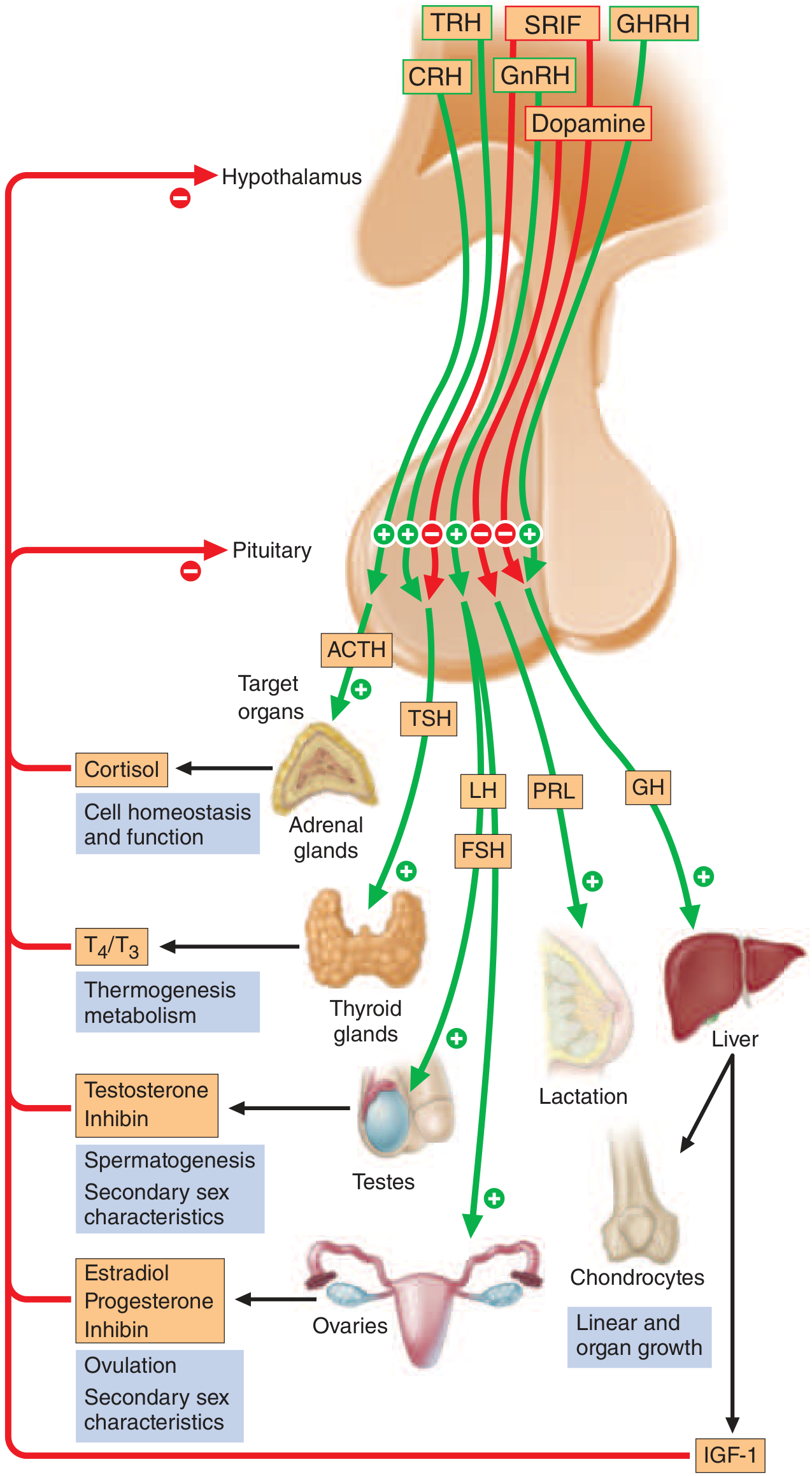
Anterior Pituitary Cell Types and Hormones
| Cell Type | Hormone | Hypothalamic Stimulator | Hypothalamic Inhibitor | Target |
|---|---|---|---|---|
| Corticotrope | ACTH (from POMC) | CRH, AVP, cytokines | Glucocorticoids | Adrenal cortex |
| Somatotrope | GH | GHRH, ghrelin | Somatostatin, IGF-1 | Liver, bone, muscle |
| Lactotrope | Prolactin (PRL) | Estrogen, TRH, VIP | Dopamine (tonic inhibition) | Breast, gonads |
| Thyrotrope | TSH | TRH | T3/T4, dopamine, somatostatin | Thyroid |
| Gonadotrope | LH, FSH | GnRH, activins, estrogen | Sex steroids, inhibin | Ovary, testes |
Key transcription factors:
- Pit-1 (Prop-1): Required for somatotrope, lactotrope, thyrotrope differentiation
- T-Pit: Corticotrope differentiation
- SF-1/DAX-1: Gonadotrope differentiation
Overview of Anterior Pituitary Disorders
Disorders fall into two major categories:
- Hypersecretion - usually caused by a functioning adenoma
- Hyposecretion (Hypopituitarism) - caused by structural damage, tumors, or developmental defects
I. Pituitary Adenomas
The most common cause of hyperpituitarism. Key pathologic features:
- Cellular monomorphism and absence of a reticulin network (distinguishing from normal gland)
- GNAS gene mutation (constitutive G-protein activation) is among the most common genetic alterations
- Microadenoma: <1 cm - usually does not cause mass effects
- Macroadenoma: ≥1 cm - may invade parasellar structures, compress optic chiasm
Mass Effects of Pituitary Adenomas
- Bitemporal hemianopia - chiasmal compression (classically superior field first)
- Headache (dural stretching)
- Cranial nerve palsies (CN III, IV, VI) from cavernous sinus invasion
- Hydrocephalus (large tumors)
- Hypopituitarism (compression of normal pituitary tissue)
- Pituitary apoplexy: acute hemorrhage/infarction into an adenoma - medical emergency with sudden headache, visual loss, ophthalmoplegia, cardiovascular collapse
Screening Tests for Functioning Adenomas
| Disorder | Screening Test | Interpretation |
|---|---|---|
| Acromegaly | Serum IGF-1; Oral GTT with GH at 0, 30, 60 min | Normal suppresses GH to <1 µg/L after glucose |
| Prolactinoma | Serum PRL | MRI if elevated; PRL >200 µg/L = likely prolactinoma |
| Cushing's disease | 24-h urinary free cortisol; 1 mg dexamethasone suppression test (11 PM) | Failure to suppress cortisol to <50 nmol/L |
| TSH-secreting | Free T4, TSH | Elevated free T4 with unsuppressed/elevated TSH |
II. Hyperprolactinemia and Prolactinoma
Physiology of Prolactin
- Normal levels: ~10-25 µg/L (women), 10-20 µg/L (men)
- Prolactin is uniquely under tonic inhibitory control by dopamine (D2 receptor-mediated)
- This explains why stalk section or dopamine-blocking drugs cause hyperprolactinemia
- PRL rises with stress, exercise, meals, coitus, chest wall injury, and markedly during pregnancy (~10-fold)
Causes of Hyperprolactinemia
I. Physiologic: pregnancy, lactation, stress, exercise, sleep
II. Hypothalamic/stalk: stalk compression by any mass (decreases dopamine delivery)
III. Pituitary tumors: prolactinoma, acromegaly (co-secretion)
IV. Systemic disorders: hypothyroidism (TRH stimulates PRL), chronic renal failure, cirrhosis
V. Drug-induced (very common - always exclude):
- Dopamine receptor blockers: antipsychotics (risperidone, haloperidol, phenothiazines), metoclopramide
- Dopamine synthesis inhibitors: alpha-methyldopa
- Opiates, H2 antagonists (cimetidine), SSRIs (fluoxetine), verapamil, estrogens
PRL >200 µg/L almost invariably indicates a prolactinoma. Values <100 µg/L may be caused by microadenomas, stalk compression, or systemic causes.
Prolactinoma: Epidemiology and Presentation
- Most common functioning pituitary tumor (~50% of all); prevalence ~30/100,000 in women, ~10/100,000 in men
- Female-to-male ratio 20:1 for microadenomas; ~1:1 for macroadenomas
- Tumor size correlates directly with PRL level
Clinical features:
- Women: amenorrhea, infertility, galactorrhea (amenorrhea-galactorrhea syndrome), loss of libido, osteoporosis
- Men: impotence, loss of libido, infertility, gynecomastia (galactorrhea less common); often present late with macroadenoma and mass effects
- Both: headache, visual field defects (macroadenoma)
Galactorrhea: Inappropriate milk discharge persisting >6 months after childbirth or cessation of breastfeeding; evaluate with mammography if bloody (rule out breast cancer).
Prolactinoma Treatment
- Asymptomatic microadenoma with no fertility desire: observe with serial PRL and MRI
- Mainstay: Dopamine agonists - suppress PRL, shrink tumor, restore gonadal function:
- Cabergoline (preferred): longer-acting, better tolerated, higher efficacy
- Bromocriptine: alternative, especially during pregnancy
- After 2 years of normoprolactinemia + significant tumor shrinkage, dopamine agonist may be withdrawn (monitor for recurrence - ~20% of patients)
- Surgery (transsphenoidal): for patients intolerant/resistant to dopamine agonists, or with acute vision loss
- PRL suppression by dopamine agonists does not always confirm prolactinoma (may suppress stalk-compression hyperprolactinemia too without shrinking the mass)
III. Growth Hormone Disorders
Acromegaly (GH excess in adults) / Gigantism (GH excess in children)
Pathophysiology: Somatotroph adenoma secretes excess GH → liver generates excess IGF-1 → promotes tissue overgrowth
Clinical Features of Acromegaly:
- Soft tissue changes: coarsening of facial features, enlarged nose, lips, ears; macroglossia; deep voice
- Acral changes: enlargement of hands and feet (patients note increasing ring/shoe size)
- Prognathism, widened tooth spacing, malocclusion
- Hypertension, cardiomegaly, cardiomyopathy (leading cause of death)
- Diabetes mellitus / insulin resistance (GH is a counterregulatory hormone)
- Sleep apnea, arthropathy, carpal tunnel syndrome
- Colonic polyps (increased cancer risk)
- Gigantism (pre-epiphyseal closure): linear growth acceleration
Diagnosis:
- Elevated age- and sex-matched serum IGF-1 (best screening test)
- Oral glucose tolerance test (OGTT): GH should suppress to <1 µg/L; failure to suppress confirms autonomous GH secretion
- MRI of pituitary for tumor characterization
Treatment:
- Transsphenoidal surgery (first-line for most)
- Somatostatin analogs (octreotide, lanreotide): inhibit GH secretion; used for residual/recurrent disease or as primary therapy if surgery is high-risk
- Pegvisomant: GH receptor antagonist - normalizes IGF-1 in resistant cases
- Dopamine agonists (cabergoline): modest efficacy
- Radiotherapy: adjunct for persistent disease
GH Deficiency (Adults - AGHD)
- Features: increased body fat (particularly visceral), decreased lean mass, reduced bone mineral density, impaired quality of life, increased cardiovascular risk
- Diagnosis: provocative GH stimulation tests (insulin tolerance test is gold standard; alternatives: arginine, glucagon, GHRH, macimorelin oral test)
- Treatment: GH replacement, starting 0.1-0.2 mg/day, titrated up to 1.25 mg/day; maintain IGF-1 in mid-normal range
- Side effects (~30%): fluid retention, joint pain, carpal tunnel; contraindicated in active neoplasm or uncontrolled diabetes with retinopathy
IV. ACTH Deficiency / Secondary Adrenal Insufficiency
Causes:
- Glucocorticoid withdrawal (most common - suppression of HPA axis)
- Pituitary adenoma mass effect
- Post-surgical (resection of ACTH-secreting adenoma - indicates cure)
- Rarely: TPIT or POMC mutations
Presentation - similar to Addison's but KEY differences:
- No hyperpigmentation (requires excess ACTH/MSH - absent in secondary)
- No mineralocorticoid deficiency (aldosterone regulated by renin-angiotensin, preserved)
- Fatigue, weakness, anorexia, nausea, vomiting, hypoglycemia
- Hyponatremia (dilutional - from ADH dysregulation)
Diagnosis:
- Low morning cortisol + low/inappropriately normal ACTH
- Short Synacthen (cosyntropin) test: impaired cortisol response
- Insulin tolerance test (ITT): most reliable - measures both ACTH and cortisol response to hypoglycemia (contraindicated in seizure disorder, ischemic heart disease, elderly)
V. Cushing's Disease (ACTH-Secreting Adenoma)
Cushing's disease = pituitary ACTH-secreting adenoma (corticotroph adenoma) → one cause of Cushing's syndrome
Clinical Features of Cushing's Syndrome:
- Central obesity, moon face, buffalo hump
- Proximal myopathy, easy bruising, striae (violaceous)
- Hypertension, hyperglycemia/diabetes
- Osteoporosis, poor wound healing
- Hirsutism, menstrual irregularity
- Psychological: depression, cognitive impairment
- Hyperpigmentation (if ectopic ACTH source with very high ACTH levels)
Diagnosis (3-step approach):
- Confirm hypercortisolism: 24-h urinary free cortisol, late-night salivary cortisol, 1 mg overnight dexamethasone suppression test (>50 nmol/L = failure to suppress)
- Confirm ACTH-dependency: plasma ACTH level
- Localize: High-dose dexamethasone suppression test (pituitary adenoma suppresses; ectopic source does not); CRH stimulation test; pituitary MRI; inferior petrosal sinus sampling (IPSS) - gold standard for confirming pituitary origin and lateralizing tumor
Treatment:
- Transsphenoidal surgery (primary treatment)
- Bilateral adrenalectomy (if pituitary surgery fails - risk: Nelson syndrome with ACTH hypersecretion and hyperpigmentation)
- Medical: ketoconazole, metyrapone, pasireotide (somatostatin analog with affinity for SST5 receptors)
VI. TSH-Secreting Adenoma (Rare)
- Causes central hyperthyroidism: elevated free T4/T3 with unsuppressed or elevated TSH
- Distinguishable from thyroid resistance by MRI showing pituitary mass + elevated alpha subunit
- Treatment: surgery + somatostatin analogs
VII. Gonadotroph Adenomas
- Most common type of clinically non-functioning pituitary adenoma
- Secrete FSH, LH, or alpha subunits inefficiently - usually present with mass effects, not hormonal excess
- Detected incidentally (incidentaloma) or with visual field defects
- Treatment: surgery if symptomatic; observation if small incidentaloma
VIII. Hypopituitarism
Definition: Deficient production of one or more anterior pituitary trophic hormones
Etiology
| Category | Examples |
|---|---|
| Tumors | Pituitary adenoma (most common), craniopharyngioma, meningioma, metastases |
| Pituitary surgery/radiation | Post-operative, post-irradiation (may be delayed years) |
| Ischemic/infarction | Sheehan syndrome (postpartum pituitary infarction from obstetric hemorrhage); pituitary apoplexy |
| Infiltrative/granulomatous | Sarcoidosis, histiocytosis X, tuberculosis, hemochromatosis |
| Autoimmune | Lymphocytic hypophysitis (especially peripartum in women) |
| Developmental/genetic | Prop-1, Pit-1, HESX1 mutations; combined pituitary hormone deficiency (CPHD) |
| Empty sella | Herniation of subarachnoid space into sella; usually incidental; pituitary function often preserved |
| Traumatic/infectious | Head trauma, meningitis, encephalitis |
Sheehan syndrome: postpartum ischemic necrosis of the pituitary due to severe intrapartum hemorrhage and hypotension. Presents as failure to lactate (first sign - PRL deficiency), then failure to resume menses.
Order of Hormone Loss in Hypopituitarism (typical sequence)
GH → LH/FSH → TSH → ACTH → PRL (last to be lost)
Clinical Manifestations by Hormone
| Deficient Hormone | Clinical Effects |
|---|---|
| GH | Growth failure (children); ↑ body fat, ↓ lean mass, reduced QoL (adults) |
| LH/FSH | Hypogonadotropic hypogonadism: amenorrhea, infertility, ↓ libido (women); impotence, infertility, ↓ testosterone (men); delayed puberty |
| TSH | Secondary hypothyroidism: fatigue, cold intolerance, weight gain (but TSH is LOW - differs from primary hypothyroidism) |
| ACTH | Secondary adrenal insufficiency: weakness, hypoglycemia; NO hyperpigmentation, NO mineralocorticoid loss |
| PRL | Failure of lactation (postpartum) |
Pituitary Apoplexy
Acute hemorrhage or infarction of a pituitary adenoma:
- Presentation: thunderclap headache, meningism, bitemporal visual field defects, ophthalmoplegia, altered consciousness, cardiovascular collapse
- Management: High-dose glucocorticoids immediately (to treat adrenal crisis); urgent neurosurgical decompression if significant or progressive visual loss/CN palsies; visual recovery inversely correlated with time to surgery
Biochemical Diagnosis of Hypopituitarism
- Low target gland hormone + low or inappropriately normal trophic hormone
- Low FT4 + low/normal TSH = central hypothyroidism
- Low testosterone + low/normal LH/FSH = hypogonadotropic hypogonadism
- Provocative tests for GH reserve: ITT, macimorelin test, arginine, glucagon
- Cortisol reserve: ITT (gold standard), CRH test, cosyntropin stimulation
Treatment of Hypopituitarism
Hormone replacement therapy tailored to each deficiency:
- ACTH deficiency: hydrocortisone (15-25 mg/day in divided doses)
- TSH deficiency: levothyroxine (start low, titrate to FT4 in upper-normal range; do NOT use TSH as target)
- LH/FSH deficiency: testosterone (men), estrogen-progesterone (women); gonadotropins for fertility
- GH deficiency: recombinant GH (adults with confirmed deficiency and symptoms)
- Always replace cortisol before thyroid hormone (thyroid hormone increases metabolic rate → can precipitate adrenal crisis if cortisol deficiency uncorrected)
Summary Table: Anterior Pituitary Disorders
| Disorder | Hormone | Key Clinical Features | First-line Treatment |
|---|---|---|---|
| Prolactinoma | ↑ PRL | Amenorrhea, galactorrhea, infertility; mass effects if macro | Cabergoline/bromocriptine |
| Acromegaly | ↑ GH → ↑ IGF-1 | Acral enlargement, coarse facies, DM, cardiomegaly | Transsphenoidal surgery |
| Gigantism | ↑ GH (pre-pubertal) | Linear overgrowth, large stature | Transsphenoidal surgery |
| Cushing's disease | ↑ ACTH | Central obesity, striae, HTN, DM, myopathy | Transsphenoidal surgery |
| TSHoma | ↑ TSH | Central hyperthyroidism (elevated FT4 + unsuppressed TSH) | Surgery + octreotide |
| Gonadotropinoma | FSH/LH/alpha | Usually non-functioning; mass effects | Surgery if symptomatic |
| Hypopituitarism | ↓ (multiple) | Depends on which hormones lost; fatigue, infertility, etc. | Targeted hormone replacement |
| Sheehan syndrome | ↓ (multiple) | Postpartum: failure of lactation, then amenorrhea | Hormone replacement |
| Pituitary apoplexy | Acute loss | Thunderclap headache, visual loss, ophthalmoplegia | Glucocorticoids + urgent surgery |
Source: Harrison's Principles of Internal Medicine 22E (2025), Ch. 390-392 - Shlomo Melmed & J. Larry Jameson; Robbins & Kumar Basic Pathology; Goldman-Cecil Medicine.
This is a shared conversation. Sign in to Orris to start your own chat.