
Infective endocarditis harrison based
Reading File
Searching sources...
Finding Sources
Searching PubMed
"infective endocarditis"[MeSH Terms] AND ("diagnosis" OR "management")
The user specifically requested Harrison's. Let me find the Harrison's section on infective endocarditis.
Finding Sources
Finding Sources
Finding Sources
Reading File
Reading File
Reading File
I now have comprehensive Harrison's content. Let me also fetch the vegetation image to include in the answer.
Loading Image
Loading Image
I now have all the key content from Harrison's 22nd edition. Here is a comprehensive, well-organized summary:
Infective Endocarditis - Harrison's Principles of Internal Medicine (22nd Ed., 2025)
Authors: Sara E. Cosgrove, Michael T. Melia | Chapter 133
Definition and Pathology
The prototypic lesion of IE, the vegetation, is a mass of platelets, fibrin, microorganisms, and scant inflammatory cells. Infection most commonly involves heart valves but may also occur on:
- Low-pressure side of a ventricular septal defect
- Mural endocardium damaged by aberrant blood jets or foreign bodies
- Intracardiac devices
The analogous process involving AV shunts, arterio-arterial shunts (patent ductus arteriosus), or coarctation of the aorta is called infective endarteritis.
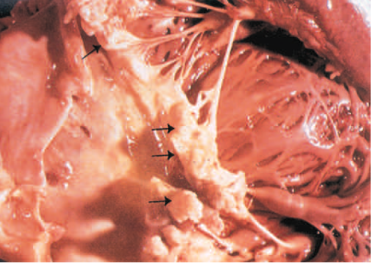
FIGURE 133-1: Vegetations (arrows) due to viridans streptococci endocarditis involving the mitral valve
Classification
| Type | Features |
|---|---|
| Acute IE | Hectically febrile, rapidly damages cardiac structures, seeds extracardiac sites, death within weeks if untreated |
| Subacute IE | Indolent course, slow structural damage, rarely metastasizes, gradually progressive unless complicated by embolic event or mycotic aneurysm rupture |
Epidemiology
- Incidence: ~15 cases per 100,000/year in the United States (increasing over recent decades)
- Traditional predisposition (chronic rheumatic heart disease) has shifted in developed countries to:
- Injection drug use (PWID)
- Degenerative valve disease
- Intracardiac devices
- 25-35% of native-valve endocarditis (NVE) in developed countries is health care-associated
- 16-30% of all cases are prosthetic-valve infections (PVE)
- Risk of PVE is greatest in the first year after valve replacement
- CIED-IE (cardiovascular implantable electronic devices): 0.5-1.14 cases per 1000 recipients
- Acceleration in mortality in ages 25-44 years linked to opioid use disorder (OUD)
Etiology
| Organism | Notes |
|---|---|
| S. aureus | Most common in developed countries; causes acute, destructive IE |
| Viridans streptococci | Oral cavity portal; subacute IE |
| Enterococci | GI/GU portal |
| Coagulase-negative staphylococci | Especially PVE |
| HACEK organisms | Haemophilus spp., Aggregatibacter spp., Cardiobacterium hominis, Eikenella corrodens, Kingella spp. - normal oropharyngeal flora |
| Fungi | Candida, Aspergillus - especially in PWID, immunocompromised, or after cardiac surgery |
| Blood culture-negative IE | Coxiella burnetii (Q fever), Bartonella spp., Tropheryma whipplei |
Pathogenesis
- Endothelial injury - turbulent blood flow, jet lesions, or foreign body → platelet-fibrin thrombus (nonbacterial thrombotic endocarditis, NBTE)
- Bacteremia - even transient (dental work, IV drug use, skin breaks) → organisms adhere to NBTE
- Vegetation growth - organisms proliferate within the platelet-fibrin matrix, protected from host defenses and antibiotics
- Most bacteria (S. aureus) can directly invade intact endothelium; viridans streptococci require pre-existing valve damage
Clinical Features
Symptoms and Signs
| Feature | Details |
|---|---|
| Fever | Most common finding; may be absent in elderly or immunocompromised |
| Heart murmur | Present in ~85% |
| New/changing murmur | Suggests valvular destruction |
| Splenomegaly | Common in subacute IE |
| Clubbing | In prolonged cases |
Peripheral Stigmata (classic but now less common)
| Sign | Description |
|---|---|
| Osler nodes | Painful, tender nodules on finger/toe pads - immune complex mediated |
| Janeway lesions | Non-tender, erythematous/hemorrhagic macules on palms and soles - septic emboli |
| Splinter hemorrhages | Linear dark streaks under fingernails |
| Roth spots | Oval retinal hemorrhages with pale center |
| Petechiae | Conjunctival, mucosal |
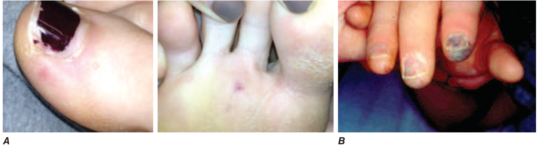
FIGURE 133-2: A. Janeway lesions on the toe and plantar surface (subacute Neisseria mucosa IE). B. Septic emboli with hemorrhage and infarction (acute S. aureus IE)
Complications
Cardiac
- Congestive heart failure - most common; due to valvular destruction (especially aortic valve perforation or rupture), intracardiac fistulae, or myocarditis
- Perivalvular infection/abscesses - especially in PVE and aortic IE; may cause new conduction abnormalities (new PR prolongation = aortic root abscess until proven otherwise)
- Pericarditis, mycotic coronary artery aneurysms
Embolic
- Arterial emboli: clinically apparent in up to 50% of patients; one-half precede diagnosis
- S. aureus IE, mobile vegetations >10 mm, and mitral anterior leaflet involvement independently increase embolization risk
- Cerebrovascular emboli (stroke/encephalopathy): 15-35% of cases; MRI shows asymptomatic emboli in 30-65%
- Stroke frequency: 8/1000 patient-days pre-diagnosis → drops to 1.7/1000 patient-days in second week of therapy
- Only 3% of strokes occur after 1 week of effective therapy
Neurologic
- Aseptic or purulent meningitis
- Intracranial hemorrhage (hemorrhagic infarcts or ruptured mycotic aneurysms)
- Seizures
- Microabscesses (common in S. aureus IE)
Renal
- Immune complex glomerulonephritis (diffuse, hypocomplementemic) - typically improves with therapy
- Embolic renal infarcts (flank pain, hematuria)
Pulmonary (right-sided IE)
- Septic pulmonary emboli: cough, pleuritic chest pain, nodular infiltrates, empyema
Special Predisposing Conditions
| Condition | Features |
|---|---|
| PWID (IV drug users) | 35-60% limited to tricuspid valve; fever, faint/no murmur, no peripheral signs; septic pulmonary emboli common |
| Prosthetic valve (PVE) | Paravalvular infection common; early PVE symptoms masked by recent surgery |
| CIED-IE | Fever, sepsis, minimal murmur, septic pulmonary emboli; may have cryptic generator pocket infection |
| TAVR-PVE | Similar incidence/decline to bioprosthetic aortic valves; TEE may miss 6-18% initially |
Diagnosis
Blood Cultures
- 3 sets from different sites before starting antibiotics
- For fastidious organisms (HACEK), most labs use extended automated culture systems (5-7 days)
- Culture-negative: hold bottles 3 weeks and use serologic tests (Coxiella, Bartonella PCR/IgG)
Duke-ISCVID Diagnostic Criteria
Definite IE: 2 major OR 1 major + 3 minor OR 5 minor criteria
Possible IE: 1 major + 1 minor OR 3 minor criteria
Rejected IE: Does not meet criteria, firm alternative diagnosis, resolves in <4 days of antibiotics, or no evidence at autopsy
Major Criteria
- Positive blood cultures (typical organisms in ≥2 separate cultures, persistently positive, or single positive for Coxiella burnetii IgG ≥1:800)
- Evidence of endocardial involvement on imaging (echo/CT/PET-CT) - vegetation, abscess, new valvular regurgitation, prosthetic valve dehiscence
Minor Criteria
- Predisposing heart condition or PWID
- Fever ≥38°C
- Vascular phenomena (emboli, septic pulmonary infarcts, mycotic aneurysm, Janeway lesions)
- Immunologic phenomena (glomerulonephritis, Osler nodes, Roth spots, RF positive)
- Positive blood culture not meeting major criteria
Non-Culture Tests (added to microbiologic major criteria in Duke-ISCVID)
- PCR/nucleic acid tests for C. burnetii, Bartonella spp., T. whipplei from blood
- Indirect immunofluorescence for Bartonella henselae/quintana IgG ≥1:800
- Next-generation metagenomic sequencing (emerging; non-C. burnetii/Bartonella results count as minor criteria for now)
Cardiac Imaging
| Modality | Details |
|---|---|
| TTE | Highly specific; inadequate images in 20%; misses 20-35% of vegetations (especially <2 mm); not optimal for prosthetic valves |
| TEE | Detects vegetations in >90%; initial false-negative 6-18% (especially TAVR-PVE); repeat in 7-10 days if initial negative and IE still suspected |
| Cardiac CT angiography | Less sensitive for vegetations than TEE; superior for pseudoaneurysm, fistula, periannular extension, intracardiac device assessment |
| 18F-FDG PET/CT | Useful for CIED-IE; detects extracardiac foci; helps avoid false-negative echo in early PVE |
| MRI/CT brain/spine | Should be performed in neurologically symptomatic patients |
Treatment
Empirical Therapy
- Stable patients with suspected subacute IE: withhold antibiotics if cultures not yet obtained (especially if antibiotics received in prior 2 weeks)
- Sepsis/hemodynamic deterioration or urgent surgery needed: treat immediately after 3 blood cultures
Antimicrobial Regimens (Key Principles)
S. aureus (native valve)
- MSSA: Nafcillin/oxacillin 2g IV q4h x 6 weeks (or cefazolin as alternative)
- MRSA: Vancomycin 15-20 mg/kg IV q8-12h x 6 weeks; target AUC/MIC 400-600
S. aureus (right-sided, uncomplicated PWID)
- Short-course IV (2 weeks) with nafcillin/cefazolin may suffice for MSSA
- Oral step-down with daptomycin or newer agents under investigation
Viridans streptococci / S. bovis (susceptible, MIC ≤0.12)
- Penicillin G 12-18 MU/day IV continuously or in 6 divided doses x 4 weeks
- Or ceftriaxone 2g IV/IM once daily x 4 weeks
Enterococcus
- Ampicillin + gentamicin (synergy) x 4-6 weeks
- High-level aminoglycoside resistance: ampicillin + ceftriaxone (double beta-lactam) x 6 weeks
HACEK organisms
- Ceftriaxone 2g IV/IM once daily x 4 weeks
Fungal IE
- Antifungal therapy (usually amphotericin B or an echinocandin) + valve surgery; long-term suppression often required
Role of Surgery
Indications (generally accepted):
- Valve dysfunction causing heart failure
- Perivalvular extension (abscess, fistula)
- Persistent bacteremia after 5-7 days of appropriate antibiotics
- Relapsing PVE after completing antibiotics
- Fungal IE or highly resistant organisms
- Recurrent emboli despite therapy or large vegetations >10 mm (especially if additional embolic risk factors)
- S. aureus PVE (high mortality; surgery often needed)
Neurologic compromise and surgery:
- Embolic stroke without hemorrhage and without coma: surgery can proceed without delay (benefit likely outweighs risk if cardiac indication is strong)
- Hemorrhagic stroke or intracranial hemorrhage: delay surgery ≥4 weeks if possible
Prevention
- The American Heart Association (AHA) limits prophylaxis recommendations to patients at highest risk for adverse outcomes from IE:
- Prosthetic heart valves (including transcatheter)
- Previous IE
- Congenital heart disease (unrepaired cyanotic CHD; repaired with prosthetic material within 6 months; residual defects adjacent to prosthetic material)
- Cardiac transplant recipients with valvulopathy
- Prophylaxis regimen for dental procedures: Amoxicillin 2g PO 30-60 minutes before procedure (clindamycin no longer recommended as an alternative due to C. difficile risk)
- Routine prophylaxis is NOT recommended for GI or GU procedures
Prognosis
- In-hospital mortality: ~15-20% for NVE; ~20-40% for PVE
- Worse prognosis with: S. aureus, aortic valve involvement, HF, paravalvular extension, neurologic events, older age
- Mortality from PWID-associated right-sided IE is lower (~5%) but recurrence is high without address of underlying drug use disorder
Source: Harrison's Principles of Internal Medicine, 22nd Edition (2025, McGraw Hill), Chapter 133 - Infective Endocarditis
This is a shared conversation. Sign in to Orris to start your own chat.