I now have comprehensive content from the leading surgical and medical textbooks. Let me compile the full clinical masterclass on shock for final year MBBS/surgery students.
SHOCK - Complete Clinical Masterclass for Surgery Final Year MBBS
Sources: Bailey & Love's Surgery 28e, Sabiston Textbook of Surgery, Robbins & Cotran Pathologic Basis of Disease, Harrison's Principles of Internal Medicine 22e (2025)
1. DEFINITION
Shock = Systemic state of low tissue perfusion that is inadequate for normal cellular respiration.
"A manifestation of the rude unhinging of the machinery of life." - Gross, 1872
More precisely: circulatory failure that impairs tissue perfusion → cellular hypoxia → initially reversible cellular injury → if prolonged, irreversible tissue death.
The key equation to remember:
Oxygen Delivery (DO₂) = Cardiac Output × Oxygen Content of Blood
Shock occurs when DO₂ fails to meet cellular metabolic demand.
2. CLASSIFICATION
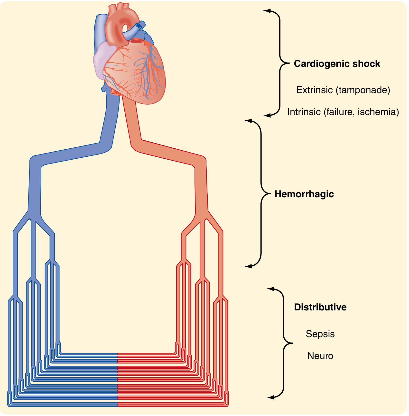
The 4-Type Schema (DCHO - most exam-relevant)
| Type | Mechanism | Examples | CO | SVR | CVP |
|---|
| Hypovolemic | ↓ blood/plasma volume | Hemorrhage, burns, vomiting, diarrhea, DKA | ↓ | ↑ | ↓ |
| Cardiogenic | Pump failure | MI, arrhythmia, tamponade, PE | ↓ | ↑ | ↑ |
| Distributive | Vasodilation/maldistribution | Sepsis, anaphylaxis, neurogenic | ↑ (initially) | ↓ | ↓ |
| Obstructive | Outflow obstruction | Tension pneumothorax, cardiac tamponade, massive PE | ↓ | ↑ | ↑ |
Harrison's 22e classifies distributive causes as: septic shock, pancreatitis, severe burns, anaphylactic shock, neurogenic shock, endocrine shock, adrenal crisis.
Distributive is UNIQUE - it is the only type where CO is compensatorily increased (because SVR is low). - Harrison's Principles of Internal Medicine 22e
3. PATHOPHYSIOLOGY
A. Cellular Level
(Bailey & Love's Surgery 28e)
- Reduced perfusion → cells deprived of O₂ → switch from aerobic → anaerobic metabolism
- Anaerobic product = lactic acid (not CO₂) → systemic metabolic acidosis
- As intracellular glucose is exhausted → failure of Na⁺/K⁺ ATPase pumps
- Intracellular lysosomes release autodigestive enzymes → cell lysis
- Intracellular K⁺ released → hyperkalaemia
B. Microvascular Level
- Hypoxia + acidosis → activate complement, prime leukocytes
- → oxygen free radicals + cytokine release
- → capillary endothelial injury → "leaky" capillaries → tissue oedema
- → exacerbates cellular hypoxia (vicious cycle)
C. Systemic Organ Responses
| System | Response |
|---|
| Cardiovascular | Baroreceptor → ↑ sympathetic activity → tachycardia + vasoconstriction (except in sepsis) |
| Respiratory | Metabolic acidosis + sympathetic → ↑ RR, ↑ minute ventilation → compensatory respiratory alkalosis |
| Renal | ↓ perfusion pressure → ↓ GFR → ↓ urine output; RAAS activated → more vasoconstriction + Na⁺/H₂O retention |
| GI/Hepatic | Gut ischaemia → bacterial translocation → perpetuates SIRS |
D. Irreversible ("Unresuscitatable") Shock
- Myocardial cell death from poor coronary perfusion
- Myocardial depression from severe acidaemia + hyperkalaemia
- Peripheral loss of vasomotor tone → no response to vasopressors
- Death is inevitable at this stage - Bailey & Love's Surgery 28e
4. STAGES OF SHOCK
| Stage | Features |
|---|
| Compensated (Early) | Tachycardia, ↑ RR, cool peripheries, normal BP, ↓ urine output |
| Decompensated (Progressive) | Hypotension, ↑ HR, oliguria, confusion |
| Irreversible (Terminal) | Profound hypotension, anuria, unconscious, MOF |
5. ATLS CLASSIFICATION OF HAEMORRHAGIC SHOCK (THE EXAM TABLE)
(Sabiston Textbook of Surgery - Advanced Trauma Life Support)
| Parameter | Class I | Class II | Class III | Class IV |
|---|
| Blood loss (%) | 0-15% | 15-30% | 30-40% | >40% |
| Blood loss (mL) | <750 | 750-1500 | 1500-2000 | >2000 |
| CNS | Slightly anxious | Mildly anxious | Anxious/confused | Confused/lethargic |
| Pulse (bpm) | <100 | >100 | >120 | >140 |
| Blood pressure | Normal | Normal | Decreased | Decreased |
| Pulse pressure | Normal | Decreased | Decreased | Decreased |
| RR | 14-20 | 20-30 | 30-40 | >35 |
| Urine output (mL/h) | >30 | 20-30 | 5-15 | Negligible |
| Fluid | Crystalloid | Crystalloid | Crystalloid + Blood | Crystalloid + Blood |
High-yield points: Hypotension appears only in Class III (not Class I or II). Pulse pressure narrows first (Class II) - a very early sign. Urine output is a reliable perfusion marker. Class IV = imminent death.
Important caveat: These classes are "problematic because they were not rigorously tested" (Sabiston). Children compensate longer and can lose large volumes before BP drops. Elderly patients may have a "normal for population" BP that is hypotensive for them. Patients on β-blockers cannot mount a tachycardia - the classic signs may be absent.
6. HAEMORRHAGE TYPES (Surgical Classification)
(Bailey & Love's Surgery 28e)
| Type | Timing | Cause |
|---|
| Primary | Immediately at time of injury | Direct vessel injury |
| Reactionary | 24-48 hours post-op | Blood pressure recovers, dislodges clot; ligature slippage |
| Secondary | 7-14 days post-op | Vessel wall erosion by infection, pressure necrosis, or malignancy |
Surgical vs Non-surgical haemorrhage:
- Surgical - discrete bleeding amenable to suture ligation or angioembolisation
- Non-surgical - diffuse bleeding from raw surfaces due to coagulopathy - cannot be stopped surgically (except packing); requires coagulation factor correction
7. FLUID RESPONSE CATEGORIES (Critical for Management)
| Response | Meaning | Action |
|---|
| Responder | Good + sustained BP improvement after fluid bolus | No active bleeding; continue resuscitation |
| Transient responder | BP improves but not sustained | Rate of haemorrhage < rate of fluid given; still bleeding - find and control source |
| Non-responder | No BP improvement to bolus | Rate of haemorrhage > rate of fluid; immediate surgical control |
8. SEPTIC SHOCK - DETAILED (Very High-Yield)
(Robbins & Cotran Pathologic Basis of Disease + Harrison's 22e)
Definition (Sepsis-3, 2016):
- Sepsis = Life-threatening organ dysfunction caused by a dysregulated host response to infection
- Septic shock = Subset of sepsis with profound circulatory, cellular, and metabolic abnormalities + greater risk of mortality
Commonest Triggers:
- Gram-positive bacteria (most common now)
- Gram-negative bacteria (classically endotoxin/LPS)
- Fungi
- Viral (e.g., SARS-CoV-2)
Pathogenesis (the molecular cascade):
- Microbial PAMPs (e.g., LPS, peptidoglycan) + host DAMPs → bind Toll-like receptors (TLRs)
- → Activate NF-κB → upregulate TNF-α, IL-1, IL-6, IL-12, IL-18, IFN-γ
- → Endothelial activation → ↑ adhesion molecules, cytokine/chemokine production
- Complement cascade activated → C3a, C5a (anaphylatoxins), C3b (opsonin)
- Coagulation activated → DIC (Disseminated Intravascular Coagulation)
- Counter-regulatory immunosuppression occurs simultaneously → oscillation between hyperinflammatory and immunosuppressed states
Haemodynamic Profile ("Warm Shock"):
- Early/Hyperdynamic: ↑ CO, ↓ SVR, warm peripheries, bounding pulse, ↓ BP
- Late/Hypodynamic: ↓ CO, MOF, cold and mottled skin
SIRS Criteria (context):
Systemic Inflammatory Response Syndrome (SIRS) - sepsis-like state triggered by non-microbial insults (burns, trauma, pancreatitis) - requires 2 of 4: fever/hypothermia, tachycardia, tachypnoea, WBC abnormality.
9. ANAPHYLACTIC SHOCK
- Type I IgE-mediated hypersensitivity
- Massive histamine release → profound vasodilation + ↑ capillary permeability
- Treatment: IM Adrenaline (Epinephrine) 0.5 mg (1:1000) immediately - the most important step
- Also: IV fluids, antihistamines, steroids, bronchodilators
10. NEUROGENIC SHOCK vs SPINAL SHOCK (Commonly Confused)
| Neurogenic Shock | Spinal Shock |
|---|
| Definition | Loss of sympathetic tone → vasodilation | Transient loss of all neurological function below injury |
| Mechanism | ↓ SVR, ↓ HR (paradoxical bradycardia with hypotension) | Flaccid paralysis, areflexia, urinary retention |
| Cause | Spinal cord injury at T6 or above | Any acute spinal cord injury |
| Haemodynamics | Hypotension + bradycardia | Not primarily haemodynamic |
| Peripheries | Warm (vasodilated) | - |
| Treatment | IV fluids + vasopressors (noradrenaline) | Corticosteroids controversial; supportive |
11. OBSTRUCTIVE SHOCK - THE SURGICAL EMERGENCIES
| Cause | Mechanism | Key Sign | Immediate Treatment |
|---|
| Tension pneumothorax | Air accumulates in pleural space → mediastinal shift → ↓ venous return | Tracheal deviation, absent breath sounds, JVP ↑ | Immediate needle decompression (2nd ICS, MCL) then chest drain |
| Cardiac tamponade | Blood in pericardium → cardiac compression → ↓ SV | Beck's triad: hypotension + muffled heart sounds + JVP ↑; pulsus paradoxus | Pericardiocentesis (emergency); surgical drainage |
| Massive PE | Obstruction of pulmonary outflow → acute RV failure | Hypoxia, hypotension, ↑ JVP, right heart strain on ECG | Thrombolysis / surgical embolectomy |
12. MONITORING IN SHOCK
| Parameter | Target | Significance |
|---|
| Urine output | >0.5 mL/kg/h (adults) / >1 mL/kg/h (children) | Best bedside perfusion marker |
| MAP | ≥65 mmHg | Minimum for organ perfusion |
| Lactate | <2 mmol/L | Marker of anaerobic metabolism; lactate clearance = resuscitation goal |
| CVP | 8-12 mmHg | Volume status guide (not perfect) |
| ScvO₂ | ≥70% | Central venous O₂ saturation - adequacy of delivery |
| Capillary refill time | <2 seconds | Peripheral perfusion (unreliable in isolation) |
Remember: Hypotension is a late sign of shock. By the time BP drops, significant compensatory mechanisms have already been exhausted.
13. PRINCIPLES OF MANAGEMENT
The ABC Framework + Simultaneous Resuscitation
Step 1 - Airway + Breathing
- Secure airway, high-flow O₂ (15 L/min via non-rebreather mask)
- Target SpO₂ >94%
Step 2 - Two large-bore IV cannulae (14-16G)
- Antecubital fossa preferred
- Send: FBC, U&E, LFTs, coagulation, group + crossmatch, lactate, ABG, blood cultures (before antibiotics in sepsis)
Step 3 - Fluid Resuscitation
| Shock Type | Initial Fluid | Volume | Notes |
|---|
| Haemorrhagic | Crystalloid (0.9% NaCl or Hartmann's) | 1-2 L bolus | Then blood - target 1:1:1 ratio (pRBCs: FFP: platelets) in massive haemorrhage |
| Septic | Balanced crystalloid (Hartmann's/Plasmalyte) | 30 mL/kg in first hour | Evidence supports balanced over 0.9% NaCl (avoids hyperchloraemic acidosis) |
| Cardiogenic | Cautious fluids | Small boluses | Avoid fluid overload; need inotropes |
| Anaphylactic | IV crystalloid | 1-2 L | After adrenaline |
| Neurogenic | IV crystalloid | As needed | Then vasopressors |
Damage Control Resuscitation (DCR): In massive haemorrhage, use permissive hypotension (target SBP 80-90 mmHg) until surgical haemostasis achieved, to avoid diluting clotting factors. Exception: head injury - maintain normal perfusion.
Step 4 - Vasopressors (when fluids insufficient)
| Agent | Receptor | Use |
|---|
| Noradrenaline (Norepinephrine) | α₁ > β₁ | First line in septic and neurogenic shock |
| Vasopressin | V1 | Second agent in septic shock; may be renoprotective |
| Epinephrine (Adrenaline) | α + β | Third line septic shock; first line anaphylaxis (IM) |
| Dobutamine | β₁ | Inotrope for cardiogenic shock / low LVEF in sepsis |
Step 5 - Source Control (Septic Shock)
- Antibiotics within 1 hour of recognition (broad spectrum, then de-escalate)
- Surgical drainage of abscess, removal of infected prosthesis, debridement of necrotising fasciitis
- Hydrocortisone 200 mg/day if vasopressors still required after adequate fluid + noradrenaline
14. SEPTIC SHOCK ORGAN DYSFUNCTION (Harrison's 22e Management Table)
| Organ | Incidence | Management |
|---|
| Neurologic (encephalopathy) | 54% | EEG, MRI; limit neurotoxic agents |
| Cardiovascular (shock) | 25% | 30 mL/kg crystalloid; noradrenaline → vasopressin → epinephrine |
| Respiratory (ARDS) | 7% | SpO₂ 90-96%; plateau pressure <30 cmH₂O; TV <8 mL/kg |
| Renal (AKI) | 67% | Avoid nephrotoxins; RRT if progressive AKI/hyperkalaemia |
15. COMPLICATIONS OF SHOCK
(Bailey & Love's Surgery 28e)
Ischaemia-Reperfusion Injury + SIRS
When circulation is restored after shock:
- Intracellular contents + K⁺/acid flood into systemic circulation
- Activate leukocytes, complement, neutrophils → remote organ injury
- Acute Lung Injury (ARDS)
- Acute Kidney Injury
- Cerebral oedema
- DIC
Multiple Organ Failure (MOF)
- Defined as ≥2 failed organ systems
- No specific treatment - only organ support (ventilation, cardiovascular support, haemofiltration/dialysis)
- Mortality: ~60%
- Prevention by early, aggressive reversal of shock is the only strategy
| Organ Failure | Manifestation |
|---|
| Heart | Cardiovascular failure |
| Lung | ARDS |
| Kidney | Acute renal failure |
| Liver | Liver failure + coagulopathy |
| Brain | Cerebral swelling, encephalopathy |
16. HIGH-YIELD EXAM PEARLS
| Fact | Detail |
|---|
| First sign of shock | Tachycardia + narrowed pulse pressure |
| Last sign of shock | Hypotension (late) |
| Best bedside monitor | Urine output (>0.5 mL/kg/h) |
| Shock with bradycardia | Neurogenic shock (T6 and above), also Bezold-Jarisch reflex in young trauma patients |
| Warm peripheries in shock | Septic (distributive) shock |
| Beck's Triad | Cardiac tamponade: hypotension + muffled heart sounds + ↑ JVP |
| "Obstructive" = surgical emergency | Tension pneumothorax, tamponade, massive PE |
| ATLS Class II key finding | BP still normal BUT pulse pressure narrowed |
| ATLS Class III | BP drops, crystalloid + blood needed |
| First-line vasopressor in septic shock | Noradrenaline |
| First-line treatment anaphylaxis | IM Adrenaline 0.5 mg (1:1000) |
| Target MAP in resuscitation | ≥65 mmHg (80-85 mmHg in neurogenic shock) |
| Lactate in shock | Marker of anaerobic metabolism; serial lactate = resuscitation guide |
| Haemoglobin in early haemorrhage | Unreliable - represents concentration, not absolute loss |
| Secondary haemorrhage timing | 7-14 days post injury |
| Reactionary haemorrhage timing | 24-48 hours |
| DIC in sepsis | Coagulation activated by endotoxin + thrombin via PAR receptors |
| Damage control resuscitation target SBP | 80-90 mmHg (permissive hypotension, NOT if head injury) |
| MOF definition | ≥2 organ systems failing; mortality ~60% |
17. QUICK DIFFERENTIATOR: HAEMODYNAMIC PROFILES
| Shock Type | HR | BP | CO | SVR | CVP/PCWP | Skin |
|---|
| Hypovolemic | ↑ | ↓ | ↓ | ↑ | ↓ | Cold, pale, clammy |
| Cardiogenic | ↑ | ↓ | ↓ | ↑ | ↑ | Cold, pale, clammy |
| Septic (early) | ↑ | ↓ | ↑ | ↓ | ↓ | Warm, flushed |
| Obstructive | ↑ | ↓ | ↓ | ↑ | ↑ | Cold, pale |
| Neurogenic | ↓ (bradycardia) | ↓ | ↓ | ↓ | ↓ | Warm, dry |
| Anaphylactic | ↑ | ↓ | ↑/↓ | ↓ | ↓ | Warm, urticaria |
Sources: Bailey and Love's Short Practice of Surgery 28e; Sabiston Textbook of Surgery (Biological Basis of Modern Surgical Practice); Robbins, Cotran & Kumar Pathologic Basis of Disease; Harrison's Principles of Internal Medicine 22e (2025); Robbins & Kumar Basic Pathology