
Synthetic mid urethral slings diagrams
Reading File
Synthetic mid-urethral slings (MUS) are minimally invasive procedures for stress urinary incontinence, with the two main types being retropubic (TVT) and transobturator (TOT/TVT-O) approaches.
Searching sources...
Finding Sources
Finding Sources
Reading File
Reading File
Reading File
I found references to key diagrams. Let me now fetch the images from the textbooks.
Loading Image
Reading File
Now let me get the retropubic and transobturator approach diagrams:
Reading File
Reading File
Loading Image
Reading File
Let me now find the single-incision sling diagram and the retropubic approach image:
Finding Sources
Reading File
The Fig. 125.11 image didn't render inline (it just shows "A" and "B" markers without a URL). Let me search for the retropubic approach image URL and the single incision diagram:
Searching Content
"cdn.orris.care" in /textbooks/9780323546423
Finding Sources
The images for Fig. 125.9 (retropubic) and Fig. 125.11 (single-incision) appear to be embedded in the text as anatomical labels rather than as URL-linked images in that block. The two main diagrams I've already retrieved (the trocars/prosthetics grid and the transobturator approach anatomy) are the key visual assets. Let me now compile everything into a comprehensive answer with the images and clinical detail.
Synthetic Mid-Urethral Slings (MUS) - Diagrams & Overview
Synthetic mid-urethral slings are the most commonly performed surgical procedure for stress urinary incontinence (SUI). Here is a structured overview with the key textbook diagrams.
Device Types and Trocars
Fig. 125.8 - MUS trocars and prosthetics (from Campbell-Walsh-Wein Urology):
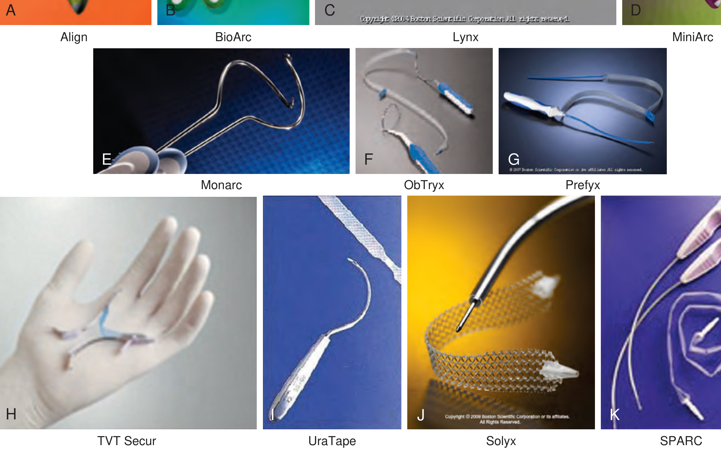
This figure shows the variety of sling systems: A. Align, B. BioArc, C. Lynx, D. MiniArc, E. Monarc (transobturator outside-in), F. ObTryx, G. Prefyx, H. TVT Secur (single-incision), I. UraTape, J. Solyx, K. SPARC. The standard sling consists of two curved 5-mm insertion trocars and a 40-cm long, 1.1-cm wide polypropylene mesh strip.
1. Retropubic Approach (TVT - Tension-Free Vaginal Tape)
Fig. 125.9 - Retropubic MUS placement:
The retropubic sling passes through the space of Retzius (retropubic space), exiting via two small suprapubic stab incisions ~2 cm lateral to the midline.
Key anatomical landmarks:
- The trocar passes from the vaginal incision (1.5 cm from the external meatus) upward through the endopelvic fascia
- Traverses the space of Retzius, stays in close contact with the inferior pubic bone surface
- Exits suprapubically just above the symphysis pubis
- Sling anchors to the endopelvic fascia
Technique steps:
- Two suprapubic stab incisions, 2 cm lateral to midline just above the symphysis
- Midline vaginal incision 1.5 cm, 1.5 cm proximal to the urethral meatus
- Dissection laterally to the pubocervical (endopelvic) fascia bilaterally (not perforated)
- Trocar passed bottom-up (vagina to abdomen) or top-down with a catheter guide deflecting the bladder
- Cystoscopy mandatory with 70-degree lens to exclude bladder/urethral perforation
- Sling placed loosely; tension adjusted with a clamp between sling and urethra
2. Transobturator Approach (TOT / TVT-O)
Fig. 125.10 - Transobturator MUS placement:
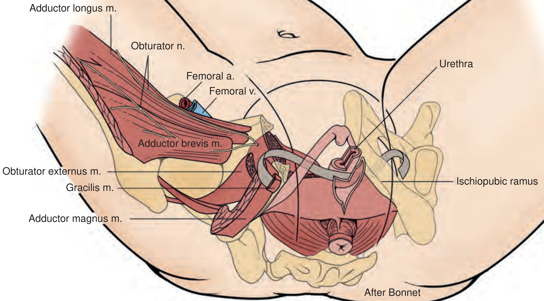
The sling passes through the obturator foramen, anchoring in the obturator internus and externus muscle/fascia, and exits via stab incisions in the groin crease at the level of the clitoris.
Key structures transited (visible in diagram):
- Adductor longus muscle (medial thigh)
- Obturator nerve (at risk)
- Obturator externus muscle
- Adductor brevis and gracilis muscles
- Ischiopubic ramus (bony landmark)
Two sub-techniques:
- Outside-in (TOT): Puncture incision in the groin crease at the inferior pubic ramus / adductor longus junction, trocar perforates obturator membrane and exits the vaginal incision - guided by the surgeon's index finger
- Inside-out (TVT-O): Vaginal incision first, introducer passed at 45 degrees through the obturator membrane, helical passer rotated to exit the interthigh stab incision
3. Single-Incision Sling (SIS / Mini-sling)
Fig. 125.11 - Single-incision MUS, two configurations:
- (A) Hammock position - anchors into the obturator internus muscle bilaterally
- (B) U-shaped position - anchors into connective tissue of the urogenital diaphragm (endopelvic fascia) behind the pubic bone
Short polypropylene mesh segment with pointed self-anchoring ends, placed through a single vaginal incision only - no abdominal or thigh exit incisions. Tension set when a hemostat tip passes easily between sling and urethra.
Material
All synthetic MUS are made of type 1 macroporous polypropylene mesh (pore size >75 microns), which allows tissue ingrowth, resists infection, and avoids the problems of microporous mesh.
Mechanism of Action
The MUS works by:
- Impeding posterior urethral wall movement during stress maneuvers (Valsalva), directing it anteriorly
- Compressing the urethral lumen by inward movement of the posterior wall under the sling
- Dynamic urethral kinking during stress events (Lo et al., 2001)
- The sling is anchored broadly - not just at the midline - which explains why continence is maintained even after sling incision for obstruction in many patients
Outcomes at a Glance
| Type | ~10-year objective cure | Key complication |
|---|---|---|
| Retropubic (TVT) | 84-91% | Bladder perforation (trocar passage), voiding dysfunction |
| Transobturator (TOT/TVT-O) | Similar efficacy | Groin/thigh pain, obturator nerve injury |
| Single-incision | Lower (variable) | Less data; early versions had higher failure rates |
Overall ~90% cure rate reported at 17-year follow-up (Bailey & Love's Surgery, p. 6105). Cystoscopy is mandatory after all approaches to exclude bladder and urethral injury.
- Campbell-Walsh-Wein Urology, 3-Volume Set, Chapter 125 (Mid-Urethral Slings)
- Bailey and Love's Short Practice of Surgery 28th Edition, Chapter 83
This is a shared conversation. Sign in to Orris to start your own chat.