
Cervical neck conditions
I'll start by reading the medical library skill, then search for cervical neck conditions comprehensively.
Quick Answer: Cervical neck conditions include a wide spectrum - from degenerative disc disease, cervical spondylosis, and herniated discs, to spinal stenosis, radiculopathy, myelopathy, whiplash injuries, and inflammatory conditions like rheumatoid arthritis affecting the cervical spine. Symptoms range from neck pain and stiffness to radiating arm pain, numbness, and in severe cases, spinal cord dysfunction.
Searching sources...
Reading File
Now I'll run parallel searches across the medical library and image database for comprehensive coverage.
Finding Sources
Finding Sources
Searching Images
cervical spine anatomy disc herniation radiculopathy
Good results. Now let me read from the most comprehensive sources - Miller's Orthopaedics and Campbell's Operative Orthopaedics for cervical spine conditions, and run more targeted FTS searches.
Reading File
Finding Sources
Finding Sources
Excellent data. Now let me read the detailed Miller's orthopaedics section and get clinical details on radiculopathy, whiplash, and management.
Reading File
Finding Sources
Finding Sources
Excellent. I have rich data. Let me now fetch the key cervical spine image and read the radiculopathy/myelopathy clinical details plus management.
Loading Image
Reading File
Reading File
Now let me get the myelopathy details and the MRI image, plus the nerve root table.
Reading File
Loading Image
I now have comprehensive, well-sourced data from multiple authoritative textbooks. Let me compile the full answer.
Cervical Neck Conditions
The cervical spine (C1-C7) is one of the most clinically active regions in the body, giving rise to a spectrum of conditions ranging from purely axial pain to devastating spinal cord injury. Here is a comprehensive breakdown organized by category.
Anatomy Relevant to Disease
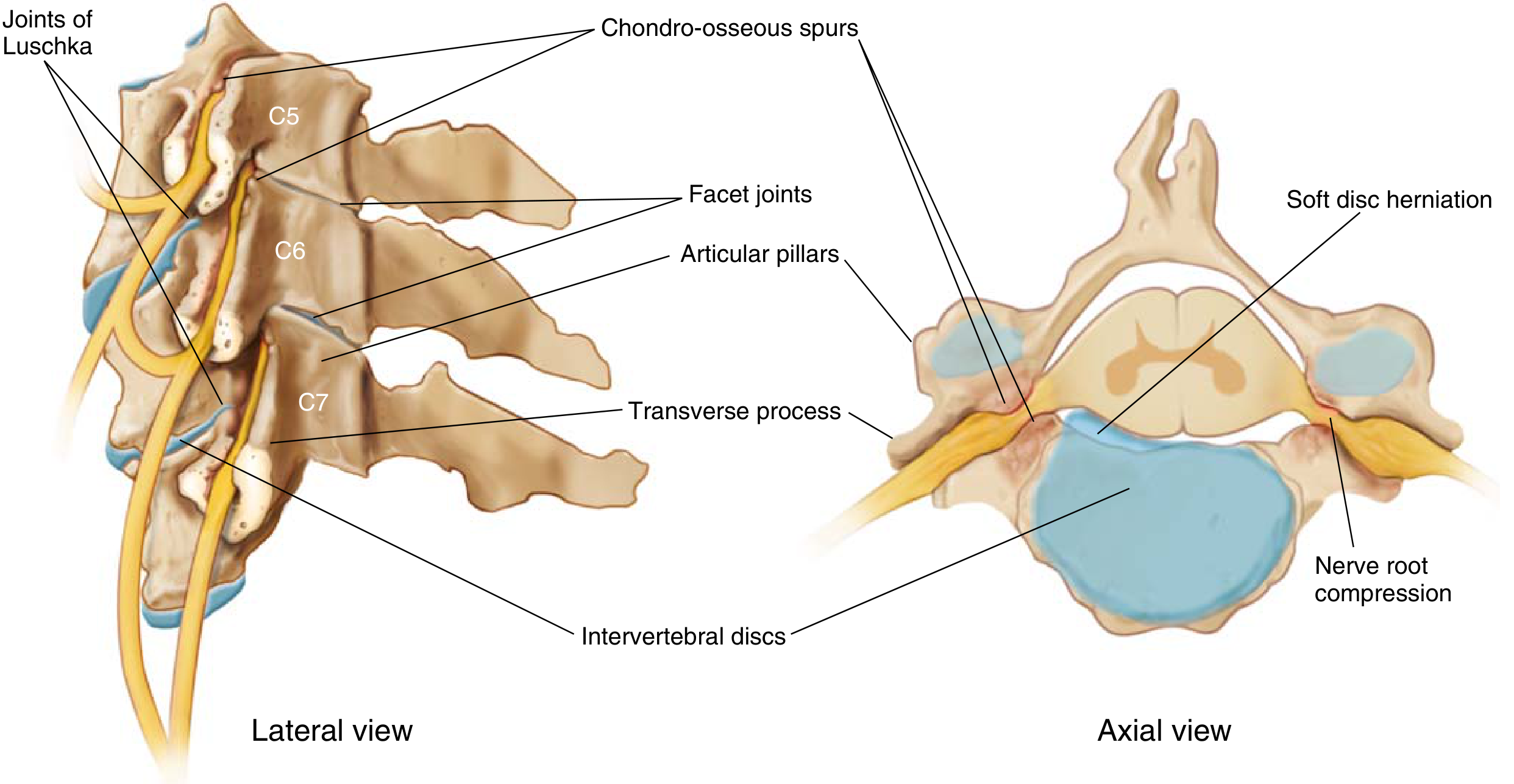
The cervical motion segment involves five key articulations per level: the intervertebral disc, two uncovertebral joints (joints of Luschka), and two facet joints. This interdependence means degeneration at one articulation stresses the others. Cervical nerve roots exit above their corresponding vertebrae - the C6 root exits at the C4-C5 foramen, so a C5-6 disc herniation compresses the C6 root.
1. Cervical Degenerative Disc Disease (Cervical Spondylosis)
The umbrella term for chronic disc degeneration with associated facet arthropathy. It produces four distinct clinical entities:
| Entity | Key Feature |
|---|---|
| Discogenic neck pain | Axial pain only, no neuro compromise |
| Radiculopathy | Nerve root compression |
| Myelopathy | Spinal cord compression |
| Myeloradiculopathy | Combined cord + root involvement |
Epidemiology
- Peaks between age 40-50 years
- Men > women
- C5-6 most commonly involved, followed by C6-7
- Risk factors: frequent lifting, cigarette smoking, excessive driving
Pathoanatomy (Degenerative Cascade)
- Disc dehydration and height loss
- Progressive collapse causes loss of normal lordosis and anterior cord compression via chondro-osseous/disc-osteophytic spurs
- Facet and uncovertebral joint loading causes foraminal narrowing
- Neck extension worsens cord compression (ligamentum flavum infolding posteriorly + spondylotic bar anteriorly); flexion slightly relieves it
Disc Types
-
"Soft" disc herniation - nucleus pulposus herniation without osteophytes; usually posterolateral; causes acute radiculopathy; rarely anterior (can cause dysphagia)
-
"Hard" disc herniation - herniation with associated disc-osteophytic spur; similar symptomatology; more chronic
-
Miller's Review of Orthopaedics 9th Edition, pp. 758-762
2. Discogenic Neck Pain (Axial Pain)
- Insidious onset of neck pain without neurologic signs
- Exacerbated by excessive vertebral motion
- No radicular or myelopathic component
- Treatment: NSAIDs, physiotherapy, cervical collar short-term, epidural steroid injections
3. Cervical Radiculopathy
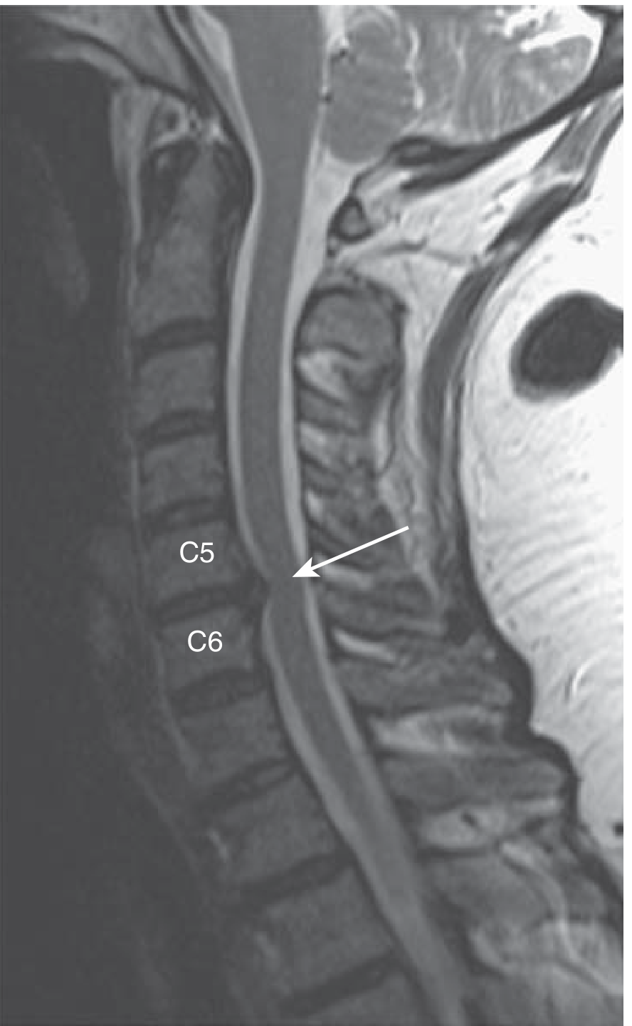
Nerve root compromise from either foraminal narrowing (spondylosis) or disc herniation.
Key clinical features:
- Neck pain that evolves into arm pain/paresthesias in a dermatomal distribution
- Motor: weakness in the corresponding myotome (less common)
- Sensory: numbness/dysesthesias in dermatomal territory (more common)
- Reflexes: typically hyporeflexic at the affected level
Dermatomal root map:
| Disc Level | Root Affected | Sensation | Reflex Lost | Motor Deficit |
|---|---|---|---|---|
| C4-5 | C5 | Lateral arm/shoulder | Biceps (partial) | Deltoid, supraspinatus |
| C5-6 | C6 | Lateral forearm, thumb/index | Biceps, brachioradialis | Biceps, wrist extensors |
| C6-7 | C7 | Middle finger, dorsal hand | Triceps | Triceps, wrist flexors |
| C7-T1 | C8 | Ring/little finger, medial forearm | None reliable | Intrinsics, finger flexors |
Special tests:
- Spurling's test - ipsilateral rotation + lateral bend + axial compression reproduces radicular symptoms; highly specific for cervical radiculopathy
- Shoulder abduction sign (Bakody sign) - relief of radicular pain when hand is placed on top of head; suggests cervical etiology
Treatment:
-
Conservative (first-line): NSAIDs, cervical epidural steroid injections, isometric exercises, traction, temporary collar immobilization
-
Surgical indications: progressive motor weakness, persistent disabling pain despite ≥6 weeks conservative treatment
-
Surgical options: ACDF (anterior cervical discectomy and fusion), cervical total disc replacement (motion-preserving), posterior foraminotomy
-
Miller's Review of Orthopaedics 9th Edition, pp. 760-763
4. Cervical Spondylotic Myelopathy (CSM)
The most serious consequence of cervical spondylosis - spinal cord compression causing upper motor neuron (UMN) signs. Harrison's 22E calls it a "comprehensive term for degenerative diseases causing symptomatic cervical spine narrowing."
Spinal canal measurements (lateral radiograph):
- Normal canal diameter: ≥14 mm
- Relative stenosis: 10-13 mm
- Absolute stenosis: <10 mm
- Pavlov (Torg) ratio (canal/vertebral body width): normal = 1.0; <0.8 = abnormal/risk factor
Clinical features:
- Gait disturbance (wide-based, spastic) - often the earliest sign
- Bilateral hand clumsiness, loss of fine motor control
- Hyperreflexia, clonus, positive Babinski sign (UMN)
- Hoffman's sign (finger flexion reflex) - flicking the middle fingernail causes reflex thumb flexion
- Lhermitte's sign - electric shock sensation down the spine on neck flexion
- Bladder dysfunction (frequency, urgency)
- Weakness in lower extremities
Myelopathy scoring: Modified Japanese Orthopaedic Association (mJOA) scale is used to grade severity (0-18; higher = better function).
Treatment:
-
Surgical decompression is the mainstay for moderate-severe CSM
-
Anterior options: ACDF, ACCF (anterior cervical corpectomy and fusion)
-
Posterior options: laminectomy + fusion, laminoplasty (contraindicated in fixed kyphosis)
-
Combined (circumferential) for complex multilevel disease
-
Most common postoperative complication: C5 nerve root palsy
-
Miller's Review of Orthopaedics 9th Edition, p. 761; Goldman-Cecil Medicine
5. Rheumatoid Cervical Spine Disease
Cervical involvement occurs in up to 90% of RA patients, more common with long-standing disease. The occipitoatlantoaxial complex (O-C2) is the primary site.
Three sequential stages:
- Atlantoaxial subluxation (AAS) - first and most common; C1 slides on C2 due to transverse ligament erosion
- Atlantoaxial invagination (AAI) - dens migrates upward into the foramen magnum (basilar invagination)
- Subaxial subluxation (SAS) - step-ladder deformity of lower cervical spine
Symptoms:
- Axial neck pain and stiffness
- Occipital headaches (C2 nerve compression - pain at base of skull relieved by traction)
- Progressive myelopathy/radiculopathy
Imaging: Flexion/extension X-rays mandatory before any elective surgery; MRI to assess cord compression. Anterior atlantodens interval (ADI) and posterior ADI (space available for cord) are key measurements.
Management: Disease-modifying antirheumatic drugs (DMARDs) have reduced the incidence; surgical stabilization (posterior C1-C2 fusion or occiput-to-cervical fusion) for progressive neurologic compromise.
- Miller's Review of Orthopaedics 9th Edition, pp. 762-764
6. Whiplash / Cervical Acceleration-Deceleration Injury
Definition: Indirect neck injury from acceleration-deceleration mechanism (typically motor vehicle accidents) causing flexion-extension injury of the cervical spine without direct trauma.
Pathology: Disruption of facet joints, anterior longitudinal ligament, intervertebral discs, and cervical muscles. Radiographs often show flattening of normal cervical lordosis.
Symptoms: Neck pain, stiffness, headache, occipital neuralgia, possible radicular pain. Without treatment, a large proportion improve within 6 months.
Treatment: Analgesia, early mobilization (collars now discouraged long-term), physiotherapy.
- Goldman-Cecil Medicine; Swanson's Family Medicine Review
7. Cervical Spinal Stenosis
- Narrowing of the cervical canal (congenital or acquired/degenerative)
- Congenital short pedicles predispose to early symptomatic stenosis with even minor disc bulging
- Presents as myelopathy (see above) or neurogenic claudication of the arms
8. Ossification of the Posterior Longitudinal Ligament (OPLL)
- Common in East Asian populations
- Calcified OPLL narrows the canal and causes myelopathy
- Harrison's 22E classifies it under "degenerative cervical myelopathy" alongside spondylosis
- Treatment: surgical decompression (often posterior laminoplasty preferred)
9. Cervical Fractures and Dislocations (Traumatic)
| Injury Pattern | Mechanism | Key Concern |
|---|---|---|
| Jefferson fracture (C1 burst) | Axial load | Transverse ligament rupture |
| Odontoid fractures (C2) | Flexion/extension | Atlantoaxial instability |
| Hangman's fracture (C2 pars) | Hyperextension | C2-3 instability |
| Bilateral facet dislocation | Flexion-distraction | Complete cord injury |
| Clay-shoveler's fracture | Sudden flexion | C7/T1 spinous avulsion |
Immobilization is essential to prevent further cord injury. Unstable fractures require surgical stabilization.
10. Other Notable Conditions
| Condition | Key Points |
|---|---|
| Thoracic outlet syndrome | Brachial plexus/vascular compression between scalene muscles and 1st rib; vague shoulder pain, ipsilateral 4th-5th digit numbness; cervical rib or scalene hypertrophy |
| Brachial neuritis (Parsonage-Turner) | Acute onset severe shoulder/arm pain followed by weakness; viral/postviral; EMG confirms; 80% recover in 2 years |
| Cervical vertigo | Spondylosis-related proprioceptive disruption or vertebrobasilar insufficiency |
| Cervical lymphadenopathy | Inflammatory, infectious (EBV, CMV, cat-scratch), or malignant (lymphoma, metastases) |
| Torticollis | Involuntary head tilt; congenital (SCM fibrosis), spasmodic, or dystonic |
| Ankylosing spondylitis | Can involve cervical spine causing rigid kyphosis and fracture risk |
Diagnostic Approach Summary
- Plain X-rays - Assess alignment, disc heights, osteophytes, canal diameter, ADI
- MRI - Investigation of choice for soft tissue, disc, cord signal change; essential for radiculopathy and myelopathy
- CT/CT myelography - Superior for bony detail, OPLL, foraminal osteophytes
- EMG/NCS - Differentiate radiculopathy from peripheral neuropathy (e.g., carpal tunnel coexistence)
- Flexion/extension films - Assess instability (RA, trauma)
Sources: Miller's Review of Orthopaedics 9th Edition | Firestein & Kelley's Textbook of Rheumatology | Harrison's Principles of Internal Medicine 22E | Goldman-Cecil Medicine | Bradley and Daroff's Neurology in Clinical Practice | Goldman-Cecil Medicine
This is a shared conversation. Sign in to Orris to start your own chat.