
Teach me enteric fever in microbiology second year everything about it from the standard textbooks sufficient for 10 marks
Salmonella typhi typhoid fever pathogenesis rose spots
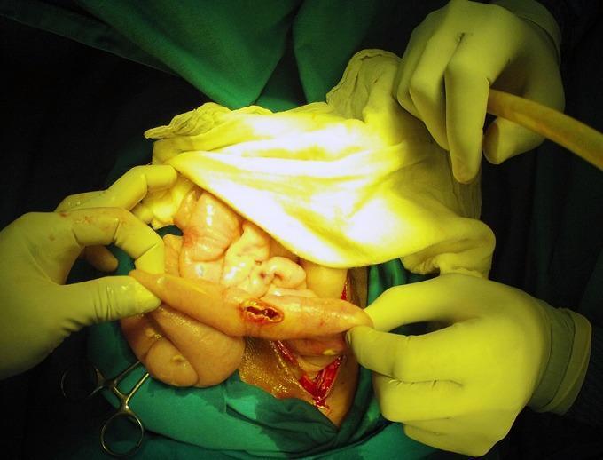
This clinical photograph shows an intraoperative view of an exploratory laparotomy, demonstrating a bowel perforation in the context of typhoid fever (Salmonella Typhi). The image focuses on a loop of small intestine held by a surgeon's gloved hands, revealing a discrete, longitudinal perforation with hyperemic and slightly everted edges. The surrounding serosa exhibits generalized inflammation and congestion. In the background, other segments of the intestine appear edematous and pale-pink, with visible white-yellowish peritoneal nodules and purulent exudate suggestive of concurrent miliary peritoneal tuberculosis and secondary peritonitis. Surgical instruments, suction tubing, and green sterile drapes define the surgical field. This image serves as an educational example of the severe gastrointestinal complications of typhoid fever, specifically intestinal perforation, and illustrates the challenging clinical scenario of co-infection with tuberculosis in an acute surgical setting.
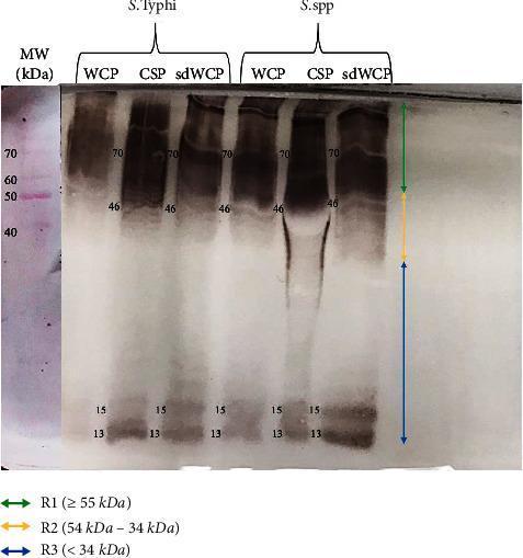
A diagnostic Western blot (WB) analysis image showing the immunoreactivity of antigens derived from Salmonella typhi and Salmonella spp. The image displays six experimental lanes representing three different protein extraction methods—Whole Cell Protein (WCP), Cell Surface Protein (CSP), and sodium deoxycholate-extracted WCP (sdWCP)—for each bacterial species. A molecular weight (MW) marker lane on the far left, stained with Ponceau S, shows reference bands from 40 kDa to 70 kDa. The WB profile is categorized into three vertical regions: R1 (≥ 55 kDa), R2 (54–34 kDa), and R3 (< 34 kDa). Prominent, high-intensity immunoreactive bands are observed in both species at approximately 70 kDa and 46 kDa, with a noticeable smear in the CSP lanes. Low molecular weight bands are consistently identified at 13 kDa and 15 kDa across all preparations. This visual provides comparative data on the antigenic profiles and antibody isotype reactivity against somatic and flagellar antigens, relevant for typhoid fever serodiagnosis research.
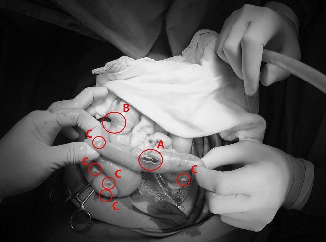
This monochrome clinical photograph captures an intraoperative view of the abdominal cavity during a surgical procedure for bowel perforation. The image illustrates two distinct infectious pathologies concurrently affecting the intestines and peritoneum. Label 'A' identifies a typhoid intestinal perforation, appearing as an irregular, longitudinal breach in the seromuscular layer of the small bowel, typical of advanced Salmonella typhi infection. Label 'B' shows a non-perforated typhoid lesion, characterized by localized inflammation and thickening without a full-thickness defect. Surrounding these intestinal segments, multiple small, discrete, whitish nodules are indicated by label 'C', representing miliary peritoneal tuberculosis. These nodules are scattered across the serosal surfaces and mesentery, demonstrating a grain-like distribution characteristic of disseminated mycobacterial infection. The image serves as a high-level educational example of acute surgical abdomen complications resulting from co-infection of typhoid fever and miliary tuberculosis, highlighting diagnostic landmarks such as transmural perforation and widespread peritoneal seeding.
Enteric Fever (Typhoid Fever) - Complete Microbiology Notes
1. DEFINITION
2. THE ORGANISM - Salmonella Typhi
Morphology
- Gram-negative, non-spore-forming, facultatively anaerobic bacilli measuring 2-3 µm x 0.4-0.6 µm
- Motile with peritrichous flagella (except S. Gallinarum-Pullorum)
- Belongs to family Enterobacteriaceae
Cultural Characteristics
- Grows readily on simple media
- Does NOT ferment lactose or sucrose (non-lactose fermenter - pale/colourless colonies on MacConkey agar)
- Ferments glucose with acid but no gas (unlike most other Salmonella serovars which produce gas)
- Produces H₂S (black precipitate on TSI agar / HE agar)
- Oxidase negative, urease negative
- On TSI agar: Alkaline/Acid (K/A), H₂S+, gas - (for Typhi)
- On HE (Hektoen Enteric) agar: Black colonies due to H₂S production
- Resistant to brilliant green, sodium tetrathionate, sodium deoxycholate (used as selective agents in isolation media)
Antigenic Structure (Kauffmann-White Scheme)
| Antigen | Type | Nature | Significance |
|---|---|---|---|
| O antigen (somatic) | 9, 12 | Lipopolysaccharide (cell wall) | Basis of serogroup D classification |
| H antigen (flagellar) | d | Protein (flagellin) | Phase 1 only (monophasic) |
| Vi antigen | Surface/capsular | Polysaccharide | Virulence antigen; restricted to S. Typhi and S. Paratyphi C |
- Antigenic formula of S. Typhi: 9, 12 (Vi) : d : -
- The Vi antigen is used in phage typing (>80 phage types), useful for epidemiological tracing
- High Vi antibody titers suggest chronic carrier state
3. EPIDEMIOLOGY
Global Burden
- ~27 million cases and >200,000 deaths annually worldwide (Medical Microbiology 9e)
- Endemic in South Asia, Africa, Latin America; declining in developed nations due to sanitation
Reservoir and Source
- Man is the ONLY reservoir - cases and carriers
- Primary sources: faeces and urine of cases or carriers
- Secondary sources: contaminated water, food, fingers, flies ("5 Fs" - food, fluid, fingers, flies, fomites)
- No animal reservoir - unlike non-typhoidal Salmonella
Carriers
- Convalescent carrier: excretes bacilli for 6-8 weeks post-illness
- Chronic carrier: excretion >1 year; develops in 2-5% of cases; organism persists in gallbladder (especially with gallstones) and biliary tract
- "Typhoid Mary" (Mary Mallon) - classic chronic carrier who caused >1300 cases over her lifetime
- Faecal carriers are more common than urinary carriers
Transmission
- Faecal-oral route (also urine-oral)
- Via contaminated water, food (milk multiplies the organism without altering taste), flies
- Infective dose: 10⁵-10⁶ bacteria (intermediate; lower than most Salmonella, higher than Shigella)
- Vi antigen may reduce the infective dose further
Incubation Period
- Usually 10-14 days (range: 3 days to 3 weeks), depending on inoculum size
Peak incidence: July-September (rainy season, increased fly population)
4. PATHOGENESIS
Step-by-step mechanism:
- Inhibiting the oxidative metabolic burst of macrophages
- The Vi (virulence) polysaccharide blocks neutrophil phagocytosis by interfering with complement deposition, favouring uptake by macrophages
Key: Fever = LPS-driven; diarrhoea is late and due to bowel reinfection, not primary enterocyte destruction.
5. CLINICAL FEATURES
Classic Course (untreated):
- Insidious onset; gradually rising step-ladder fever (39-40°C), headache, malaise, myalgia, anorexia
- Initial constipation (common), dry cough
- Relative bradycardia (Faget's sign) - pulse rate slower than expected for the fever level
- High sustained fever, "typhoidal" toxic state
- Rose spots - 1-4 mm blanching pink maculopapular rash, 2-4 crops, typically on trunk/chest; seen in ~30% of patients; can culture S. Typhi from biopsy
- Hepatosplenomegaly (3-6%)
- "Pea-soup" diarrhoea or continued constipation
- Abdominal distension
- Leukopenia (WBC normal or LOW - unlike other bacterial infections)
- Defervescence begins OR complications occur
- Intestinal haemorrhage (6%) and perforation (1%) - most dangerous
- Neuropsychiatric: "muttering delirium," "coma vigil" (picking at bedclothes)
Paratyphoid Fever
- Similar but milder than typhoid
- Caused by S. Paratyphi A (most common), B, C
- Shorter incubation, less severe
6. COMPLICATIONS
| System | Complication |
|---|---|
| GI (most common) | Intestinal perforation (terminal ileum), haemorrhage (weeks 3-4) |
| Neurological | Meningitis, Guillain-Barré syndrome, neuritis, typhoid encephalopathy |
| Cardiovascular | Endocarditis, myocarditis, pericarditis |
| Haematological | DIC, haemolytic-uraemic syndrome |
| Others | Hepatitis, cholecystitis, osteomyelitis, orchitis, pneumonia |
7. LABORATORY DIAGNOSIS
A. Culture (Gold Standard)
| Specimen | Week | Yield | Notes |
|---|---|---|---|
| Blood | Week 1-2 | 80-90% | Best in 1st week; 10-15 mL blood in 100 mL broth (1:10 ratio) |
| Bone marrow | Any week | 90% | Remains positive even after antibiotics - most sensitive |
| Stool | Week 2-3 | Positive from 2nd week | Also positive in carriers |
| Urine | Week 3 | Variable | Less reliable |
| Rose spot biopsy | Week 1-2 | Good | Direct culture positive |
- Selective/differential: MacConkey agar, Hektoen Enteric (HE) agar, SS (Salmonella-Shigella) agar
- Enrichment: Selenite F broth, Tetrathionate broth
- Non-lactose fermenting colonies + H₂S production = presumptive positive
B. Serological - Widal Test (Felix-Widal Test)
- O antibodies: appear on day 6-8 (early, short-lived, indicate current/recent infection)
- H antibodies: appear on day 10-12 (persist longer, may indicate past infection or vaccination)
- O antigen titre ≥ 1:160 (or 1:320) - considered positive
- H antigen titre ≥ 1:640 - considered positive
- Rising titre (4-fold) in paired sera (7-10 days apart) = stronger evidence
- Can be negative in up to 30% of culture-proven cases (especially with prior antibiotics)
- False positives: cross-reaction with other Salmonella serotypes, Enterobacteriaceae; also in malaria, typhus, bacteraemia, cirrhosis, vaccination
- Not reliable as sole diagnostic test
C. Newer/Rapid Diagnostic Tests
- Typhidot®: detects IgM and IgG against a 50 kDa antigen of S. Typhi; takes 3 hours
- Typhidot-M®: detects IgM only (more specific for acute infection)
- IDL Tubex® test: detects IgM O9 antibodies; results in minutes
- Dipstick test: detects S. Typhi-specific IgM against LPS antigen
- NAATs/PCR: increasingly used in reference/public health labs
- Blood cultures remain gold standard wherever available
D. Haematology
- Leucopenia with relative lymphocytosis (characteristic - distinguishes from other bacteraemias)
- Anaemia, raised ESR, mildly elevated LFTs
8. TREATMENT
Uncomplicated Enteric Fever:
- Azithromycin 1 g once, then 500 mg daily × 7 days (oral; now preferred for uncomplicated)
- Fluoroquinolones (ciprofloxacin): first-line for susceptible strains; avoid if fluoroquinolone-resistant (especially South Asian strains)
- Third-generation cephalosporins (ceftriaxone, cefixime): mainstay for MDR strains
Complicated/Severe Disease:
- Parenteral ceftriaxone or fluoroquinolone for ≥10 days; admit to hospital
Antibiotic Resistance:
- Chloramphenicol (historical first-line) → replaced due to resistance
- Multi-drug resistant (MDR) S. Typhi: resistant to chloramphenicol, ampicillin, TMP-SMX
- XDR (extensively drug resistant) S. Typhi: also resistant to fluoroquinolones and 3rd-gen cephalosporins (emerging from Pakistan)
- Treatment with antibiotics reduces mortality from >10% to <1%; fever resolves in 3-5 days
Chronic Carrier Treatment:
- High-dose fluoroquinolone for 4-6 weeks
- Often requires cholecystectomy if gallstones present (organisms form biofilm on stones)
9. PREVENTION AND CONTROL
Three Lines of Defence (Park's):
- Early diagnosis, isolation, treatment of cases
- Identification, surveillance, and treatment of carriers
- Carriers should not handle food
- Safe water supply (chlorination, boiling)
- Proper sewage disposal; fly control
- Pasteurisation of milk and dairy products
- Safe food handling - cooking, refrigeration
| Vaccine | Type | Route | Protection | Duration |
|---|---|---|---|---|
| Ty21a (Vivotif) | Live attenuated oral | Oral (4 capsules over 7 days) | ~70% | 5-7 years |
| Vi capsular polysaccharide (Typhim Vi) | Purified Vi antigen | Single IM injection | ~70-75% | 2-3 years (booster q2y) |
| Typhoid conjugate vaccine (TCV) | Vi conjugated to carrier protein | IM | Better immune memory | Preferred by WHO; single dose from 6 months |
- Killed whole-cell vaccine (old) is no longer recommended due to adverse reactions
- Vaccines are only moderately effective (~70%); sanitation remains key
- TCV is now preferred by WHO for all ages as it generates immunological memory
10. IMMUNITY
- Natural infection confers immunity; reinfection is rare unless treatment was early
- Both Th1 and Th2 cell-mediated immunity are stimulated
- Circulating antibodies to O and Vi antigens are related to resistance
- Secretory IgA prevents attachment to intestinal epithelium
- Relapses possible in 2-3 weeks despite antibodies (in ~10% of untreated cases)
- Patients with sickle cell disease are particularly susceptible to severe Salmonella infections (osteomyelitis)
QUICK REVISION SUMMARY (Memory Aids)
| Feature | Key Point |
|---|---|
| Organism | S. Typhi - Gram -ve, non-lactose fermenter, H₂S+, peritrichous flagella |
| Antigenic formula | 9, 12 (Vi) : d : - |
| Only reservoir | Man (cases + carriers) |
| Infective dose | 10⁵-10⁶ organisms |
| Incubation | 10-14 days |
| Pathognomonic sign | Rose spots (30%), relative bradycardia, step-ladder fever |
| Lab gold standard | Blood culture (week 1), Bone marrow (any time) |
| WBC finding | Leucopenia (not leucocytosis) |
| Widal O titre | ≥1:160-1:320 |
| Widal H titre | ≥1:640 |
| Vi antibody | High titre = chronic carrier |
| Drug of choice | Azithromycin (uncomplicated); Ceftriaxone/FQ (complicated) |
| Danger complication | Intestinal perforation (week 3-4) |
| Chronic carrier | Organisms in gallbladder; treat with cholecystectomy + antibiotics |