
Hypokalemia approach
Reading File
Now let me search the medical library for hypokalemia content in parallel:
Finding Sources
Finding Sources
Excellent. Now let me read the most comprehensive sources in parallel - Brenner & Rector (nephrology gold standard), Goldman-Cecil, and Comprehensive Clinical Nephrology:
Reading File
Reading File
Reading File
Good content. Now let me read the Brenner & Rector stepwise diagnostic approach block and the Comprehensive Nephrology hypokalemia section for the algorithmic approach:
Reading File
Reading File
Finding Sources
There's a diagnostic diagram in Frameworks for Internal Medicine. Let me read that section and also the Comprehensive Nephrology diagnostic approach, and fetch the key image:
Reading File
Reading File
Loading Image
Now let me get the diagnostic step-by-step approach from Brenner & Rector block 41 (which has the best stepwise clinical approach) and the Frameworks for Internal Medicine excess loss section:
Reading File
Reading File
Finding Sources
Let me fetch the diagnostic flowchart and read a couple more key sections:
Loading Image
Finding Sources
Reading File
Now let me get the key diagnostic workup section from Brenner & Rector block 14 (stepwise approach by acid-base status) and also the ECG/treatment details from Tietz:
Finding Sources
Reading File
Excellent - there's a key diagnostic algorithm image. Let me fetch that and also get more of the Tietz content on ECG changes and treatment:
Loading Image
Reading File
Searching PubMed
hypokalemia diagnosis treatment
I now have comprehensive content from multiple authoritative textbooks. Let me compile the full answer.
Hypokalemia: A Complete Clinical Approach
Definition: Serum K+ < 3.5 mEq/L. Severe hypokalemia is generally defined as < 2.5 mEq/L.
Pathophysiology: Three Core Mechanisms

Hypokalemia arises from three mechanisms (often overlapping):
1. Low Intake
- Poor oral intake (alcoholism, starvation, postoperative K+-poor IV fluids)
- When dietary K+ is zero, obligatory renal losses still occur (~5-10 mEq/day minimum)
- Average daily K+ intake: ~75 mEq/day (men), ~55 mEq/day (women); ~85-90% is absorbed in the GI tract
2. Transcellular Shift (Redistribution)
K+ moves from ECF into ICF - total body K+ is normal, serum K+ falls:
| Cause | Mechanism |
|---|---|
| Insulin (exogenous or endogenous) | Activates Na+/K+-ATPase |
| Metabolic alkalosis | K+ enters cells as H+ exits via Na+-H+ antiporter |
| Beta-2 adrenergic excess (albuterol, stress, pheochromocytoma) | Stimulates Na+/K+-ATPase |
| Hypokalemic periodic paralysis | Episodic intracellular shift |
| Hypothermia | Redistribution |
| Pseudohypokalemia | Very high WBC (>100,000/µL) - cells in tube take up K+ at room temperature |
Redistributive hypokalemia is transient and reverses when the underlying condition corrects. Watch for rebound hyperkalemia if over-treated.
3. Excess Loss
Subdivided into renal vs extrarenal loss - the key distinction in workup.
Causes by Category
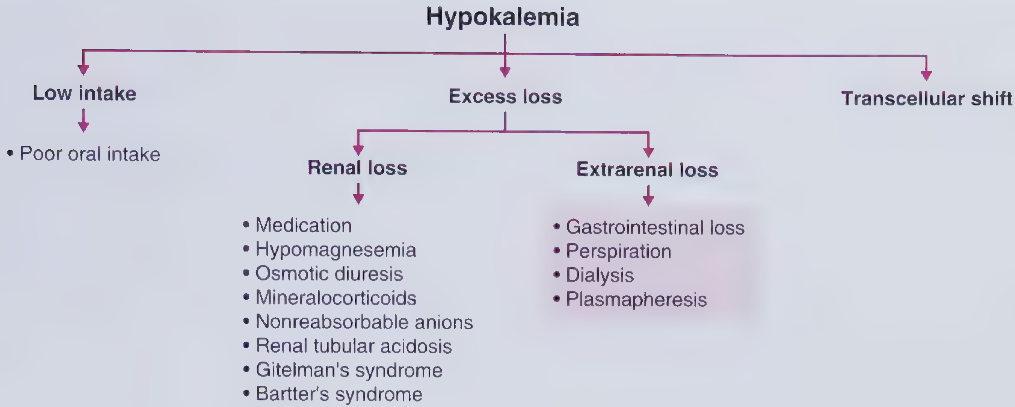
Renal losses (urine K+ >25-30 mmol/day):
- Diuretics (loop and thiazides - most common cause; affects up to 50% of patients)
- Mineralocorticoid excess: primary hyperaldosteronism, secondary hyperaldosteronism, Cushing's, ectopic ACTH, apparent mineralocorticoid excess, Liddle syndrome
- Renal tubular acidosis (type 1 and type 2)
- Bartter syndrome, Gitelman syndrome (hereditary tubular disorders)
- Hypomagnesemia (reduces inhibitory effect on luminal K+ channels)
- Osmotic diuresis (DKA, uncontrolled DM)
- Nonreabsorbable anions in tubule (bicarbonate in vomiting, beta-hydroxybutyrate in DKA, penicillin antibiotics)
- Nephrotoxins: amphotericin B, aminoglycosides, cisplatin, ifosfamide
Extrarenal losses (urine K+ <25 mmol/day):
- GI losses: diarrhea (K+-rich stool), vomiting/NG suction (volume depletion → secondary hyperaldosteronism)
- Excessive perspiration
- Dialysis, plasmapheresis
Diagnostic Algorithm
The approach follows a stepwise process:
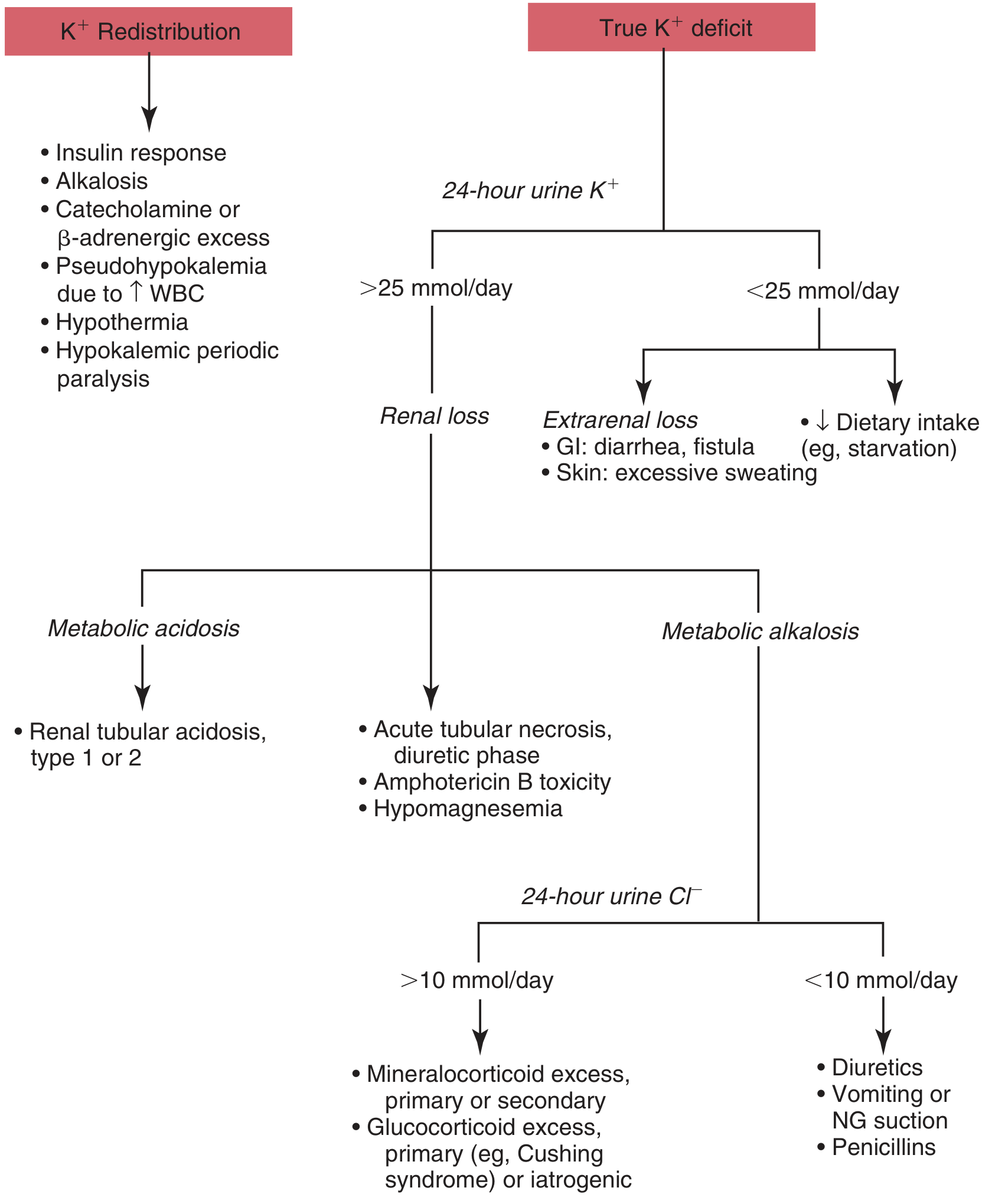
Step 1: Rule out pseudohypokalemia
- Check if WBC is markedly elevated; if so, process sample on ice immediately
Step 2: Is there transcellular redistribution?
- History: insulin given recently, beta-agonist use, periodic paralysis, alkalosis
- If yes, treat the underlying cause; do not aggressively supplement (risk of rebound hyperkalemia)
Step 3: True K+ deficit - check 24-hour urine K+ (or spot urine K+/Cr ratio)
| Urine K+ | Interpretation |
|---|---|
| < 25-30 mmol/day (or spot K+/Cr <1.5 mEq/mmol) | Renal conservation is intact → extrarenal loss or low intake |
| > 25-30 mmol/day | Inappropriate renal wasting → renal cause |
Step 4: If renal loss - check acid-base status
Renal loss + Metabolic ACIDOSIS:
- Renal tubular acidosis type 1 (distal) or type 2 (proximal)
- DKA (osmotic diuresis + beta-hydroxybutyrate as non-reabsorbable anion)
Renal loss + Metabolic ALKALOSIS - check urine Cl-:
| Urine Cl- | Interpretation |
|---|---|
| < 10 mmol/day ("Cl--responsive") | Diuretics (past use), vomiting/NG suction, penicillins |
| > 10 mmol/day ("Cl--resistant") | Mineralocorticoid or glucocorticoid excess |
Step 5: If mineralocorticoid excess - check blood pressure
| BP | Think |
|---|---|
| Hypertension + hypokalemia | Primary hyperaldosteronism (check plasma aldosterone:renin ratio), Cushing's, ectopic ACTH, Liddle syndrome, apparent mineralocorticoid excess, renovascular hypertension |
| Normal BP + hypokalemia + metabolic alkalosis | Bartter syndrome (presents like loop diuretic use) or Gitelman syndrome (presents like thiazide use - hypomagnesemia, hypocalciuria) |
Clinical Manifestations
Severity correlates roughly with degree of hypokalemia, though acute drops are more dangerous than chronic equivalent levels.
Neuromuscular:
- Skeletal muscle weakness (proximal > distal), myalgia
- Flaccid paralysis (respiratory failure if severe)
- Rhabdomyolysis (reduced skeletal muscle blood flow from impaired nitric oxide release)
- Paralytic ileus
Cardiac:
- ECG changes: flattening/inversion of T waves, prominent U waves, ST depression, widened QRS (severe)
- Ventricular tachycardia, ventricular fibrillation, sudden cardiac death
- Particularly dangerous in patients on digoxin (digoxin toxicity potentiated by hypokalemia) and those with coronary artery disease
Metabolic/Endocrine:
- Impaired insulin secretion + insulin resistance → worsened glycemic control
- Metabolic alkalosis (K+ exits cells in exchange for H+)
Renal:
- Nephrogenic diabetes insipidus (polyuria, polydipsia)
- Hypokalemic nephropathy/tubulointerstitial fibrosis (outer medulla most affected)
- Increased blood pressure (K+ depletion promotes Na+ retention via NCC and ENaC upregulation)
Treatment
General Principles
- Oral route is preferred whenever possible
- IV is reserved for: unable to take oral, symptomatic hypokalemia (arrhythmia, paralysis), K+ < 2.5-3.0 mEq/L
- Always check and replete magnesium first/simultaneously - hypomagnesemia causes ongoing urinary K+ wasting and will prevent K+ correction
- Avoid glucose-containing IV fluids (insulin spike will further lower K+)
Oral Replacement
- KCl is the preferred salt in most situations - also corrects coexisting metabolic alkalosis
- Potassium citrate - preferred in metabolic acidosis (e.g., RTA)
- Potassium phosphate - when concurrent phosphate deficiency (refeeding, DKA)
- Avoid potassium gluconate - a non-reabsorbable anion that may be ineffective
- Dose: 40-100 mmol/day depending on deficit
IV Replacement
- Usual concentration: 20-40 mmol K+ in 1 L NS
- Max peripheral rate: 10-20 mmol/hour
- If K+ < 3.0 mmol/L or rate > 10 mmol/hour: continuous cardiac monitoring required
- Central line needed for higher concentrations (extravasation of KCl causes tissue necrosis)
- Do NOT use dextrose-containing solutions for K+ replacement
Ongoing/Maintenance
- Treat the underlying cause (stop offending drugs if possible)
- For diuretic-induced hypokalemia requiring continued diuretics: add potassium-sparing diuretics (spironolactone, eplerenone, amiloride, triamterene)
- For primary hyperaldosteronism: mineralocorticoid receptor antagonist or surgical resection
- Target K+ ≥ 4.0 mEq/L especially in high-risk patients (cardiac disease, digoxin use)
Special Situations
- Prerenal azotemia + hyperglycemia or metabolic alkalosis: give K+ prophylactically with saline resuscitation - volume expansion alone can cause life-threatening kaliuresis
- DKA: K+ often appears normal or high initially but is severely depleted; begin K+ replacement when serum K+ < 5.0 mEq/L (and hold insulin until K+ ≥ 3.5 mEq/L)
- Refeeding syndrome/alcoholism: often have concurrent hypophosphatemia, hypomagnesemia - all three must be repleted
- Cancer patients: hypokalemia common from multiple overlapping causes - check for cisplatin nephrotoxicity, ectopic ACTH, lysozymuria (AML subtypes M4/M5)
Key Pearls
- Always check Mg2+ - refractory hypokalemia is usually due to uncorrected hypomagnesemia
- Vomiting causes hypokalemia via secondary hyperaldosteronism (volume depletion), not primarily from gastric K+ loss (gastric K+ content is low ~10 mEq/L); the urine Cl- will be low (<10 mmol/day)
- Diarrhea causes hypokalemia via direct K+ loss in stool (stool K+ content is high); urine K+ will be appropriately low
- Bartter vs Gitelman: Bartter (furosemide-like: hypercalciuria, normal Mg) vs Gitelman (thiazide-like: hypocalciuria, hypomagnesemia)
- RTA type 4 (hypoaldosteronism) causes hyperkalemia, not hypokalemia - unlike types 1 and 2
- For every 0.3 mEq/L fall in serum K+ below 3.5, approximately 100 mEq of total body K+ deficit exists (rough guide, varies with acid-base status)
- Avoid administering K+ too fast - potassium traverses the extracellular space before entering intracellular stores, making rapid IV infusion dangerous
Sources: Comprehensive Clinical Nephrology 7e, p.162-165 | Brenner and Rector's The Kidney | Tietz Textbook of Laboratory Medicine 7e, p.1942-1943 | Goldman-Cecil Medicine, p.549-557 | Frameworks for Internal Medicine, pp.497-500 | Miller's Anesthesia 10e
This is a shared conversation. Sign in to Orris to start your own chat.