
Layers of knee
layers of the knee anatomy cross section
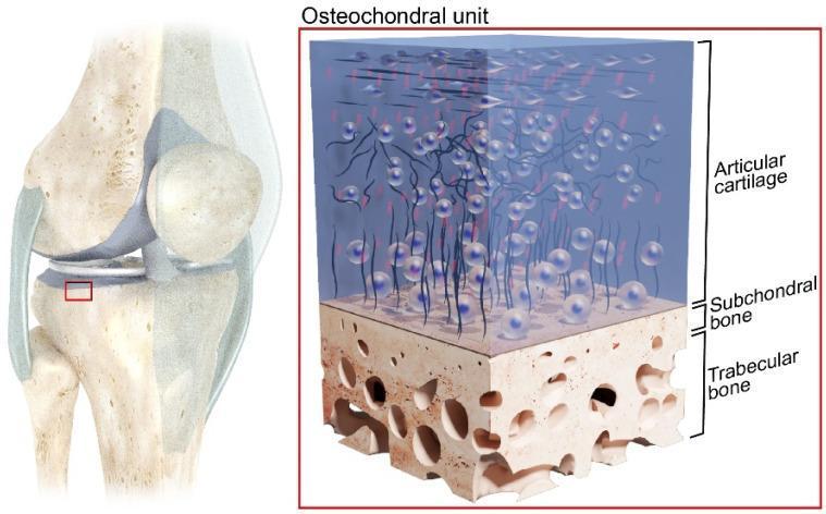
This medical illustration depicts the anatomical structure of the human osteochondral unit, with a contextual focus on the knee joint. The image consists of two parts: a macro-level schematic of the knee joint and a micro-level cross-sectional diagram of the osteochondral tissues. The knee schematic highlights the proximal tibia as the source of the tissue sample. The detailed cross-section identifies three primary layers: the Articular Cartilage (AC), the Subchondral Bone (SB), and the Trabecular Bone (TB). The Articular Cartilage is shown as a thick, blue-tinted translucent layer containing chondrocytes organized within an extracellular matrix, transitioning from horizontally oriented cells at the surface to vertically aligned cells in the deeper zones. Below this lies the Subchondral Bone, a thin, dense, beige layer that serves as the interface between cartilage and bone. The base layer is the Trabecular Bone (cancellous bone), characterized by its highly porous, honeycomb-like structure with irregular voids. This diagram illustrates the integrated mechanical system responsible for load distribution and low-friction motion within musculoskeletal joints.
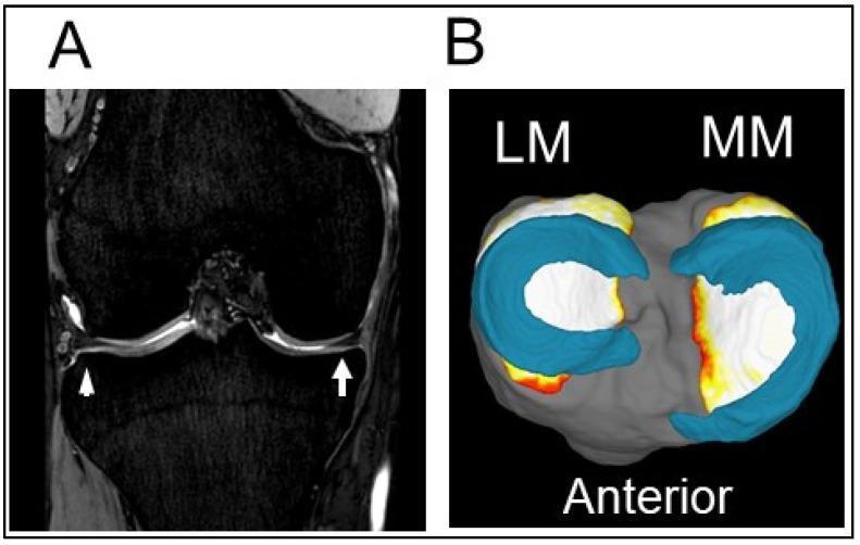
This composite image illustrates the normal anatomy of the knee menisci using two different Magnetic Resonance Imaging (MRI) perspectives. Figure A presents a coronal MRI cross-section of a right knee, where the medial meniscus (MM) and lateral meniscus (LM) appear as low-signal (dark), wedge-shaped fibrocartilaginous structures situated within the joint space between the femoral condyles and the tibial plateau. The medial meniscus is indicated by a white arrow, and the lateral meniscus is marked with a white arrowhead. Figure B displays a 3D MRI reconstruction of the same structures viewed from a superior perspective. In this 3D model, the menisci are color-coded in teal blue, highlighting their characteristic C-shaped (medial) and more circular (lateral) morphology sitting atop the gray-shaded tibial plateau. The 'Anterior' label provides orientation. These images serve as an educational baseline for identifying healthy meniscal tissue and its positioning, which is critical for evaluating pathologies such as meniscal tears or extrusions that contribute to the development of osteoarthritis.
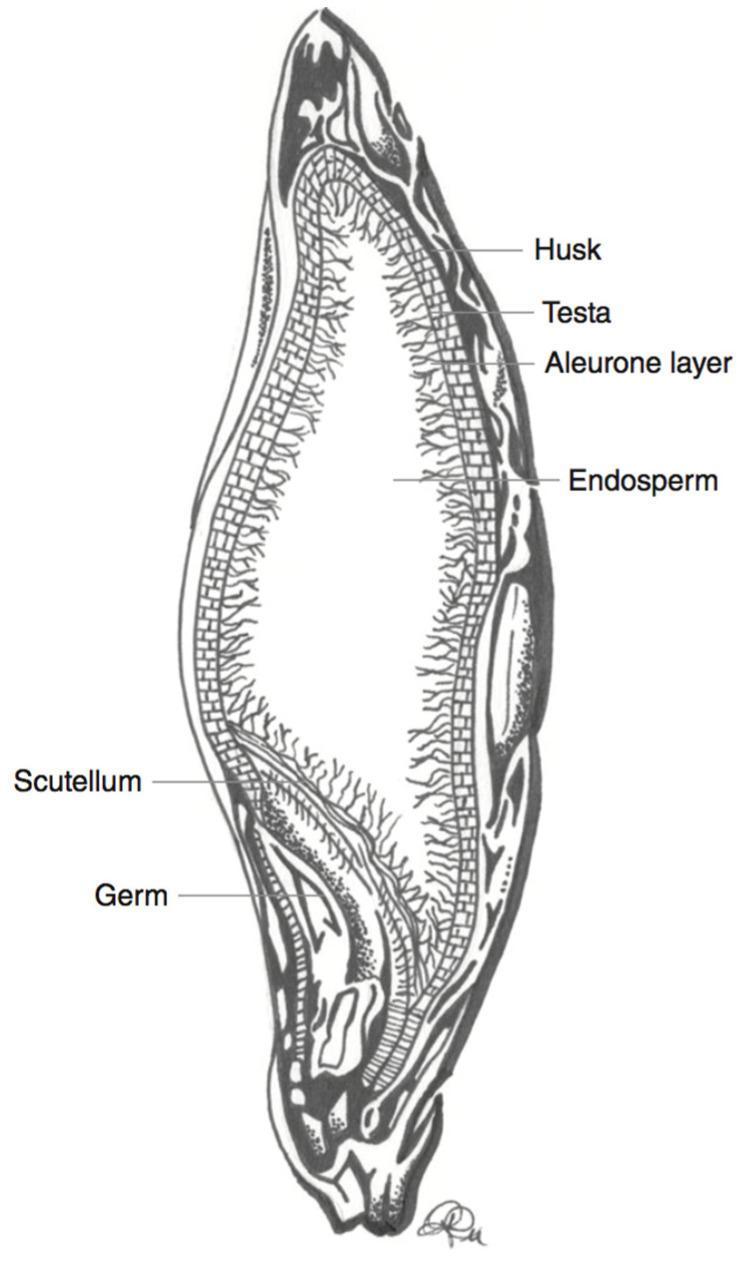
This medical-botanical schematic illustration depicts a longitudinal cross-section of a barley kernel (Hordeum vulgare), serving as a model for cereal grain anatomy relevant to human nutrition and dietetics. The diagram identifies key structural components involved in nutrient distribution, specifically the localization of anthocyanins and fiber. The anatomical layers are labeled from the periphery to the interior: the outermost fibrous Husk, the thin protective Testa, and the Aleurone layer composed of structured, brick-like cells. The largest central region is the Endosperm, characterized by a grainy, fibrous texture. At the base of the kernel, the embryo-related structures, the Scutellum and the Germ, are clearly demarcated. This diagram is educationally significant for understanding the localization of bioactive compounds like phenolic pigments, which are primarily concentrated in the outer layers (pericarp and aleurone) and are often lost during commercial milling processes.
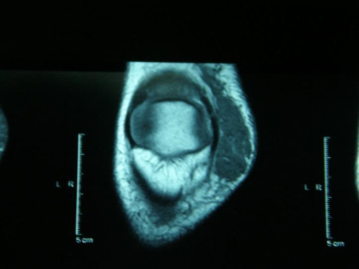
This diagnostic image is an axial magnetic resonance imaging (MRI) scan of the right knee joint. The cross-section clearly demonstrates the bony anatomy, including the distal femoral metaphysis or proximal tibia, characterized by a well-defined cortical rim and internal cancellous bone signal. Surrounding soft tissue structures are visible, with varying signal intensities distinguishing muscle groups, subcutaneous fat, and connective tissues. A 5 cm scale is provided on both the left and right sides of the image frame for anatomical measurement. This imaging modality is essential for evaluating intra-articular pathologies such as meniscal tears, ligamentous injuries (ACL/PCL), and osteochondral defects. The scan utilizes a standard clinical gray-scale display to represent tissue density and proton distribution, serving as a primary tool for musculoskeletal radiology education and orthopedic diagnostic planning.
knee joint layers medial lateral structures anatomy diagram
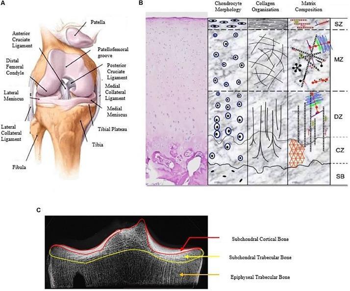
A multi-panel educational figure illustrating the macro and micro-anatomical structures of the human knee joint and articular cartilage. Panel A is an anatomical diagram of the right knee joint in an anterior view, highlighting skeletal and ligamentous structures including the femur (distal femoral condyle), tibia, fibula, and patella. Key ligaments shown include the anterior and posterior cruciate ligaments, and the medial and lateral collateral ligaments, along with the medial and lateral menisci and the patellofemoral groove. Panel B provides a histological and schematic cross-section of hyaline articular cartilage organized into four zones: Superficial (SZ), Middle (MZ), Deep (DZ), and Calcified (CZ), transitioning to Subchondral Bone (SB). It details variations in chondrocyte morphology (flattened to columnar), collagen orientation (tangential to vertical), and extracellular matrix composition across these layers. Panel C is a diagnostic radiographic image (CT/X-ray) of the tibial plateau, segmenting the subchondral bone into three regions: the subchondral cortical bone (end-plate), subchondral trabecular bone, and epiphyseal trabecular bone. This figure serves as a comprehensive reference for orthopedic anatomy and cartilage pathophysiology.
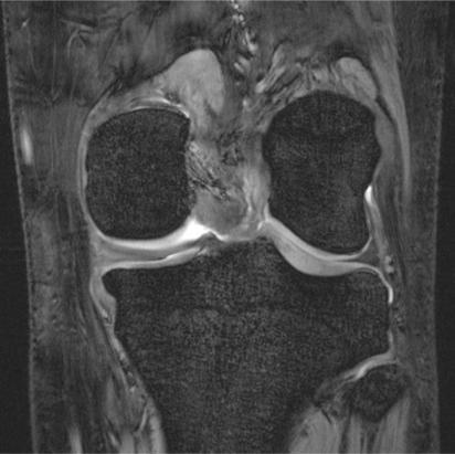
This diagnostic image is an ultra-high resolution, coronal plane 7 Tesla (7T) gradient-echo Magnetic Resonance Image (MRI) of the human knee joint. The imaging demonstrates exceptional detail of the musculoskeletal anatomy, specifically targeting the tibiofemoral joint. Key visible structures include the medial and lateral femoral condyles and the tibial plateau, both showing a dark signal intensity with a highly defined, interconnected network of bright and dark lines representing the fine trabecular bone architecture. The hyaline cartilage layers covering the articular surfaces appear as bright, smooth, and continuous hyperintense bands, allowing for precise assessment of thickness and surface integrity. The medial and lateral menisci are clearly visualized as dark, low-signal, wedge-shaped structures situated within the joint space. This ultra-high-field modality provides the spatial resolution necessary to detect subtle chondral defects, meniscal micro-tears, and changes in subchondral bone density, making it a valuable tool in both advanced clinical diagnostics and osteoporosis research.
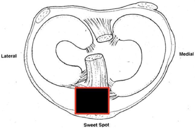
This medical anatomical diagram displays an axial cross-section of the human knee joint, focusing on the tibial plateau and its associated soft tissue structures. The illustration highlights the medial and lateral menisci as crescent-shaped fibrous structures resting on the articular surface. Centrally, the anterior and posterior cruciate ligaments are depicted, showing their relative spatial orientations. Soft tissue attachments, including the capsule and ligamentous fibers, are shown peripherally. A region labeled 'Sweet Spot' is highlighted with a red box at the anterior-inferior aspect of the joint space, corresponding to the clinical target for safe entry during suprapatellar tibial nailing (SPTN) procedures. This site is anatomically significant as the ideal starting point for intramedullary nail insertion, typically located on the medial aspect of the lateral tibial spine to minimize articular damage and optimize fracture reduction in proximal third tibial fractures. The diagram serves as an educational tool for orthopedic surgical planning and biomechanical understanding of the knee joint.
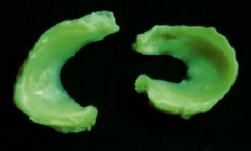
A clinical photograph of human medical specimens featuring the medial and lateral menisci of a right knee joint. The image displays two distinct, semi-lunar fibrocartilaginous structures set against a dark background. On the left, the medial meniscus is characterized by a larger, more open 'C' shape with a wider radius, reflecting its anatomical attachment to the tibial plateau. On the right, the lateral meniscus exhibits a more circular, closed 'O' or tight 'C' morphology, where the anterior and posterior horns are positioned closer together. Both specimens appear to have been treated or dyed for contrast, showing a uniform light green color with a smooth, glistening surface texture. These specimens are typical of those used in orthopedic research and biomechanical studies to evaluate meniscal anatomy, tissue repair using nano-biomaterials, and the mechanical properties of the anterior horn, body, and posterior horn regions.
medial side knee three layers anatomy Warren Marshall
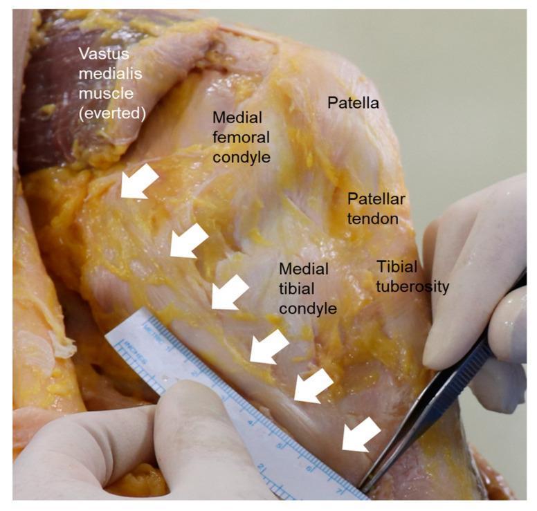
This clinical photograph displays a gross anatomical dissection of the medial aspect of a human knee joint, demonstrating the primary capsuloligamentous structures. The Medial Collateral Ligament (MCL) is highlighted by a series of white arrows, tracing its path from the medial femoral condyle to its distal insertion on the medial tibial condyle. The surrounding anatomy is clearly labeled: the everted vastus medialis muscle is visible superiorly, alongside the patella and the patellar tendon extending toward the tibial tuberosity. The image demonstrates the layered anatomical organization of the knee (Warren and Marshall’s layers), specifically focusing on the Layer II superficial MCL. In the foreground, surgical instrumentation including a metric ruler and fine-tipped forceps are used to measure the longitudinal dimension of the ligament fibers. This visual serves as an educational reference for orthopedic surgery and anatomy, illustrating the relationship between the major medial stabilizers of the knee and their bony landmarks during a surgical or cadaveric study.
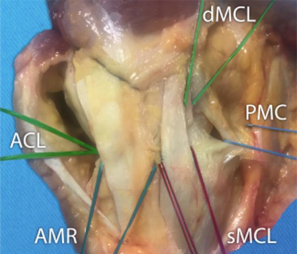
A clinical photograph of a cadaveric dissection showing the medial aspect of a right knee, focusing on the stabilizing ligamentous and capsular structures. The visual is divided into three functional zones. The middle third highlights the medial collateral ligament (MCL), clearly differentiated into its superficial layer (sMCL) and deep layer (dMCL). The superficial MCL is shown as a distinct longitudinal band, while the deep MCL appears closely associated with the joint capsule. The anterior third demonstrates the anteromedial retinaculum (AMR), which comprises the fascia and capsule. The posterior third shows the posterior medial capsule (PMC), where the deep layers merge into a thicker capsular complex that includes the posterior oblique ligament. Internally, the anterior cruciate ligament (ACL) is identified within the joint space. The image illustrates the complex layered anatomy of the medial knee, useful for understanding orthopedic surgical approaches and biomechanical stability. The structures are marked with color-coded lines and anatomical abbreviations for educational clarity.
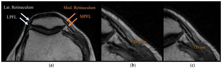
This diagnostic image consists of three axial MRI slices (a-c) of the human knee, focusing on the patellofemoral joint anatomy. Panel (a) provides a labeled overview of the medial and lateral stabilizers. On the lateral side, the lateral retinaculum is shown as a superficial layer, with the lateral patellofemoral ligament (LPFL) located deeper and indicated by white arrows. On the medial side, the medial retinaculum and the medial patellofemoral ligament (MPFL) are identified by orange arrows. Panels (b) and (c) provide magnified views of the medial compartment to demonstrate biometric thickness measurements. In (b), the medial retinaculum is measured at 1.26 mm. In (c), the MPFL is measured at 1.61 mm. The images highlight the distinct layered morphology of these fascial and ligamentous structures, which appear as low-signal (dark) bands against the adjacent tissues. This visualization is clinically relevant for assessing patellar stability, ligamentous tears, or chronic remodeling in conditions such as patellar dislocation or patellofemoral pain syndrome.
Layers of the Knee
Medial Side - Three Layers (Warren and Marshall)
| Layer | Name | Contents |
|---|---|---|
| Layer I | Deep (crural) fascia | Most superficial layer; the investing fascia of the leg |
| Layer II | Superficial MCL layer | Superficial medial collateral ligament (sMCL), structures anterior to it, and ligaments of the posteromedial corner (including the posterior oblique ligament - POL) |
| Layer III | Capsular layer | Joint capsule of the knee + deep MCL (meniscofemoral and meniscotibial portions) |
- Superficial MCL (sMCL) - primary stabilizer against valgus stress; originates on the medial epicondyle, inserts 7-10 cm below the joint line on the posterior tibial metaphysis, deep to pes anserinus
- Deep MCL - thickening of the midmedial capsule; divided into meniscofemoral and meniscotibial portions
- Posterior Oblique Ligament (POL) - posteromedial corner stabilizer
Lateral Side - Three Layers (Seebacher et al.)
| Layer | Contents |
|---|---|
| Layer I (most superficial) | (1) Iliotibial tract (IT band) + its anterior expansion; (2) Superficial portion of biceps femoris + its posterior expansion. The common peroneal nerve lies on the deep side of Layer I, just posterior to the biceps tendon |
| Layer II | Retinaculum of the quadriceps - fibers descend anterolaterally adjacent to the patella |
| Layer III | Knee joint capsule + key lateral ligamentous structures: Fibular collateral ligament (FCL/LCL), popliteofibular ligament (PFL), fabellofibular ligament, anterolateral ligament (ALL), and popliteus tendon |
- FCL (LCL) - primary static stabilizer against varus stress at 0-30° flexion; runs from lateral femoral epicondyle to fibular head (~70 mm long)
- Anterolateral Ligament (ALL) - resists internal tibial rotation, particularly in ACL-deficient knees; insertion halfway between Gerdy's tubercle and fibular head
- IT Band (3 layers within it): superficial layer, deep layer (Kaplan fibers - restrains internal tibial rotation), and capsule-osseous layer
Articular Cartilage Layers (Zones)
| Zone | Features |
|---|---|
| Superficial zone (SZ) | Flattened chondrocytes; tangentially oriented collagen fibers; low friction surface |
| Middle zone (MZ) | Round chondrocytes; randomly oriented collagen |
| Deep zone (DZ) | Columnar chondrocytes; vertically oriented collagen; highest proteoglycan content |
| Calcified zone (CZ) | Transitional zone; tide mark separates it from deep zone |
| Subchondral bone (SB) | Dense cortical bone beneath the cartilage |
Summary: Overall Knee Stabilizers
| Type | Structures |
|---|---|
| Static stabilizers | Joint capsule, menisci, cruciate ligaments (ACL/PCL), collateral ligaments (MCL/LCL), POL, ALL |
| Dynamic stabilizers | Quadriceps, hamstrings, popliteus, gastrocnemius |
- The shape of the knee joint surfaces is inherently unstable - stability depends heavily on the ligamentous and muscular layers
- The posteromedial corner (layer II/III junction) includes the POL and oblique popliteal ligament - often injured in combined MCL/ACL tears
- The posterolateral corner (layer III) includes the FCL + popliteofibular complex - the "terrible triad" of posterolateral instability involves FCL, popliteus tendon, and PFL