
Cervical spondylosis
Reading File
Now let me search the medical library for cervical spondylosis content in parallel.
Finding Sources
Finding Sources
Searching PubMed
"cervical spondylosis"[MeSH Terms] AND management
Good results. Let me read the main textbook sections in parallel.
Reading File
Reading File
Reading File
Let me read more content - the management and clinical features sections.
Reading File
Reading File
Reading File
Excellent. Now let me get management content and also read the Adams & Victor section on treatment/pathology, plus fetch the key X-ray image.
Reading File
Reading File
Loading Image
Let me get the management/treatment section and the MRI image.
Reading File
Loading Image
I now have comprehensive information to write a detailed answer. Let me compile it.
Cervical Spondylosis
Cervical spondylosis (also called cervical osteoarthritis or degenerative disc disease of the cervical spine) is a progressive degenerative condition involving the intervertebral discs, vertebral bodies, facet joints, and supporting ligaments of the cervical spine. It is the most frequently observed myelopathy in general practice.
Epidemiology
- Peaks between ages 40 and 50 years; ubiquitous with increasing age - approximately 40% of people over 50 have some clinical cervical abnormality
- Men affected more than women
- C5-C6 most frequently involved, followed by C6-C7 (greatest mobility, greatest disc degeneration)
- C5, C6, and C7 roots are those most commonly affected clinically
Risk factors: frequent lifting, cigarette smoking, history of excessive driving; patients with dystonia/cervical movement disorders are predisposed to premature degeneration
Pathoanatomy
Cervical spondylosis involves the interplay of the intervertebral disc and four other articulations:
- Two uncovertebral joints of Luschka
- Two facet joints (capsules contain sensory receptors involved in pain and proprioception)
The degenerative cascade (Kirkaldy-Willis) proceeds as:
- Progressive disc collapse → loss of normal cervical lordosis → chronic anterior cord compression across a kyphotic spine
- Subsequent loading of facet and uncovertebral joints → spondylotic foraminal narrowing → spinal cord and/or nerve root compression
"Soft" disc herniation - herniated nucleus pulposus without bony osteophytes; usually posterolateral, may cause acute radiculopathy; anterior herniation can rarely cause dysphagia
"Hard" disc herniation - HNP with disc-osteophytic spur; can cause cord/root compression
The mechanism of cord injury is intermittent compression and ischemia. During flexion/extension, the spinal cord is literally dragged over protruding osteophytes and hypertrophied ligaments. On neck extension, the cord is compressed between degenerative disc/spondylotic bar anteriorly and infolding ligamentum flavum posteriorly. A canal diameter of 7-12 mm (normal: 17-18 mm) produces symptomatic myelopathy; a congenitally narrow canal is a major predisposing factor.
- Miller's Review of Orthopaedics, 9th Ed.
- Adams and Victor's Principles of Neurology, 12th Ed.
Lateral Cervical X-ray - Typical Changes
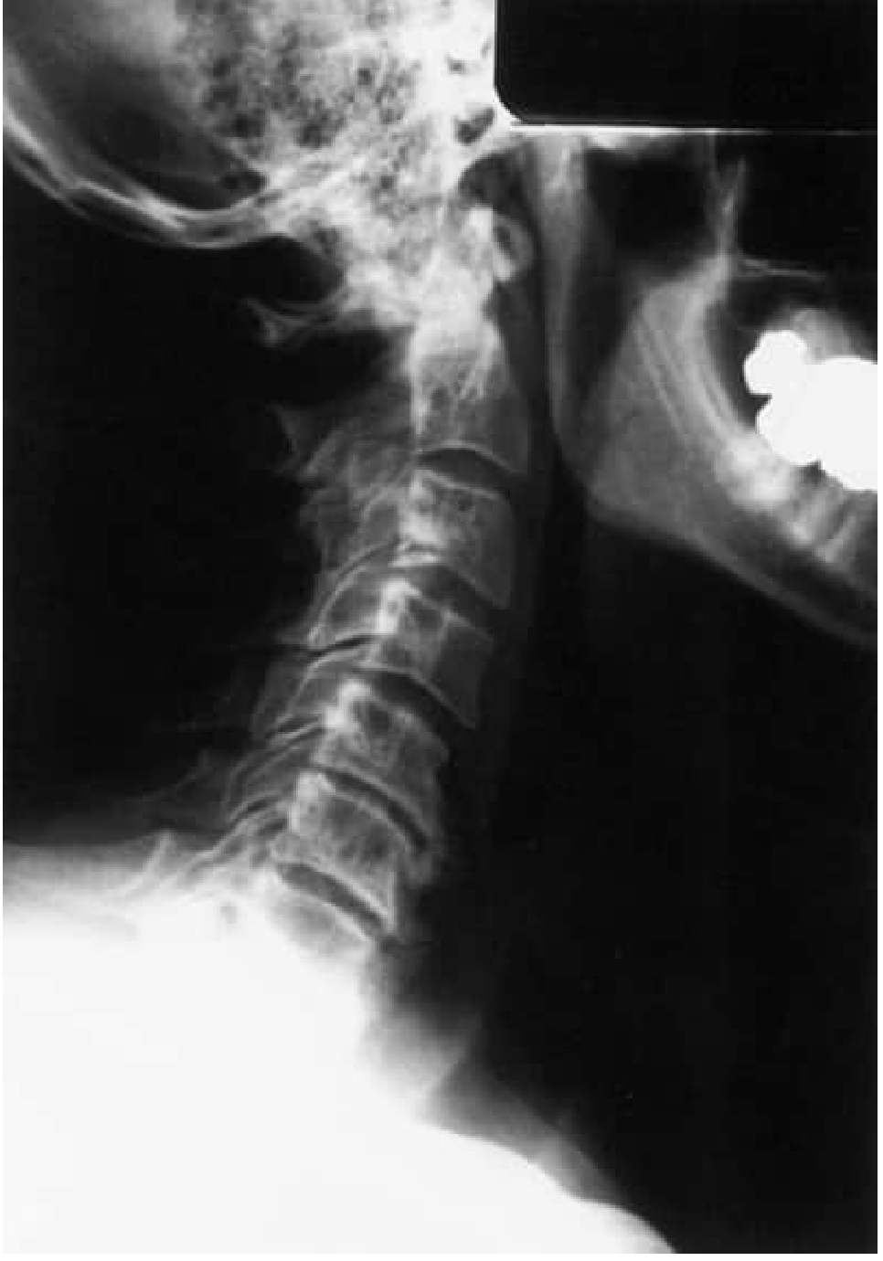
Canal diameter on plain lateral radiograph (posterior vertebral body to spinolaminar line):
| Category | Measurement |
|---|---|
| Normal | ≥14 mm |
| Relative stenosis | 10-13 mm |
| Absolute stenosis | <10 mm |
Pavlov (Torg) ratio (canal width / vertebral body width): Normal = 1.0; ratio <0.8 is abnormal and a risk factor for neurologic involvement
Four Clinical Entities
| Syndrome | Description |
|---|---|
| Discogenic neck pain | Axial pain from disc degeneration alone, no neurologic signs, occipital headache common |
| Radiculopathy | Nerve root compression - pain + sensory/motor deficits in a dermatomal/myotomal distribution |
| Myelopathy | Spinal cord compression - the most serious presentation |
| Myeloradiculopathy | Combined cord + root involvement |
Clinical Features
The Characteristic Triad of Myelopathy
- Painful, stiff neck with pain in neck, shoulders, upper arms (brachialgia) - aching or radicular, evoked by movement
- Numbness and paresthesias of the hands - "wearing gloves," "swollen," or "coated with glue"; may involve digits, palm, or a band along the forearm
- Spastic leg weakness with Babinski signs, unsteadiness of gait, and a Romberg sign
Additional sensory features: impaired vibratory sensation and diminished position sense in feet (posterior column signs), a "tabetic" unsteadiness; Lhermitte symptom (electrical feelings down the spine on neck flexion); rarely a Brown-Séquard pattern.
Bladder dysfunction (frequency, urgency, urgency incontinence) indicates need for imaging.
Radiculopathy by Level
| Disc Level | Root | Pain/Sensory Distribution | Motor Weakness | Reflex Loss |
|---|---|---|---|---|
| C4-C5 | C5 | Shoulder, outer upper arm | Deltoid, biceps, brachioradialis | Biceps, supinator |
| C5-C6 | C6 | First two digits, lateral forearm | Brachioradialis, wrist extensors | Biceps, brachioradialis (may be inverted) |
| C6-C7 | C7 | Index, middle, ring fingers | Triceps, wrist flexors, pronators | Triceps |
Note: Spread of biceps reflex to finger flexors, an increased triceps reflex, or a paradoxical biceps reflex signals concurrent myelopathy.
MRI: Spondylotic Myelopathy
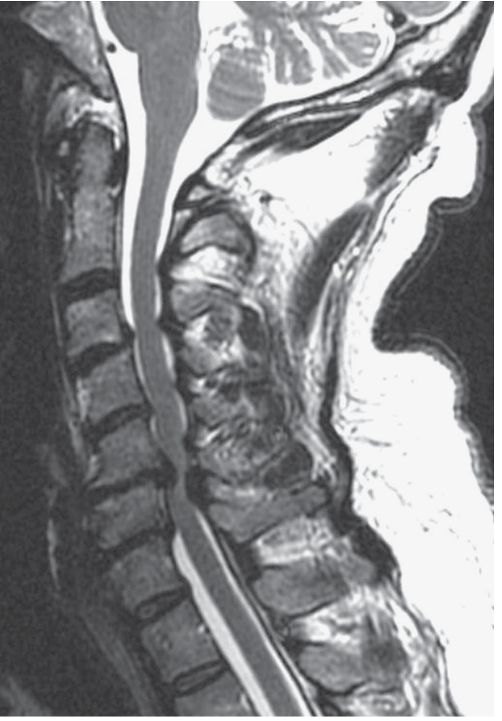
MRI features supporting spondylotic cord compression:
- Clear deformation of the cord into a "kidney bean" shape
- Obliteration of surrounding CSF spaces in transverse images
- Intrinsic T2 signal changes within the cord at or within half a segment of compression (indicates some irreversibility)
- "Pancake-like" gadolinium enhancement at/just caudal to site of maximal compression
Diagnosis
MRI is the imaging study of choice - identifies nerve root compression, causes of myelopathy, and disc herniation. Must be interpreted with caution as degenerative changes are extremely common in asymptomatic spines.
CT myelography is preferred when:
- MRI-incompatible hardware (pacemakers, SCS)
- Prior cervical fusion surgery (hardware artifact)
- Severe claustrophobia
- Need to distinguish non-calcified disc from osteophytes
EMG/NCS in cervical radiculopathy: loss of CMAP amplitude on affected side with preserved SNAP (characteristic - intraspinal lesion proximal to DRG). Useful to exclude brachial plexopathy or peripheral neuropathy.
Plain cervical radiography is of little value in diagnosing or excluding radiculopathy.
Differential diagnosis of myelopathy:
- Spinal multiple sclerosis (oligoclonal bands, optic nerve/brain lesions)
- ALS (widespread denervation, no sensory symptoms, progressive)
- Subacute combined degeneration (B12 deficiency - symmetric)
- Normal-pressure hydrocephalus (Romberg sign absent, magnetic gait)
- HTLV-I myelopathy, HIV myelopathy
- Ossification of the posterior longitudinal ligament (OPLL)
- Spinal cord tumor
Treatment
Conservative (Non-operative)
Appropriate for mild symptoms, radiculopathy without significant neurologic deficit, and discogenic neck pain:
- Soft cervical collar (limits harmful neck movement, especially extension)
- Cervical traction
- NSAIDs and analgesics
- Physical therapy - cervical strengthening, posture correction
- Fluoroscopically guided cervical nerve blocks (for radiculopathy)
- Activity modification - avoid activities that extend or compress the neck
Surgical Indications
- Cervical myelopathy - surgery is generally indicated to halt progression; conservative management alone carries risk of irreversible cord damage
- Radiculopathy not responding to conservative measures (6-12 weeks)
- Progressive neurologic deficit
- Significant cord compression on imaging with symptoms
Operative approaches:
- Anterior - Anterior cervical discectomy and arthrodesis (ACDA/ACDF): Robinson approach; ideal for 1-2 level disease, anterior compression
- Posterior - Laminectomy, laminoplasty: for multi-level disease, preserving motion; expansive laminoplasty particularly for radiculomyelopathy
- Disc arthroplasty - alternative to fusion for single-level disease, preserves motion
Important caveat: Cervical spine surgery for degenerative pathology is rarely if ever indicated purely for headache or neck pain in the absence of radiculopathy or myelopathy.
Sources: Bradley and Daroff's Neurology in Clinical Practice; Adams and Victor's Principles of Neurology, 12th Ed.; Miller's Review of Orthopaedics, 9th Ed.
This is a shared conversation. Sign in to Orris to start your own chat.