
Eosinophilia eosiphagitis and how it is different from herd md pathology exam
eosinophilic esophagitis GERD histology diagnosis
eosinophilic esophagitis vs GERD histology eosinophils comparison
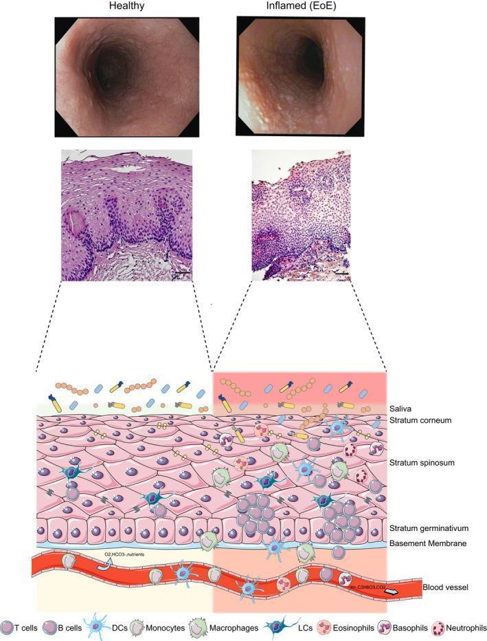
Educational comparison of healthy esophagus versus Eosinophilic Esophagitis (EoE), presented through endoscopic, histological, and schematic views. The top row features macroscopic endoscopic photographs: the healthy esophagus shows a smooth, pale pink lumen, while the EoE esophagus exhibits characteristic longitudinal furrowing and white exudates (micro-abscesses). The middle row presents H&E stained histological sections, with the EoE slide demonstrating significant intraepithelial eosinophil infiltration and basal cell hyperplasia compared to the structured healthy tissue. The bottom section is a pathophysiology diagram illustrating the esophageal layers (stratum corneum, spinosum, germinativum, and basement membrane). In the healthy state (left), immune cell presence is minimal, primarily consisting of T cells and Langerhans cells. In the inflamed EoE state (right), there is a dense recruitment of immune cells from the vasculature, including eosinophils, basophils, neutrophils, dendritic cells, B cells, macrophages, and monocytes, highlighting the Th2-mediated chronic inflammatory response.
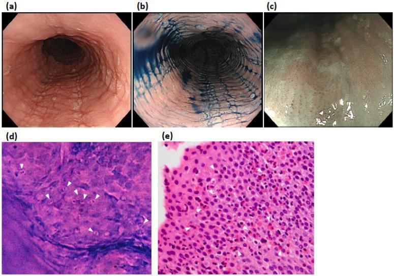
This composite educational image illustrates diagnostic modalities for Eosinophilic Esophagitis (EoE) in a 52-year-old patient. Panels (a) and (b) present White Light Endoscopy (WLE) and indigocarmine chromoendoscopy, respectively, showing classic endoscopic features: linear furrows, white exudates, and concentric rings (trachealization) within the esophageal lumen. Panel (c) utilizes Narrow-Band Imaging Magnifying Endoscopy (NBI-ME), demonstrating beige-colored mucosa and characteristic dot-shaped intrapapillary capillary loops (IPCLs) without visible cyan vessels. Panel (d) displays high-magnification endocytoscopy (x520), an in vivo imaging technique showing multiple cells with purple bilobed nuclei and nonstained peripheries (white arrowheads), characteristic of eosinophilic infiltration. Panel (e) provides histological correlation (H&E stain) from a biopsy of the same site, confirming dense eosinophilic infiltration (>50 eosinophils per high-power field) with prominent bilobed nuclei (white arrowheads). The image provides a comprehensive comparison between gross endoscopic findings, advanced in vivo microscopy, and gold-standard histopathology for gastroenterology education.
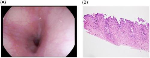
A dual-panel comparison exhibiting normal esophageal findings. Panel (A) is a clinical photograph from an esophagogastroduodenoscopy (EGD) showing a healthy esophageal lumen. The mucosa is characterized by a smooth, pink, and glistening surface without evidence of mucosal rings, linear furrows, white exudates, or strictures, indicating the absence of typical eosinophilic or lymphocytic esophagitis features. Panel (B) is a diagnostic histology image (Hematoxylin and Eosin stain) of an esophageal biopsy at high power. It demonstrates normal stratified squamous epithelium with organized cellular maturation. The basal cell layer appears healthy without hyperplasia, and the lamina propria is free of significant inflammatory cell infiltration, specifically lacking eosinophils or intraepithelial lymphocytes. Together, these images serve as a baseline for normal esophageal anatomy and histology in a pediatric gastroenterology context, often used to confirm mucosal healing following treatment for conditions such as chemical or thermal injury.
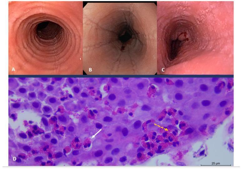
This composite educational image illustrates the diagnostic features of eosinophilic esophagitis (EoE) through endoscopy and histopathology. Upper panels (A, B, and C) display clinical endoscopic views of the esophagus. Panel A reveals trachealization, characterized by prominent circular rings. Panel B shows longitudinal linear furrows and narrowings. Panel C demonstrates mucosal fragility (crepe-paper esophagus) with visible erosions and minor surface bleeding. The lower panel (D) is a high-power light microscopy image of an H&E stained esophageal biopsy. It shows dense intraepithelial inflammation with numerous eosinophils (indicated by the dashed orange arrow), exceeding the diagnostic threshold of 15 eosinophils per high-power field. A solid white arrow points to an eosinophilic microabscess, a collection of eosinophils within the squamous epithelium. The image serves as a clinical reference for gastroenterology and pathology, highlighting the correlation between macroscopic structural changes and microscopic inflammatory infiltrates in chronic allergic esophagitis.
Eosinophilic Esophagitis (EoE) - Full Pathology Notes + Comparison with GERD
What is Eosinophilic Esophagitis (EoE)?
- Clinically: symptoms of esophageal dysfunction (dysphagia, food impaction in adults; feeding intolerance, GERD-like symptoms in children)
- Histologically: dense eosinophilic infiltration of the esophageal squamous epithelium
Pathogenesis (for exam)
| Step | Mechanism |
|---|---|
| Trigger | Food allergens (cow's milk, soy, wheat, eggs, nuts, seafood) |
| Sensitization | Through inflamed skin (atopic background) |
| Cytokines | IL-33 and TSLP activate Th2 cells -> IL-5, IL-13, eotaxin-3 |
| Result | Eosinophil recruitment into esophageal epithelium, barrier disruption, tissue remodeling |
Histological Features of EoE (KEY for path exams)
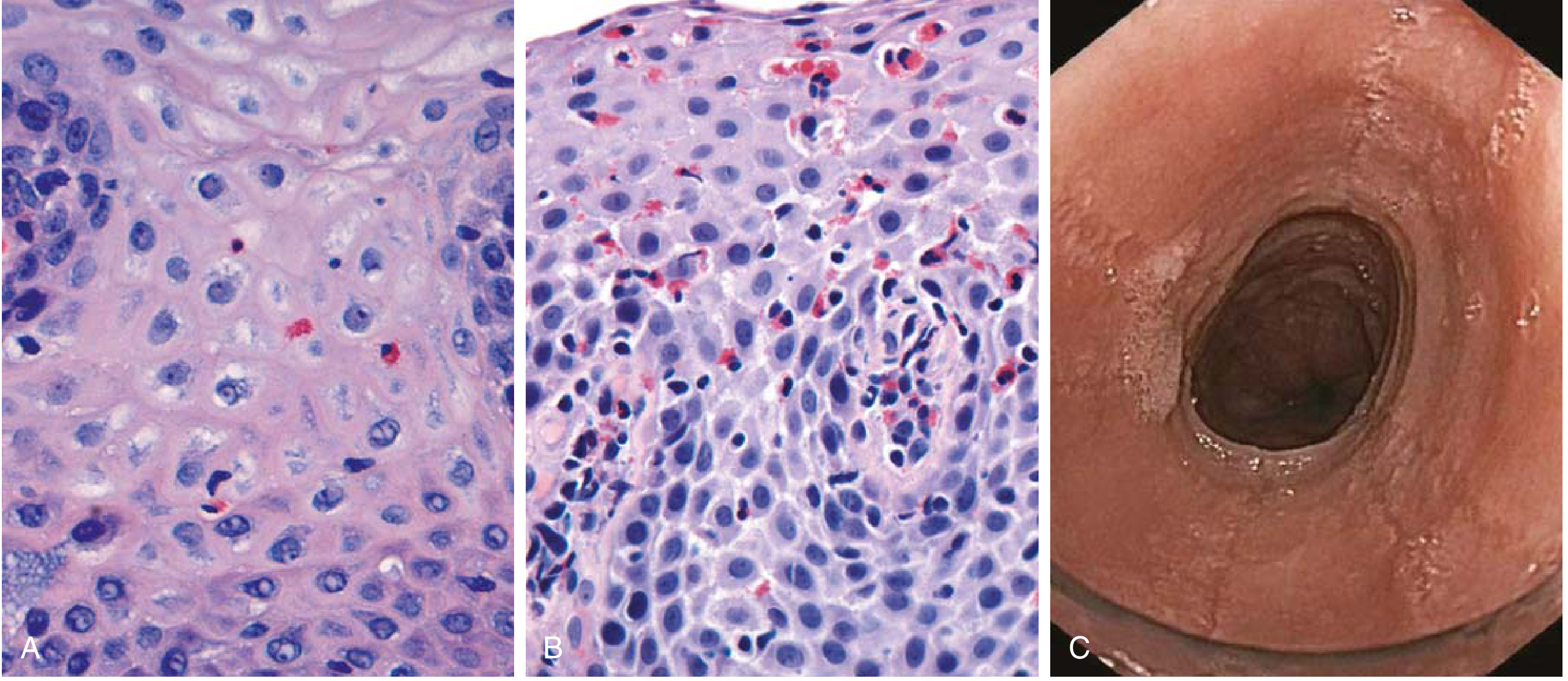
- Intraepithelial eosinophils: ≥15 per high-power field (HPF) - the diagnostic threshold
- Superficial/surface layering of eosinophils (eosinophils concentrated superficially, away from GEJ)
- Eosinophilic microabscesses - clusters of eosinophils within the epithelium (hallmark feature)
- Extracellular eosinophilic granules (degranulation)
- Basal cell hyperplasia (also seen in GERD - overlapping)
- Subepithelial fibrosis (from chronic disease/remodeling)
- Distribution: proximal, mid AND distal esophagus equally - panesophageal involvement
The Big Comparison: EoE vs GERD (Exam Favorite)
| Feature | EoE | GERD (Reflux Esophagitis) |
|---|---|---|
| Etiology | Allergic / immune-mediated (Th2, food allergens) | Acid reflux - mechanical/chemical injury |
| Demographics | Children and young adults; male predominance; atopic history | Any age; common in obese, smokers, hiatal hernia |
| Symptoms | Dysphagia, food impaction, feeding problems in kids | Heartburn, regurgitation, water brash |
| Response to PPI | Refractory to PPI (key distinguishing point!) | Responds to PPI |
| Peripheral eosinophilia | Often present (modest) | Not present |
| Endoscopy location | Proximal and mid esophagus (rings, furrows, trachealization) | Distal esophagus (erosions, erythema near GEJ) |
| Endoscopic findings | Rings, longitudinal furrows, white exudates/plaques, narrow-caliber esophagus, mucosal fragility ("crepe paper") | Erosions, erythema, linear streaks at distal esophagus |
| Eosinophil count (HPF) | ≥15 (often ≥20-30+), distributed throughout proximal-distal equally | Usually <15, predominantly in distal esophagus only |
| Eosinophilic microabscesses | Yes - characteristic | Absent |
| Surface eosinophil layering | Yes | No |
| Basal cell hyperplasia | Present | Present (overlapping feature) |
| Papillary elongation | Present | Present (overlapping feature) |
| Subepithelial fibrosis | Yes (remodeling) | Less prominent |
| Distribution of eosinophils | Panesophageal - proximal = distal | Patchy, distal >> proximal |
| Biopsy protocol | 2-4 biopsies each from proximal AND distal esophagus (panesophageal sampling) | Distal esophagus, >2 cm above GEJ |
| Treatment | Dietary elimination (milk, soy, wheat), topical swallowed corticosteroids (budesonide, fluticasone), esophageal dilation | PPI, lifestyle changes |
Endoscopy: EoE vs GERD
- Circumferential rings ("trachealization" / feline esophagus)
- Longitudinal furrows
- White exudates/plaques (eosinophilic microabscesses)
- Diffuse narrowing, mucosal fragility
- Erosions and erythema at the distal esophagus / GEJ
- Possible Barrett's esophagus (intestinal metaplasia) in chronic cases
- Normal proximal esophagus
The Critical Diagnostic Pitfall (Exam High-Yield!)
- Eosinophil count (≥15/HPF, especially in proximal biopsies = strong evidence for EoE)
- Microabscesses and surface layering = EoE-specific
- Proximal esophageal involvement = points to EoE, not GERD
- Atopic history + PPI refractoriness = clinical context
Summary for Exam Memory
EoE = Allergic esophagitis. ≥15 eos/HPF + microabscesses + surface layering + rings on scope + proximal distribution + atopic + PPI-refractory.GERD = Acid-reflux injury. <15 eos/HPF + distal distribution + basal cell hyperplasia + papillary elongation + no microabscesses + responds to PPI.
- Robbins & Kumar Basic Pathology 10e, p. 549
- Clinical Gastrointestinal Endoscopy 3e, pp. 84-85
- Sleisenger & Fordtran's Gastrointestinal and Liver Disease