Skull bone anatomy
Reading File
Finding Sources
Searching Images
skull bone anatomy cranial bones
Finding Sources
Finding Sources
Reading File
Reading File
Reading File
Reading File
Finding Sources
Reading File
Reading File
Reading File
Loading Image
Loading Image
Loading Image
Finding Sources
Reading File
Loading Image
Here is a comprehensive overview of skull bone anatomy, based on Gray's Anatomy for Students, Rosen's Emergency Medicine, and Cummings Otolaryngology.
Skull Bone Anatomy
The skull has 22 bones (excluding the ossicles of the middle ear). Except for the mandible, all skull bones are connected by sutures — immovable fibrous joints. Collectively they form the cranium.
Overview: Divisions of the Cranium
| Region | Function | Key Bones |
|---|---|---|
| Calvaria (cranial vault) | Encloses and protects the brain | Frontal, 2× parietal, 2× temporal, sphenoid, occipital |
| Cranial base | Floor of the cranial cavity | Sphenoid, temporal, occipital, frontal, ethmoid |
| Facial skeleton (viscerocranium) | Forms the face | Nasal, palatine, lacrimal, zygomatic, maxillae, inferior nasal conchae, vomer |
| Mandible | Lower jaw; only movable bone | Mandible |
Anterior View
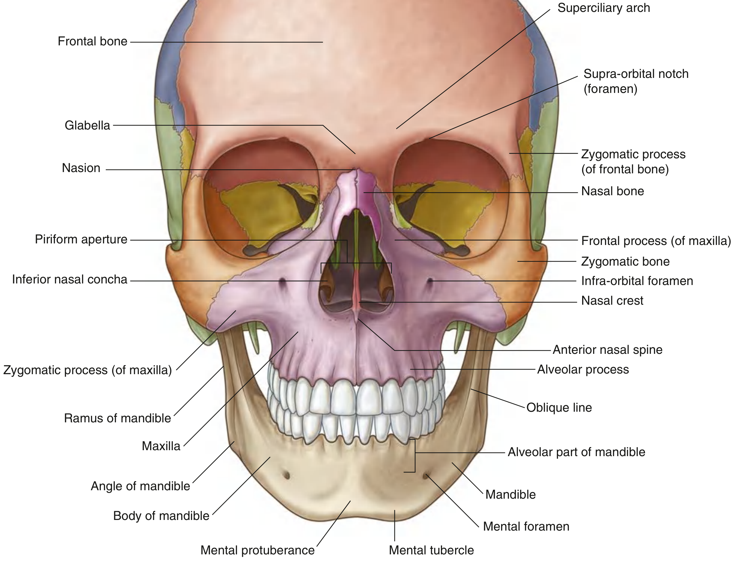
Frontal Bone
- Forms the forehead and superior orbital rim
- Bears the superciliary arches (more pronounced in males) and glabella (midline depression between them)
- The supra-orbital foramen (or notch) transmits the supra-orbital nerve and vessels
- Its zygomatic process projects laterally to form the upper lateral orbital rim, articulating with the frontal process of the zygomatic bone
Zygomatic Bone (Cheekbone)
- Forms the lower lateral orbital rim
- Its temporal process projects posteriorly to meet the zygomatic process of the temporal bone, forming the zygomatic arch
- Bears the small zygomaticofacial foramen on its lateral surface
Nasal Bones
- Paired small bones forming the bridge of the nose
Maxilla (paired)
- Forms the upper jaw with its alveolar process (tooth-bearing)
- Contributes to the inferior and medial orbital rim
- Its frontal process articulates with the frontal bone; its zygomatic process articulates with the zygomatic bone
Mandible
- The only freely movable skull bone
- Has a body (anterior, tooth-bearing), ramus (vertical posterior projection), condylar process (articulates with temporal bone at the TMJ), and coronoid process (attachment for temporalis muscle)
- The mental foramen on the lateral body surface transmits the mental nerve and vessels
Lateral View
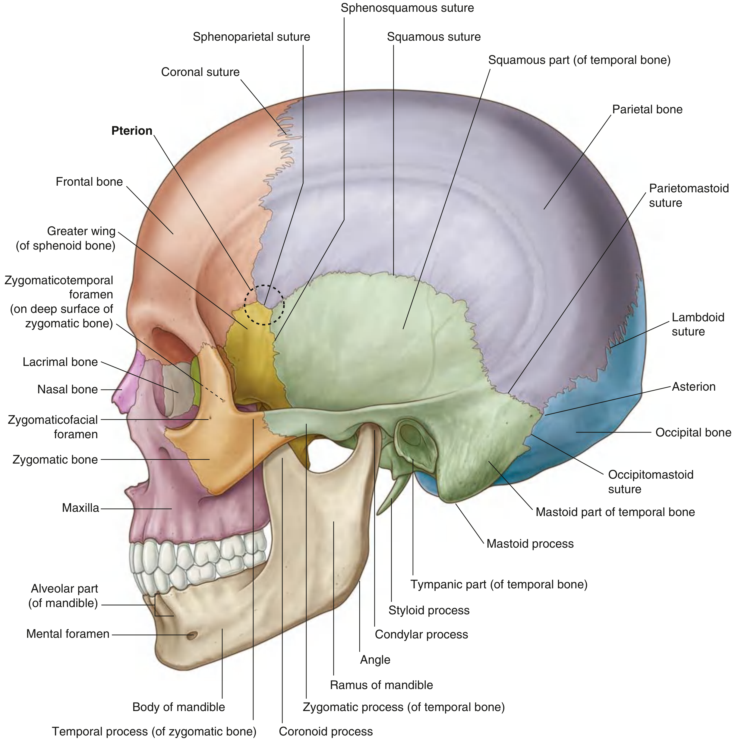
Key Sutures (Lateral View)
| Suture | Bones Joined |
|---|---|
| Coronal | Frontal ↔ Parietal |
| Squamous | Temporal (squamous) ↔ Parietal |
| Sphenoparietal | Sphenoid (greater wing) ↔ Parietal |
| Sphenosquamous | Sphenoid ↔ Temporal |
| Lambdoid | Parietal ↔ Occipital |
| Occipitomastoid | Occipital ↔ Mastoid part of temporal |
Pterion ⚠️ Clinically Important
The pterion is the H-shaped junction where the frontal, parietal, sphenoid, and temporal bones meet. The bone here is particularly thin and overlies the anterior division of the middle meningeal artery. A skull fracture here can tear this artery, causing an extradural (epidural) hematoma.
Temporal Bone (Complex, Multi-part)
- Squamous part — large flat plate forming the lateral cranial wall
- Zygomatic process — projects anteriorly to form the zygomatic arch
- Tympanic part — surrounds the external acoustic meatus (ear canal)
- Petrous part — dense pyramidal part housing the inner ear structures; forms part of the cranial base
- Mastoid part — posterior projection bearing the mastoid process (attachment for sternocleidomastoid); contains air cells
- Styloid process — slender inferior projection
Posterior View
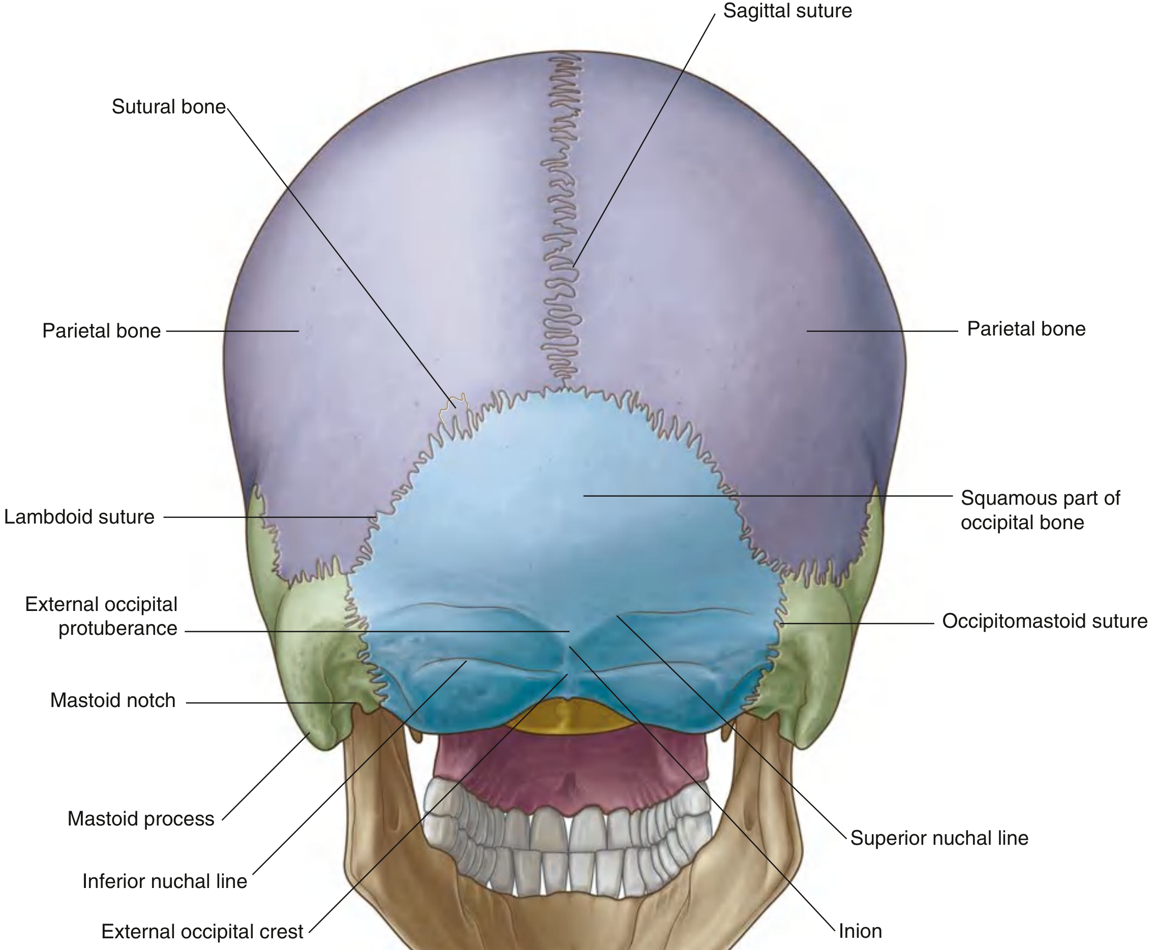
Occipital Bone
- Dominant bone of the posterior cranium
- Squamous part articulates with parietal bones at the lambdoid suture and with temporal bones at the occipitomastoid sutures
- External occipital protuberance (inion) — palpable midline bump; origin of the superior nuchal line
- Superior and inferior nuchal lines — bony ridges for posterior neck muscle attachment
- Sutural (Wormian) bones — small accessory ossification centers occasionally found along the lambdoid suture
Inferior View (Skull Base)
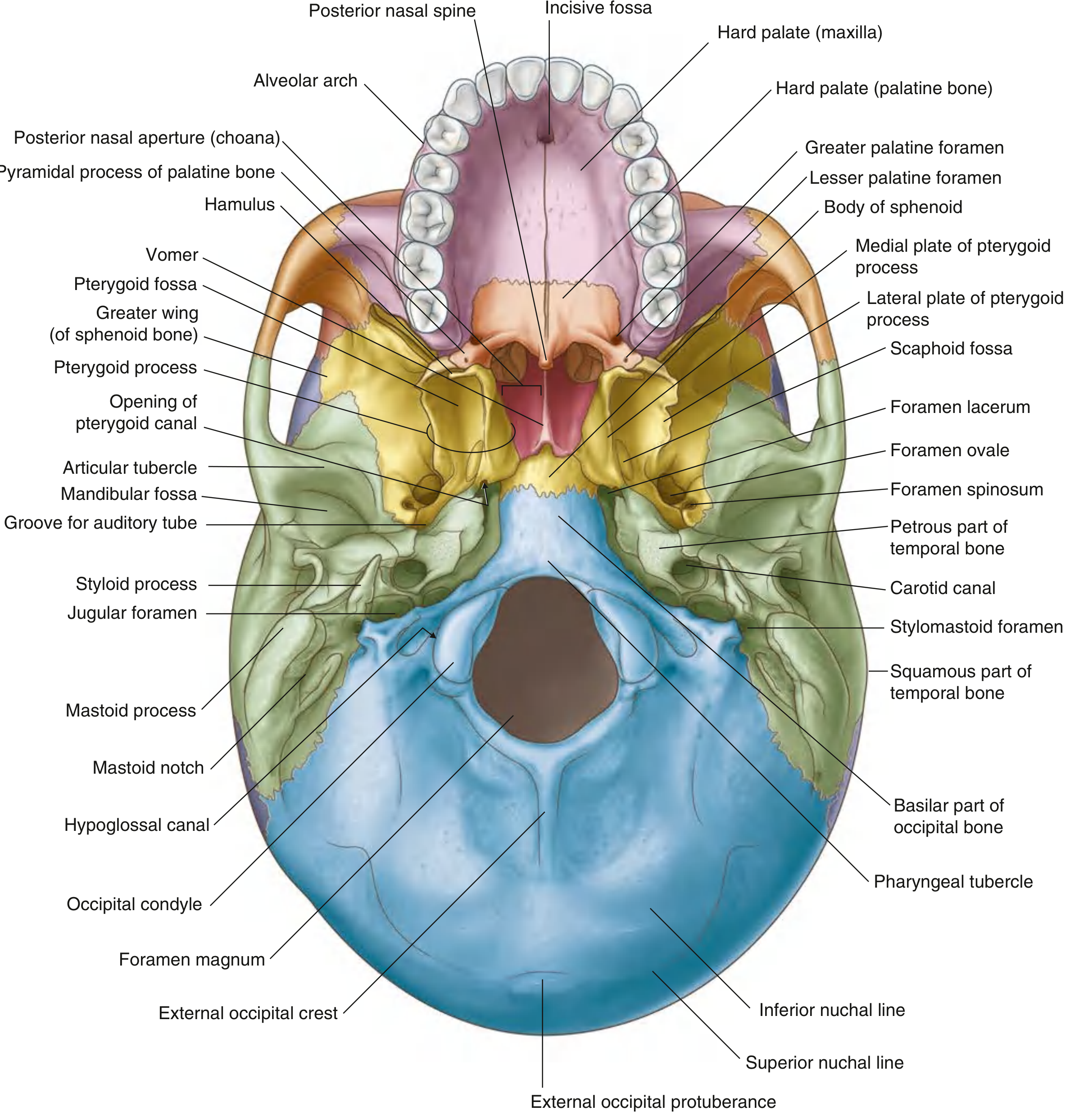
The skull base is formed by five bones: frontal, ethmoid, temporal, sphenoid, and occipital.
Sphenoid Bone
A butterfly-shaped bone at the center of the skull base. Consists of:
- Body — centrally placed, contains paired sphenoidal air sinuses
- Greater wings — project laterally; contain foramen ovale (CN V₃, lesser petrosal n.) and foramen spinosum (middle meningeal a.)
- Lesser wings — project anteriorly; not visible from below
- Pterygoid processes — project inferiorly with medial and lateral plates enclosing the pterygoid fossa; medial plate ends in the pterygoid hamulus; contains the pterygoid canal
- Foramen lacerum — filled with cartilage in life; at junction with temporal and occipital bones
Occipital Bone (Inferior Aspect)
- Basilar part — articulates with sphenoid bone at the spheno-occipital synchondrosis (fuses ~age 25)
- Occipital condyles — articulate with atlas (C1) at the atlanto-occipital joint
- Foramen magnum — large opening transmitting the brainstem/spinal cord junction, vertebral arteries, anterior and posterior spinal arteries, accessory nerve (CN XI) roots
- Hypoglossal canal — transmits CN XII
- Jugular foramen — transmits the internal jugular vein (origin), CN IX, X, XI, and inferior petrosal sinus
Vomer
- Small midline bone contributing to the bony nasal septum, resting on the sphenoid
Bone Structure
Each cranial bone consists of:
- Outer cortical table (periosteum-covered)
- Diploe (cancellous/spongy bone with red marrow)
- Inner cortical table (lined by periosteal dura)
Adult skull bones average 2–6 mm in thickness; the temporal region is the thinnest.
Major Foramina Summary
| Foramen | Transmits |
|---|---|
| Supra-orbital foramen | Supra-orbital n. & vessels |
| Infra-orbital foramen | Infra-orbital n. & vessels |
| Mental foramen | Mental n. & vessels |
| Foramen ovale | CN V₃ (mandibular n.), lesser petrosal n. |
| Foramen spinosum | Middle meningeal artery |
| Foramen lacerum | Filled with cartilage (ICA traverses superior) |
| Carotid canal | Internal carotid artery |
| Stylomastoid foramen | CN VII (facial n.) |
| Jugular foramen | IJV, CN IX, X, XI, inferior petrosal sinus |
| Hypoglossal canal | CN XII |
| Foramen magnum | Medulla/spinal cord, vertebral arteries, CN XI roots |
Sutures and Fontanelles
In fetal and neonatal skulls, large gaps called fontanelles exist between the unfused bones:
- Anterior fontanelle (bregma) — largest; junction of frontal, sagittal, and coronal sutures; closes by ~18 months
- Posterior fontanelle (lambda) — junction of sagittal and lambdoid sutures; closes by ~2–3 months
- Sphenoidal fontanelle — anterolateral
- Mastoid fontanelle — posterolateral
These allow head molding during birth and accommodate postnatal brain growth. After the first few months of life the bones progressively fuse, ultimately forming the rigid, non-expandable cranial vault.
Sources: Gray's Anatomy for Students (9780323934237); Rosen's Emergency Medicine (9780323757898); Cummings Otolaryngology Head and Neck Surgery (9780323612173)
This is a shared conversation. Sign in to Orris to start your own chat.