
Severe impetigo pediatric managment
impetigo children management treatment
impetigo pediatric skin lesions clinical photo
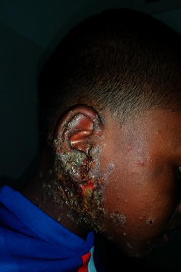
This clinical photograph displays a pediatric patient with classic signs of impetigo, a highly contagious bacterial skin infection commonly caused by Staphylococcus aureus or Streptococcus pyogenes. The visual focus is on the right auricular and preauricular regions, extending down toward the neck and onto the cheek. The primary lesions are characterized by thick, adherent, honey-colored (mellicerous) crusts overlying erythematous bases. Areas of active serosanguinous exudate and open erosions are visible, particularly around the earlobe and external auditory meatus. Surrounding the main confluent plaques, scattered satellite papules and small pustules are present on the facial skin, indicating local spread. The surrounding skin shows signs of inflammation and secondary irritation. This image serves as a hallmark educational example of non-bullous impetigo, emphasizing the distinctive crusting morphology and typical distribution in pediatric dermatology.
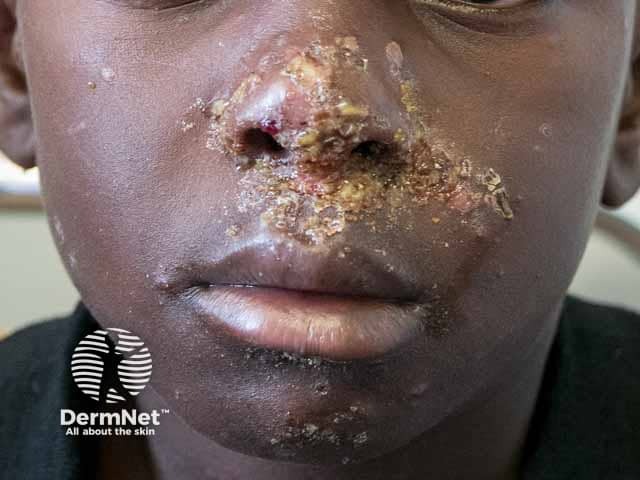
Imaging modality: Clinical photography (digital color image) of a pediatric patient's face in frontal view, focusing on the nasal and perioral regions. The image depicts honey-colored crusts and erosions centered around the nostrils and mouth with surrounding erythema and scaling, compatible with impetigo. Lesions appear superficial with crusted exudate, partial fissuring at the alar rim, and crusts coalescing into golden-brown plaques. The bilateral distribution is localized to the midface, with consented close-up representation suitable for dermatology assessment. The skin surface shows dry crusts, crusted erosions, and minimal edema; surrounding skin is mildly inflamed. The appearance is classic for non-bullous impetigo, most commonly caused by Staphylococcus aureus or Streptococcus pyogenes, though other organisms can contribute. The image helps distinguish impetigo from eczematous dermatitis, herpes simplex infection, or ecthyma by the characteristic honey crusts and focal periorificial involvement. Clinically, this finding is contagious and prompts antibiotic therapy and hygiene measures; topical agents such as mupirocin or fusidic acid may be used, with systemic antibiotics if extensive or bullous lesions are present. For education and research, this photograph serves as a reference for pattern recognition, differential diagnosis, and treatment planning in pediatric dermatology.
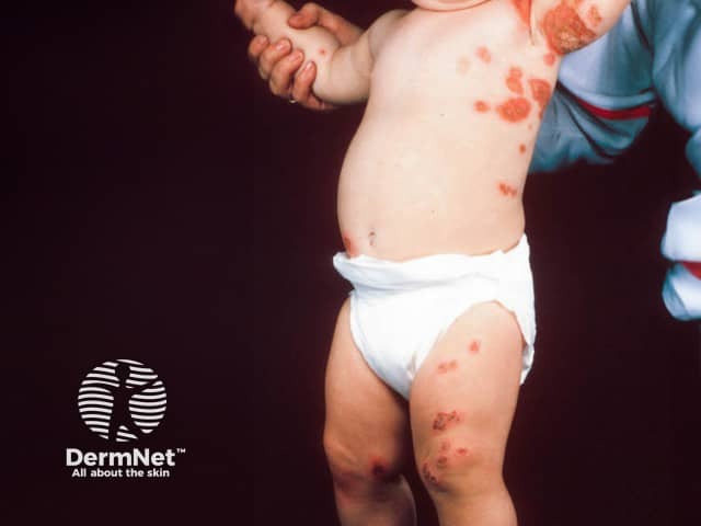
Clinical photography of a pediatric patient demonstrating bilateral axillary impetigo with kissing lesions. Imaging modality: dermatologic clinical photograph; technique: high-resolution color image under standardized frontal/anterolateral illumination; no staining or contrast employed. Anatomic focus: integumentary system, especially intertriginous axillary skin on both sides, extending to adjacent shoulder and upper chest. Visual features: multiple erythematous papules and vesicles with golden honey‑colored crusts along skin folds; some lesions show superficial crusting and erosions; bilateral symmetry with lesions meeting at the axillary creases, creating kissing lesions from close contact. The surrounding skin exhibits mild erythema and mild edema; distribution is intertriginous and continuous across bilateral axillae, with secondary spread to proximal medial arms. Pathology (clinical correlates): classic nonbullous impetigo caused by Staphylococcus aureus and/or Streptococcus pyogenes; contagious, transmissible in household contacts; result of minor trauma and moisture in skin folds. Diagnostic significance: recognition of kissing lesions in intertriginous areas supports impetigo diagnosis and informs antibiotic therapy and hygiene measures. Potential differential diagnoses include candidal intertrigo, contact dermatitis, eczema, varicella in atypical presentation, or fungal infection. Clinical use cases: educational dermatology imagery, triage reference for pediatric skin infections, infection control planning, and documentation of treatment response.
Severe Impetigo - Pediatric Management
Overview
Clinical Forms in Children
| Form | Key Features | Pathogen |
|---|---|---|
| Non-bullous (impetigo contagiosa) | Thin vesicles → pustules → honey-colored crusts; face/extremities; lymphadenopathy | S. aureus, S. pyogenes |
| Bullous impetigo | 1-2 cm flaccid bullae → thin brown crust/collarette; face, neck, extremities; seen in infants/young children | S. aureus (exfoliative toxin A) |
| Ecthyma (deep impetigo) | Punched-out ulcers with raised margins and thick crust; lower extremities; can scar | S. pyogenes, S. aureus |
| Neonatal impetigo | Highly contagious; begins day 4-10 of life; predilection for face/perineum; risk of bacteremia/pneumonia/meningitis | S. aureus |
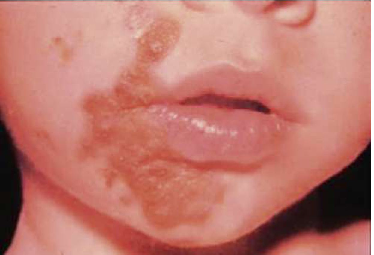
Management by Severity
1. Localized / Mild Disease
- Topical mupirocin (active against most MRSA strains) - apply 3x daily for 5 days
- Alternative topical agents: retapamulin, ozenoxacin 1% cream (approved ≥2 months of age), bacitracin, fusidic acid
- Soak off crusts before application
2. Severe / Extensive Disease (multiple lesions, bullous form, ecthyma)
- Cephalexin (first-line) or dicloxacillin - 25-50 mg/kg/day divided q6-8h (max 500 mg/dose)
- Alternatively: amoxicillin-clavulanate, cloxacillin
- TMP-SMX (trimethoprim-sulfamethoxazole) - preferred for community-associated MRSA
- Clindamycin (check local resistance; always D-test for inducible resistance)
- Doxycycline (for children ≥8 years)
- Systemic antibiotics active against both MRSA and streptococcus - cephalexin + TMP-SMX is a recommended combination
- Alternatives: erythromycin, azithromycin (for penicillin allergy / MSSA)
- Rarely caused by CA-MRSA, so cephalexin or dicloxacillin x 7 days as first-line is appropriate
3. Neonates (<6 Weeks of Age)
- Treat more aggressively - strong consideration for oral (or IV) antibiotics even for limited disease
- First-line: penicillinase-resistant beta-lactam (first- or second-generation cephalosporin)
- If MRSA suspected or failure to respond: vancomycin IV
- Rule out HSV with PCR if any suspicion (HSV in neonates can be lethal)
- Monitor for staphylococcal scalded skin syndrome (SSSS) - if SSSS develops: IV antibiotics + supportive care
Wound Care (All Severities)
- Soak off crusts with warm water/saline - essential before topical antibiotic application
- After crusts removed, apply antibacterial ointment
- Prophylactic antibiotic ointment to skin trauma sites reduces recurrence by ~47% in high-risk children (day care settings)
- Good hand hygiene; keep child's nails short and clean; avoid scratching
MRSA Considerations
- CA-MRSA impetigo is increasingly common and must be considered in treatment failures, recurrent disease, or areas with high MRSA prevalence
- Topical mupirocin retains activity against most MRSA strains and is adequate for localized disease
- For recurrent staphylococcal impetigo: culture anterior nares (nasal carriage common) - treat nasal carrier state with mupirocin ointment intranasally twice daily, or a 10-day course of rifampin combined with:
- Dicloxacillin (if MSSA)
- TMP-SMX (if MRSA)
Monitoring for Complications
| Complication | Notes |
|---|---|
| Post-streptococcal glomerulonephritis (AGN) | Occurs 10 days - 5 weeks after GAS impetigo; incidence ~2-5% (10-15% with nephritogenic strains); mostly affects children <6 yrs; nephritogenic strains include types 49, 55, 57, 60, M-type 2; antibiotic treatment does NOT prevent AGN |
| Rheumatic fever | GAS skin impetigo is NOT associated with rheumatic fever (only pharyngeal GAS causes RF) |
| SSSS (Scalded Skin Syndrome) | Rare complication of S. aureus impetigo via hematogenous spread of exfoliative toxin; more common in neonates; treat with IV antibiotics + fluid management |
| Bacteremia / Sepsis | Rare; higher risk in neonates and immunocompromised children |
| Cellulitis / Lymphangitis | Treat with systemic antibiotics; may require IV if severe |
When to Admit / IV Antibiotics
- Neonates with any skin infection
- Rapidly spreading infection with systemic signs (fever, rigors, hypotension)
- Suspected bacteremia, pneumonia, or meningitis
- Failure to respond to oral antibiotics within 48-72 hours
- Immunocompromised child
- SSSS in infants/toddlers
Key Points Summary
- Severe impetigo = systemic antibiotics (oral first-line; IV for neonates or systemic illness)
- Cover S. aureus (MSSA) as the primary pathogen in most cases
- Use MRSA-active agents (TMP-SMX or clindamycin) when MRSA is suspected or confirmed
- Bullous impetigo = always treat systemically; cephalexin + TMP-SMX covers both MRSA and streptococcus
- Neonates: treat aggressively; consider vancomycin if MRSA or treatment failure
- Monitor renal function in GAS impetigo (AGN risk)
- Nasal decolonization for recurrent disease