
Explain Vitamin A in biochemistry
Vitamin A metabolism retinol retinal retinoic acid biochemistry pathway
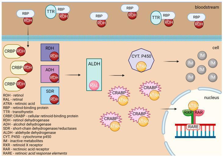
A pathophysiology diagram illustrating the metabolic pathway and cellular signaling of Vitamin A. The diagram is divided into three compartments: the bloodstream, the cytoplasm, and the nucleus. In the bloodstream, Retinol (ROH) is transported bound to Retinol-Binding Protein (RBP) and Transthyretin (TTR). Upon entering the cell, ROH binds to Cellular Retinol-Binding Protein (CRBP). It undergoes enzymatic oxidation via Retinol Dehydrogenase (RDH), Alcohol Dehydrogenase (ADH), or Short-chain Dehydrogenase/Reductases (SDR) to form Retinal (RAL). RAL is further oxidized by Aldehyde Dehydrogenase (ALDH) into All-Trans Retinoic Acid (ATRA). In the cytoplasm, ATRA binds to Cellular Retinoic Acid-Binding Protein (CRABP) or is catabolized by Cytochrome P450 into inactive metabolites (IM). Functionally, ATRA translocates to the nucleus where it binds to Retinoid X Receptor (RXR) and Retinoic Acid Receptor (RAR) complexes. This heterodimer binds to Retinoic Acid Response Elements (RARE) on DNA to regulate gene expression. The diagram uses standard abbreviations and includes a comprehensive legend for enzymes and transport proteins.
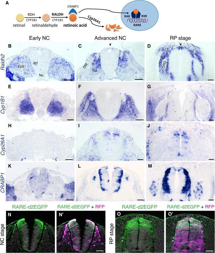
This composite educational figure illustrates the metabolic pathway and spatio-temporal dynamics of retinoic acid (RA) signaling during avian neural tube (NT) development across three stages: early neural crest (NC), advanced NC, and roof plate (RP). Panel A presents a schematic of RA metabolism, highlighting enzymes like RALDH/ALDH1 (synthesis) and CYP26A1 (degradation), carrier protein CRABP1, and the RARE-mediated transcriptional mechanism via RAR/RXR receptors. Panels B–M show in situ hybridization (ISH) results for key metabolic markers. Raldh2 transitions from paraxial mesoderm expression in early stages to strong localized expression in the nascent roof plate. Cyp1B1 remains restricted to the mesoderm. Cyp26A1 is detected in dorsal interneuron progenitors during advanced stages, while CRABP1 appears in lateral RP domains and differentiating neurons. Panels N–O' utilize fluorescence microscopy to visualize RA activity using a RARE-d2EGFP reporter. At both NC and RP stages, GFP signal (RA activity) is concentrated in the dorsal half of the neural tube, contrasted against an RFP control which shows ubiquitous transfection across the dorso-ventral axis. This visual evidence supports the role of local RA production in the transition from NC delamination to RP formation.
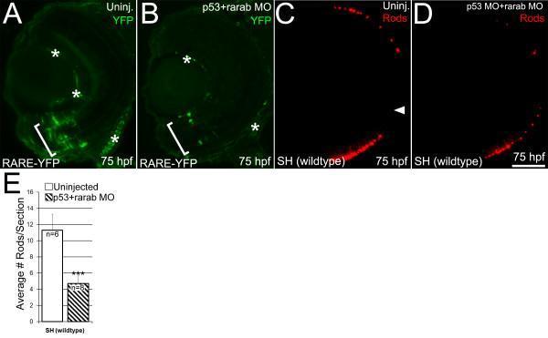
This composite figure illustrates the effect of retinoic acid receptor alpha b (RARαb) knockdown on retinal signaling and rod photoreceptor production in zebrafish embryos at 75 hours post-fertilization (hpf). Panels A and B are immunofluorescence images of RARE-YFP transgenic embryos. Panel A shows robust endogenous RA signaling (green YFP expression) in the ventral retina (bracketed), while Panel B demonstrates a marked reduction in signaling following injection of rarab/p53 morpholino (MO). Asterisks denote non-specific staining. Panels C and D show wildtype (SH) retinal sections labeled with 1D1 rod opsin antibody (red). Uninjected embryos (C) show normal rod distribution, whereas MO-injected embryos (D) exhibit a significant decrease in rod opsin expression. An arrowhead in C identifies the optic nerve head. Panel E is a bar graph quantifying these findings, showing that the average number of rods per section is significantly lower in the p53+rarab MO group (approximately 5 rods/section) compared to the uninjected control (approximately 11 rods/section), with statistical significance indicated by triple asterisks. Scale bar equals 50 μm.
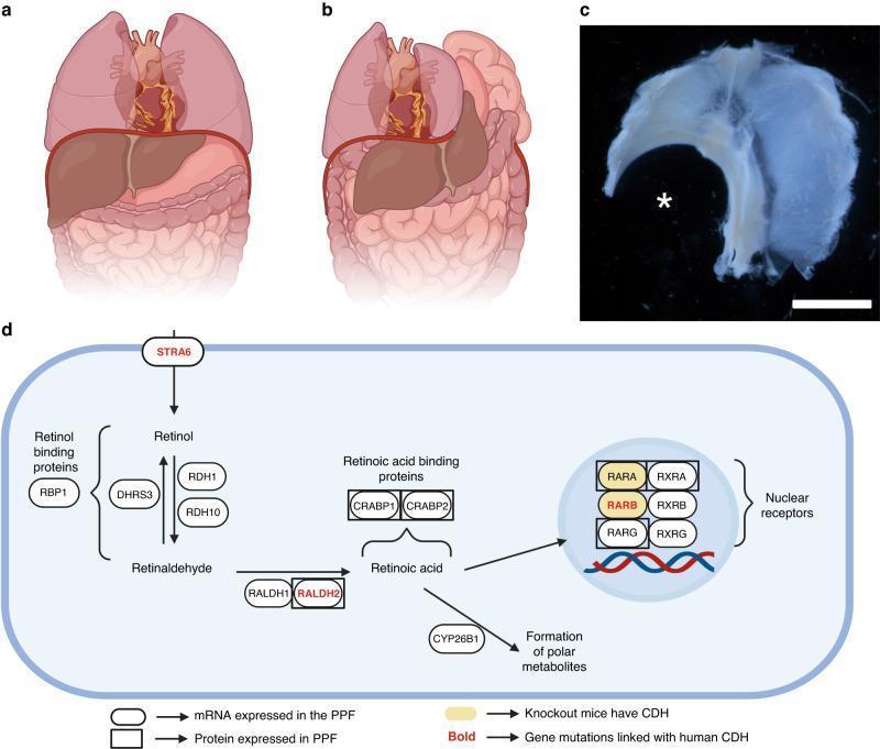
This composite educational graphic illustrates the pathogenesis of Congenital Diaphragmatic Hernia (CDH) and the role of Retinoic Acid (RA) signaling. Panel (a) is an anatomical diagram showing a normal torso with an intact diaphragm separating the thoracic cavity (lungs, heart) from the abdominal cavity (liver, stomach, intestines). Panel (b) depicts CDH, where a diaphragmatic defect allows abdominal viscera to herniate into the chest, causing lung compression and mediastinal shift. Panel (c) is a clinical photograph of a dissected mouse fetal diaphragm (superior view) following teratogen exposure; an asterisk identifies a large, left-sided posterolateral (Bochdalek) defect. Panel (d) is a pathophysiology flowchart of the RA signaling pathway within the pleuroperitoneal fold (PPF). It details biochemical transitions from Retinol to Retinaldehyde and Retinoic Acid, mediated by enzymes like RDH10 and RALDH2. The diagram highlights key molecular components (STRA6, RARB, RXRA) where mutations or knockouts are linked to human CDH or rodent models, illustrating how disrupted gene expression in the PPF leads to structural diaphragmatic failure.
Vitamin A deficiency night blindness xerophthalmia clinical signs
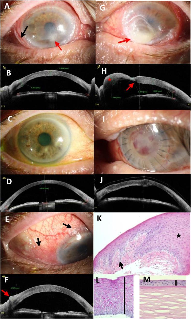
This composite figure illustrates the clinical and diagnostic progression of severe xerophthalmia and keratomalacia secondary to vitamin A deficiency in a 70-year-old male. Panels A, E, and G show clinical photographs highlighting characteristic Bitot's spots (foamy keratinized conjunctival plaques), corneal stromal infiltrates, and advanced keratomalacia with focal bulging (staphyloma formation). Panels B, D, F, H, and J utilize Anterior Segment Optical Coherence Tomography (AS-OCT) to quantify pathology, revealing conjunctival thickening (up to 737 μm), corneal stromal edema (1039 μm), and focal thinning (401 μm). Post-treatment panels (C, D, I, J) demonstrate clinical resolution after vitamin A supplementation and tectonic lamellar keratoplasty, with AS-OCT confirming successful graft adaptation. Histopathological sections (K, L) using HE and PAS stains contrast the diseased cornea against a normal control (M), showing massive epithelial hyperplasia (approximately 20 cell layers), focal keratinization, loss of Bowman's layer, and granular PAS-positive deposits, pathognomonic for squamous metaplasia in the setting of nutritional deficiency.
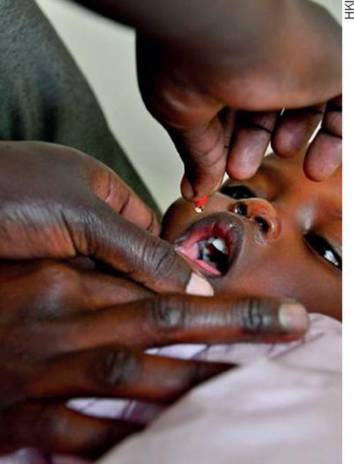
This clinical photograph demonstrates the oral administration of high-dose vitamin A supplementation to a pediatric patient. The image shows an adult health worker or caregiver using a manual technique to ensure safe delivery: one hand gently stabilizes the child's jaw and depresses the lower lip to keep the mouth open, while the other hand squeezes a small capsule or dispenser. A single, translucent red droplet of the liquid supplement is captured in mid-air, directed toward the child's open mouth. The child, appearing to be an infant or toddler, has their mouth open wide, revealing the tongue and primary dentition. This visual illustrates a common public health intervention used in nutritional programs to prevent vitamin A deficiency (VAD) and its associated complications, such as xerophthalmia and increased susceptibility to infections. The focus is on the practical application and administration technique within a community health or pediatric clinical setting.
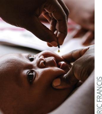
A clinical photograph depicting the oral administration of a medical supplement, specifically Vitamin A, to a four-month-old infant. The infant is positioned supine, with the head slightly elevated and stable. A healthcare worker is shown using their thumb and index finger to gently depress the chin and retract the cheek, ensuring the mouth is sufficiently open for safe delivery. The administrator's other hand holds a small dropper or capsule, from which a single yellow liquid drop is descending into the infant's oral cavity. This visual illustrates a standard public health intervention in pediatrics, aimed at preventing xerophthalmia and reducing child morbidity and mortality associated with vitamin A deficiency in low-income regions. The procedure emphasizes safe manual handling and the technique for oral liquid dosing in infants who cannot yet swallow tablets or coordinate drinking from a cup.
Vitamin A - Biochemistry
1. Overview
- Maintenance of normal vision (especially in dim light)
- Regulation of cell growth and differentiation
- Immune defense via epithelial barrier maintenance
- Reproduction
2. Chemical Forms (Structure)
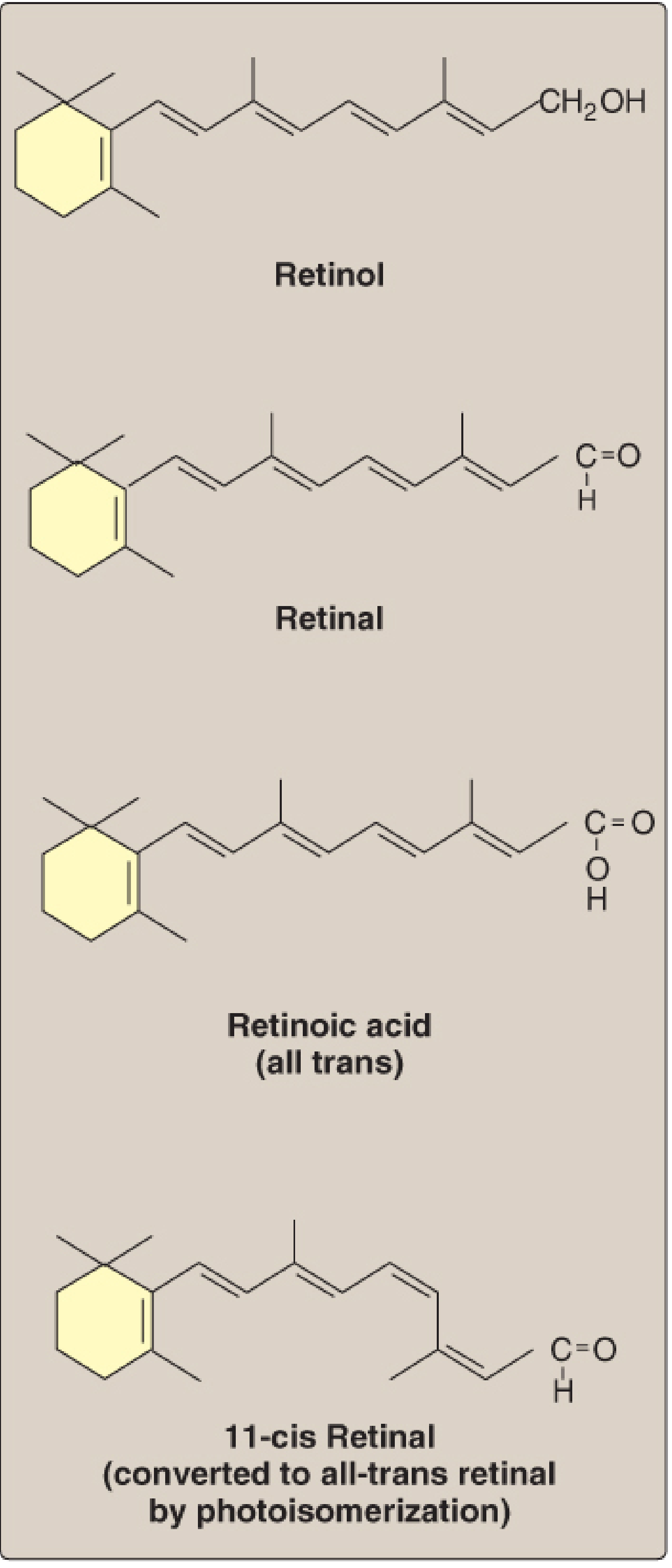
| Form | Chemical Group | Notes |
|---|---|---|
| Retinol | Primary alcohol (-CH₂OH) | Transport and storage form; found as retinyl ester in animal tissues |
| Retinal | Aldehyde (-CHO) | Oxidized from retinol; reversible conversion |
| Retinoic acid | Carboxylic acid (-COOH) | Oxidized from retinal; irreversible - cannot be reduced back |
| 11-cis Retinal | Aldehyde, cis-isomer | Critical form in the visual cycle (rhodopsin component) |
| β-Carotene | Provitamin A | Cleaved symmetrically in the intestine to yield 2 retinal molecules |
Key point: Retinoic acid cannot be reduced back to retinal or retinol, so it is inactive in the visual cycle and reproduction - it only promotes epithelial differentiation.
3. Dietary Sources
- Preformed vitamin A (retinyl esters, retinol): Animal sources - liver, fish, eggs, butter, milk, kidney
- Provitamin A (β-carotene): Plant sources - yellow/orange/dark-green vegetables (carrots, squash, spinach, broccoli)
4. Absorption, Transport, and Storage
-
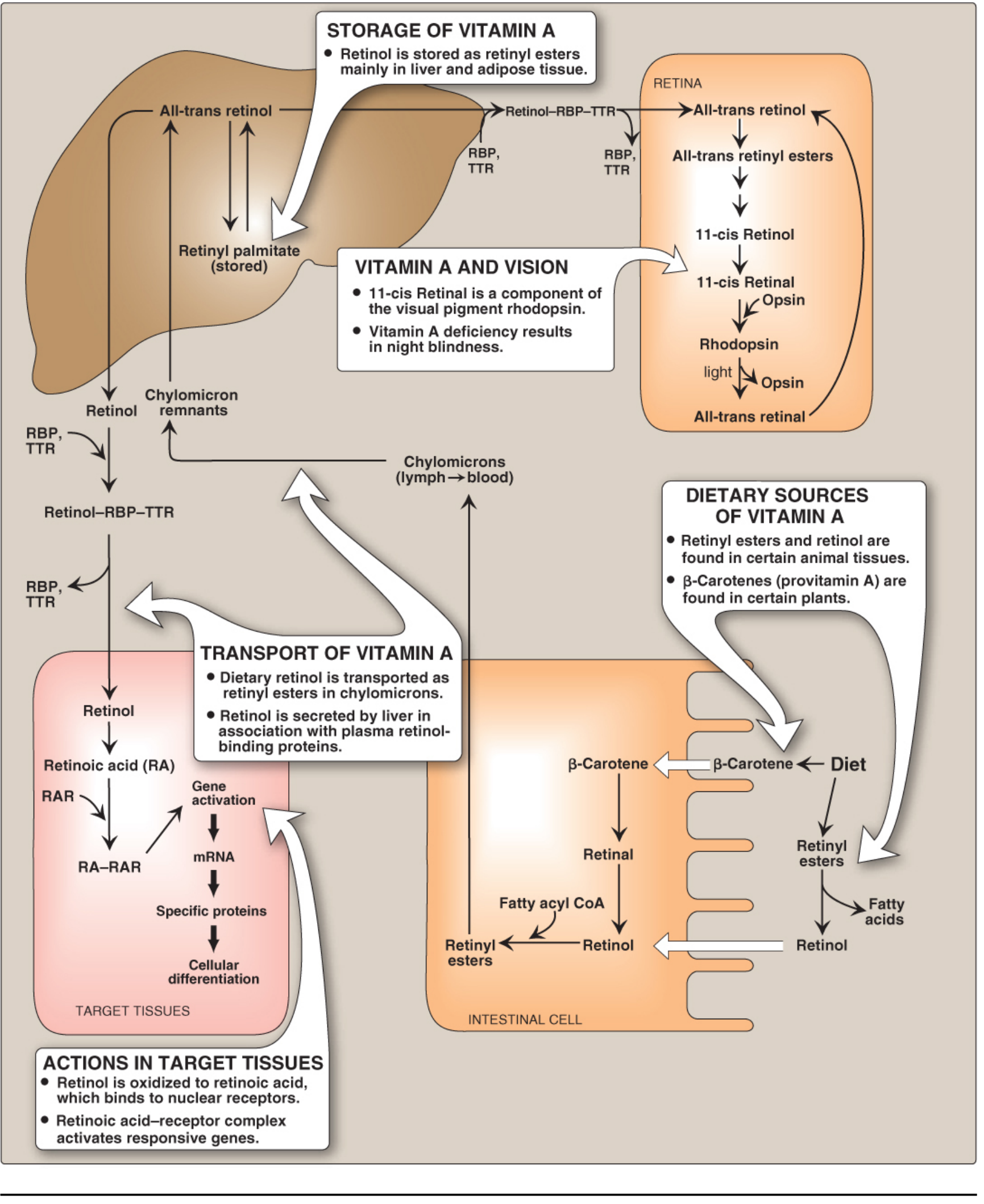
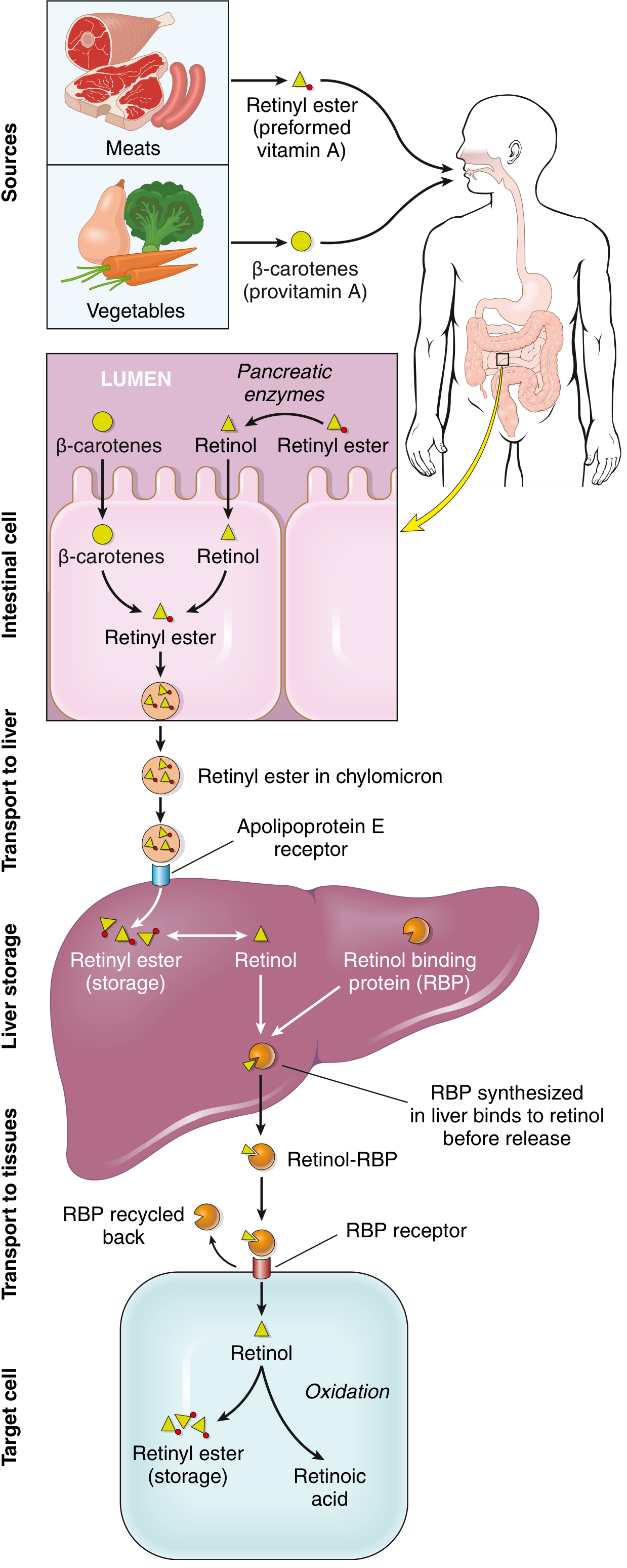
Intestinal lumen: Retinyl esters are hydrolyzed by pancreatic enzymes (requires bile salts). β-Carotene is oxidatively cleaved to retinal, then reduced to retinol.
-
Enterocytes: Retinol is re-esterified with long-chain fatty acids (fatty acyl-CoA) to form retinyl esters → packaged into chylomicrons → secreted into lymph → blood.
-
Liver uptake: Chylomicron remnants are taken up via the apolipoprotein E receptor. Over 90% of body reserves are stored in the liver, mainly in perisinusoidal stellate (Ito) cells as retinyl palmitate. These reserves last at least 6 months in healthy individuals.
-
Release from liver: When needed, retinol is bound to retinol-binding protein (RBP), which complexes with transthyretin (TTR). This retinol-RBP-TTR ternary complex travels in plasma to peripheral tissues.
-
Peripheral tissue uptake: Cells have surface RBP receptors. After retinol enters, RBP is recycled back to blood. Intracellular retinol-binding proteins carry retinol to the nucleus.
5. Mechanism of Action - Retinoic Acid and Nuclear Receptors
- In target cells, retinol is oxidized to retinoic acid (RA)
- RA binds with high affinity to nuclear Retinoic Acid Receptors (RARs)
- The RA-RAR complex forms a heterodimer with Retinoid X Receptor (RXR)
- The RAR/RXR heterodimer binds to Retinoic Acid Response Elements (RAREs) in DNA
- This recruits transcriptional activators or repressors → regulates specific mRNA synthesis → controls production of specific proteins
- Keratin gene expression in epithelial tissues
- Receptors for growth factors
- Tumor suppressor genes
- Cell-cycle regulators
- PPARs (peroxisome proliferator-activated receptors) - controlling fatty acid oxidation, adipogenesis, lipoprotein metabolism
- Vitamin D receptors - explaining the interplay between these fat-soluble vitamins
6. Functions
6a. Visual Cycle (Dim Light Vision)
- Rhodopsin (in rod cells) = 11-cis retinal + opsin protein (a G protein-coupled receptor)
- Light hits rhodopsin → photochemical isomerization → 11-cis retinal converts to all-trans retinal → rhodopsin "bleaches" → retinal dissociates from opsin
- Conformational change activates the G protein transducin → triggers a nerve impulse → optic nerve → brain
- Regeneration: all-trans retinal is reduced to all-trans retinol → esterified → isomerized to 11-cis retinol → oxidized back to 11-cis retinal → combines with opsin → rhodopsin reformed
6b. Epithelial Differentiation
- Conjunctiva and cornea (eye)
- Respiratory tract (loss of mucociliary lining)
- Urinary tract (desquamation → stones)
6c. Reproduction
- Retinol and retinal support spermatogenesis and prevent fetal resorption
- Retinoic acid supports epithelial differentiation but is inactive in reproduction and vision
6d. Immune Function
- Vitamin A supplementation reduces diarrhea morbidity by ~15% and mortality by ~30%
- Promotes regeneration of damaged epithelia (first line of defense)
- Supports optimal immune function through uncertain mechanisms
6e. Metabolic Effects
- Retinoids inhibit adipogenesis and stimulate lipid breakdown
- RXR-PPAR heterodimers mediate these metabolic effects on fat tissue
7. Deficiency
| Stage | Finding |
|---|---|
| Early | Night blindness (nyctalopia) - raised visual threshold in dim light |
| Progressive | Xerosis conjunctivae - dryness of conjunctiva |
| More advanced | Bitot spots - foamy, keratinized opaque plaques on conjunctiva |
| Severe | Keratomalacia - corneal ulceration, softening, and destruction |
| End stage | Blindness from opaque corneal scar tissue |
- Squamous metaplasia of respiratory epithelium → secondary pulmonary infections
- Metaplasia of urinary tract → renal/bladder stones
- Follicular or papular dermatosis (skin)
- Impaired immune function → increased infection mortality
8. Toxicity (Hypervitaminosis A)
9. Clinical Applications of Retinoids
| Drug | Form | Use |
|---|---|---|
| Tretinoin (all-trans retinoic acid) | Topical | Acne vulgaris, mild skin aging |
| Tretinoin (oral) | Oral | Acute promyelocytic leukemia (APL) - ATRA therapy |
| Isotretinoin (13-cis retinoic acid) | Oral | Severe cystic acne |
| Acitretin (synthetic retinoid) | Oral | Psoriasis |
Summary Pathway
Diet (retinyl esters, β-carotene)
         ↓  intestinal cleavage/hydrolysis
     Retinol (enterocyte)
         ↓  re-esterification + chylomicron
     Liver (storage as retinyl palmitate in Ito cells)
         ↓  release bound to RBP-TTR
     Target cell
      ↙              ↘
Retinal             Retinoic acid (RAR → gene regulation)
  ↓ (11-cis)
Rhodopsin (vision)
Create a study guide or cheat sheet on vitamin A metabolism
What format would you like the study guide in?