
why treating syphilis causes more harm then benifit
The phenomenon you're describing is the Jarisch-Herxheimer reaction - a well-known adverse effect when treating syphilis (and other spirochetal infections). It's not that treatment is harmful overall, but the initial treatment can cause a sudden, sometimes dangerous reaction.
Searching sources...
Reading File
Searching the Web
Jarisch-Herxheimer reaction syphilis treatment mechanism
Finding Sources
Finding Sources
Reading File
Reading File
Great - I now have excellent textbook detail from Harrison's and Creasy & Resnik's. Here's the full answer:
The Jarisch-Herxheimer Reaction (JHR) - Why Treating Syphilis Can Temporarily Feel Like It's Making Things Worse
The short answer: treating syphilis does not cause more harm than benefit overall. But a well-known reaction called the Jarisch-Herxheimer reaction (JHR) can cause a sudden, dramatic worsening of symptoms within hours of the first antibiotic dose - which is the likely source of this idea.
What is the Jarisch-Herxheimer Reaction?
When antibiotics (usually benzathine penicillin G) kill Treponema pallidum rapidly, the dying bacteria release lipoproteins and other breakdown products that trigger a massive immune (inflammatory) response. This is NOT an allergic reaction to penicillin - it is your immune system reacting to bacterial debris flooding the bloodstream.
"This reaction is thought to be a response to lipoproteins released by dying T. pallidum organisms."
- Harrison's Principles of Internal Medicine, 22E
How Common Is It?
| Stage of Syphilis | JHR Frequency |
|---|---|
| Primary syphilis | ~50% of patients |
| Secondary syphilis | ~90% of patients |
| Late/latent syphilis | Less common |
| General paresis (neurosyphilis) | Up to 75% |
Symptoms - What the Patient Experiences
Within 1-8 hours of the first dose:
- Sudden high fever and chills/rigors
- Severe headache and myalgia (muscle aches)
- Tachycardia and rapid breathing
- Hypotension (low blood pressure)
- Worsening of skin lesions (rashes look worse, become more red and swollen)
- Anxiety
These symptoms typically resolve within 12-24 hours on their own without stopping treatment.
"A dramatic although self-limited reaction consisting of fever, chills, myalgia, headache, tachycardia, and increased respiratory rate may follow the initiation of treatment for syphilis."
- Harrison's Principles of Internal Medicine, 22E
When Is It Actually Dangerous?
For most patients, JHR is unpleasant but harmless. However, two groups face real risk:
1. Pregnant Women
This is where JHR can be genuinely life-threatening to the fetus:
- In one study of 33 pregnant women, JHR complicated therapy in 100% of those with primary syphilis and 60% with secondary syphilis
- The most common findings were: fever (73%), uterine contractions (67%), and decreased fetal movement (67%)
- Transient late fetal heart rate decelerations were observed in 30% of monitored fetuses
- JHR can trigger preterm labor or fetal distress
"Obstetrical caregivers should be aware that women treated for syphilis during the second half of pregnancy are at risk for preterm labor or fetal distress if the Jarisch-Herxheimer reaction occurs."
- Creasy & Resnik's Maternal-Fetal Medicine
Despite this risk, the textbook is clear: the devastation of congenital syphilis is far worse, so treatment must not be withheld. Pregnant women in the second half of pregnancy need close fetal monitoring (ultrasound before treatment, hospital monitoring after the first dose).
2. Neurosyphilis / Late-Stage Disease
Patients with neurosyphilis or late complications can have more severe hemodynamic instability during JHR.
Why Treatment Is Still Always the Right Call
The JHR is:
- Transient - resolves within 24 hours
- Self-limited - does not require stopping antibiotics
- Manageable - with supportive care (antipyretics, fluids, monitoring)
- NOT a reason to withhold treatment
Untreated syphilis causes: cardiovascular syphilis (aortic aneurysm), neurosyphilis (dementia, paralysis, blindness), congenital syphilis (stillbirth, bone/organ destruction in the newborn), and death.
Key Takeaway
The phrase "more harm than benefit" is a misunderstanding. The JHR is a predictable, temporary immune flare that results from successful treatment working. Patients should be warned beforehand, monitored appropriately (especially if pregnant), and given symptomatic support. The benefits of curing syphilis vastly outweigh the temporary discomfort of JHR.
Management of JHR:
- Reassure the patient it is expected and temporary
- Antipyretics (e.g. paracetamol/acetaminophen) for fever and myalgia
- IV fluids if hypotensive
- Do NOT stop the antibiotic course
- For pregnant women: hospitalize, fetal monitoring, obstetric review
Sources: Harrison's Principles of Internal Medicine 22E, Creasy & Resnik's Maternal-Fetal Medicine; StatPearls - JHR
st segment elevation in pericarditis diff with stemi
Differentiating pericarditis ST elevation from STEMI is a classic ECG challenge. Key points: pericarditis causes diffuse (widespread) ST elevation in most leads, is concave ("saddle-shaped") upward, and lacks reciprocal changes - while STEMI is localized, convex ("tombstone"), and has reciprocal ST depression in opposite leads.
Searching sources...
Finding Sources
Searching the Web
pericarditis vs STEMI ECG differentiation ST elevation features 2024
Finding Sources
Finding Sources
Reading File
Reading File
Reading File
Reading File
Finding Sources
Finding Sources
Finding Sources
Reading File
Reading File
Excellent - I now have very detailed, authoritative textbook data. Let me also fetch the ECG diagram image from Rosen's.
Loading Image
Here is a complete, detailed comparison synthesized from multiple authoritative textbooks (Fuster & Hurst's The Heart, Rosen's Emergency Medicine, Goldman-Cecil Medicine, Braunwald's Heart Disease):
ST Elevation in Pericarditis vs. STEMI - ECG Differentiation
The Core Problem
Both conditions elevate the ST segment and both can cause chest pain with troponin elevation - making this one of the most clinically dangerous ECG mimics. Missing a STEMI means no reperfusion. Misdiagnosing pericarditis as STEMI leads to unnecessary catheterization and thrombolysis.
Side-by-Side ECG Comparison Table
(From Fuster & Hurst's The Heart, 15th Ed, Table 53-5)
| ECG Feature | Acute Pericarditis | STEMI |
|---|---|---|
| ST morphology | Concave upward ("saddle-shaped") | Convex ("tombstone") - upward bulge |
| Lead distribution | Diffuse / widespread - most leads (I, II, III, aVF, V2-V6) | Localized - territory of occluded artery |
| Reciprocal ST depression | Absent (only aVR shows ST depression) | Present - hallmark finding |
| PR segment depression | Yes - highly specific for pericarditis (best seen in lead II and V6) | No |
| PR elevation in aVR | Yes - mirror of PR depression | No |
| Q waves | Never develops Q waves | Q waves may develop (irreversible necrosis) |
| T-wave inversion timing | Occurs after ST segment normalizes (Stage 3) | Occurs during or alongside ST elevation |
| aVR changes | ST depression + PR elevation in aVR | ST elevation in aVR (suggests left main/proximal LAD occlusion) |
| V1 | Variable or spared | Elevated in anterior STEMI |
| ST/T ratio in V6 | >0.25 (useful to differentiate from early repolarization) | Variable |
| QT prolongation | No | Possible |
| Evolution | Gradual over days (4 stages) | Rapid - minutes to hours |
Key ECG Hallmarks - Explained
1. Shape of ST Elevation (Most Important Single Sign)
- Pericarditis: The ST segment curves upward concavely - like a "smiley face" or saddle. The J point lifts and the segment slopes gently upward.
- STEMI: The ST segment is convex - it domes or bulges upward like a tombstone. This reflects the current of injury from transmural ischemia.
"Convex or domed ST segment morphology strongly suggests STEMI."
- Rosen's Emergency Medicine
2. Lead Distribution (Extremely Useful)
- Pericarditis: ST elevation is diffuse - present in almost all leads (I, II, III, aVL, aVF, V2-V6). This is because the entire epicardium is inflamed.
- Except: ST is depressed (not elevated) in aVR - a signature finding.
- V1 is variable or spared.
- STEMI: ST elevation is localized to the leads corresponding to the culprit artery's territory:
- Inferior STEMI: II, III, aVF (RCA)
- Anterior STEMI: V1-V4 (LAD)
- Lateral STEMI: I, aVL, V5-V6 (LCX)
3. Reciprocal Changes (Most Specific for STEMI)
- STEMI: Leads opposite to the infarct zone show ST depression - this is electrical reciprocity from the vector of injury.
- e.g., Inferior STEMI (STE in II, III, aVF) → ST depression in I, aVL
- "New primary ST depression in aVL is 99% sensitive for inferior OMI" - Emergency Medicine Cases
- Pericarditis: No reciprocal changes except the expected depression in aVR. Any ST depression in limb leads (other than aVR) virtually excludes pericarditis.
4. PR Segment Depression (Pathognomonic for Pericarditis)
- Pericarditis: Atrial epicarditis causes the PR segment to deviate. PR depression is seen in inferior leads and V6, with PR elevation in aVR.
- It is insensitive (~60%) but highly specific for pericarditis.
- STEMI: PR depression does NOT occur.
"PR segment depression is an insensitive yet specific associated electrocardiographic finding in pericarditis."
- Rosen's Emergency Medicine
ECG Waveform Patterns (from Rosen's Emergency Medicine)
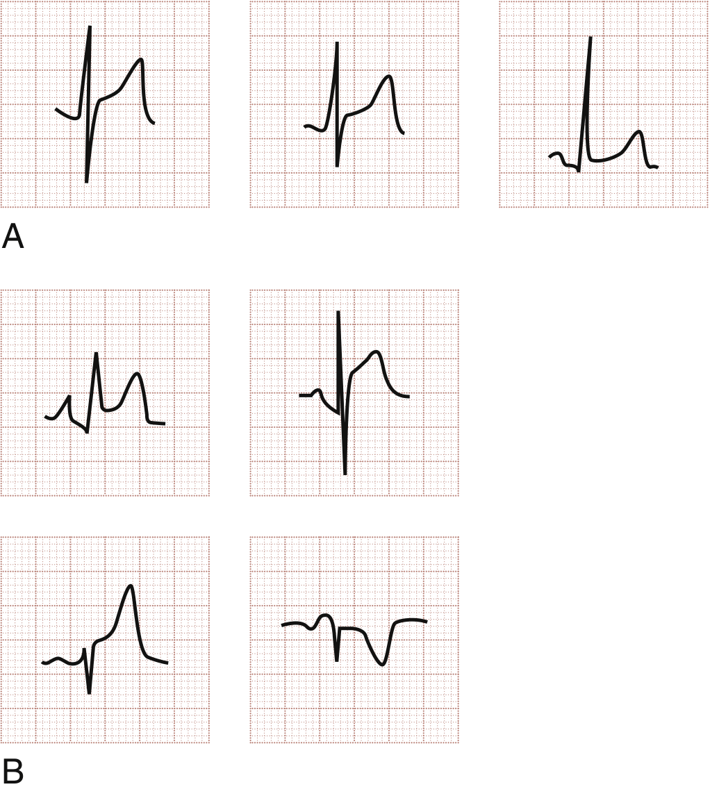
Fig: Row A = Benign early repolarization (concave). Row B = Pericarditis patterns: concave STE with PR depression (top), concave STE without PR changes (bottom left), and lead aVR showing ST depression + PR elevation (bottom right - the mirror image)
The 4 ECG Stages of Pericarditis
| Stage | Timing | Changes |
|---|---|---|
| Stage 1 | Hours to days | Diffuse concave ST elevation + PR depression (can mimic STEMI) |
| Stage 2 | Days | ST normalizes, PR returns to baseline |
| Stage 3 | Days to weeks | Diffuse T-wave inversion (after ST normalizes - key difference from STEMI) |
| Stage 4 | Weeks | ECG normalizes |
Less than 50% of patients go through all four stages. In STEMI, T-wave inversion occurs during ST elevation - in pericarditis it comes after the ST normalizes.
Clinical (Non-ECG) Features That Help Differentiate
| Feature | Pericarditis | STEMI |
|---|---|---|
| Pain character | Sharp, pleuritic (worse with inspiration, lying flat) | Crushing, pressure, squeezing |
| Position effect | Relieved by sitting forward | No positional change |
| Radiation | To trapezius ridge (phrenic nerve irritation) | Left arm, jaw, shoulder |
| Pericardial rub | May be present (scratchy, 3-component) | Absent |
| Fever/CRP | Common (viral/inflammatory etiology) | Absent initially |
| Troponin | Mildly elevated - normalizes in 1-2 weeks | Often markedly elevated, rises and falls |
| Echo | Pericardial effusion; normal wall motion | Regional wall motion abnormality |
| Response to NSAIDs | Good pain relief | No relief |
The "Spodick's Ratio" Trick
In lead V6, the ratio of ST elevation amplitude / T-wave amplitude:
- > 0.25 = suggests pericarditis
- < 0.25 = suggests benign early repolarization
This is particularly useful when differentiating pericarditis from early repolarization (both show concave ST elevation), but early repolarization does NOT have PR depression.
Clinical Bottom Line
The single most reliable rule:
Any ST depression in leads other than aVR essentially excludes pericarditis and should be treated as STEMI until proven otherwise. If the patient has reciprocal ST depression in any limb lead, activate the cath lab.
Conversely, if you see:
- Diffuse ST elevation in most leads
- Concave ("smiley face") morphology
- PR depression in II/V6, PR elevation in aVR
- No reciprocal changes
- Pleuritic chest pain relieved by leaning forward
...think pericarditis, not STEMI.
Sources: Fuster & Hurst's The Heart 15E, Rosen's Emergency Medicine, Goldman-Cecil Medicine, Braunwald's Heart Disease 2E, Symptom to Diagnosis 4E
This is a shared conversation. Sign in to Orris to start your own chat.