
Bcg vaccine in 2 months, baby development wound with mucus like pimple
- BCG is given at birth (or early infancy), and around 2-6 weeks later a small red papule forms at the injection site.
- By about 2 months, this papule often breaks down into a small ulcer or pustule that may ooze a whitish/mucus-like discharge.
- The site then crusts over and heals, leaving the characteristic small scar.
- This process is not an infection and does not need antibiotics or dressing - it should be left open to air.
BCG vaccine site reaction infant pustule nodule normal
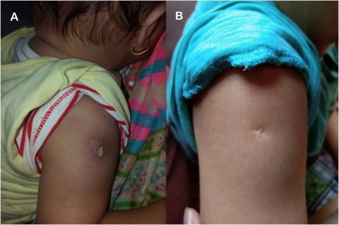
Clinical photograph comparison in a pediatric patient showing the evolution of a BCG (Bacillus Calmette-Guérin) vaccine inoculation site on the right deltoid. Figure A demonstrates a local adverse reaction characterized by an erythematous, indurated area with active purulent discharge emanating from a central pustule. This represents the acute inflammatory phase or a localized abscess. Figure B shows the same anatomical site two months later, following wound management. The inflammatory features—erythema and discharge—have completely resolved, replaced by a characteristic, small, hypopigmented atrophic scar. The images illustrate the progression from an active localized complication to a healed BCG scar, providing a visual reference for clinical monitoring of vaccination sites.
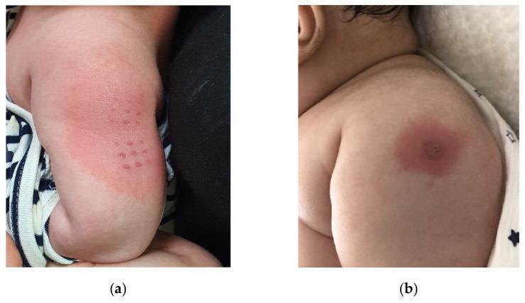
A clinical photograph comparison of two different types of local skin reactions (BCGitis) at the Bacille Calmette-Guerin (BCG) inoculation site on the upper arm of pediatric patients. Image (a) demonstrates erythema following the transdermal delivery method, characterized by a diffuse, irregular patch of redness on the deltoid region containing a grid-like pattern of multiple small, discrete erythematous papules corresponding to the multi-puncture device application. Image (b) shows the reaction following the intradermal delivery method, presenting as a localized, circular erythematous plaque with a solitary, central crusting papule or pustule, often described as a bullseye appearance. These visual manifestations are clinically significant as markers for Kawasaki disease (KD) when observed in febrile infants, representing a specific cutaneous finding known as BCG site reactivation. The image serves as an educational tool for distinguishing between normal and pathological vaccine site inflammation across different administration routes in pediatric dermatology and immunology.
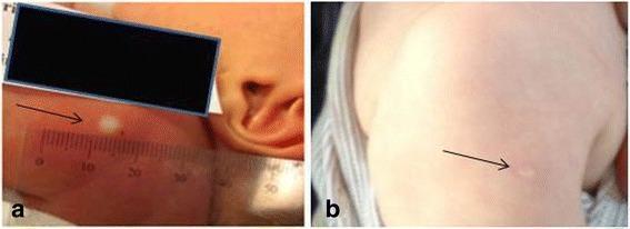
This clinical photograph set illustrates the progression of a Bacillus Calmette-Guérin (BCG) vaccination site on an infant, demonstrating the transition from an immediate skin reaction to long-term scarring. Panel (a) shows a neonatal skin reaction immediately following intradermal injection. A characteristic raised, blanched papule or 'wheal' is visible, representing the dermal deposit of the vaccine. The diameter of this wheal is being objectively measured using a transparent millimeter ruler to assess the success of the administration. Panel (b) depicts the same anatomical region—the upper left arm—at 13 months of age. It shows a definitive BCG scar, which is characterized by a round, flat, and hypopigmented area relative to the surrounding tissue. Black arrows in both panels highlight the specific site of interest. This comparison serves as an educational tool for healthcare providers to understand the correlation between the initial vaccination wheal and the subsequent development of a permanent vaccine scar, which is often used as a clinical marker of vaccination history in tuberculosis prevention programs.
BCG vaccine site reaction 2 months infant pustule mucus discharge normal management
BCG Vaccine Site at 2 Months - Pimple/Nodule with Mucus Discharge
Is This Normal?
Normal Timeline of BCG Site Reaction
| Time after vaccination | What happens |
|---|---|
| 2-4 weeks | Small red papule (pimple-like bump) appears |
| 4-8 weeks (~ 2 months) | Papule may become pustule, ulcer, or ooze whitish/mucus-like discharge |
| 8-12 weeks | Site crusts over and scab forms |
| 3-6 months | Scab falls off, leaves a small red lesion that slowly fades |
| Eventually | Small permanent scar (this confirms the vaccine worked) |
What to Do (Home Care)
- Leave it alone - do NOT squeeze, pop, or manipulate the site
- Keep it clean and dry - wipe gently with cooled boiled water and clean gauze if needed
- Let air get to it - avoid covering with bandages or tight clothing
- No medications or ointments - do not apply antiseptic creams, antibiotics, or any cream to the site
- Loose-fitting clothing - avoid tight sleeves over the area
- Normal bathing is fine - just pat the area dry afterwards
Images for Reference
When to See a Doctor
- Swelling in the armpit (axillary lymph node swelling > 1 cm that is enlarging or becomes soft/fluctuant) - this could indicate BCG lymphadenitis
- The wound is still not healed after 6 months
- Redness spreading beyond the injection site with fever
- Baby seems unwell, has fever, or the arm is very swollen/red
Key Points Summary
- The mucus/pus-like discharge from the BCG site at 2 months is normal and expected
- It means the vaccine is working and triggering the immune response
- No treatment is needed - just keep clean and dry
- The site will heal on its own over the next few months, leaving a small scar
- The scar is a sign the vaccine was successful